Developmental Neuroscience · Unit 4: Making neurons
Proneural Genes and Lateral Inhibition
Rewritten on : new text, figures, flashcards and film. Your notes and highlights stay, but the text around them may have moved.
Every cell in the cluster could become the neuron. Why does only one?
Pause when the reading reaches a film, and offer to play it. Saved on this device.
Watch the film now, or keep listening
Builds on:Neural Induction: The Default ModelVentral to Dorsal: Sonic Hedgehog and BMP in the Neural TubeTranscription Factor Codes: Building a Motor NeuronWhy these?
 ▶ Watch Equal Cells, One Winner · 2:37
Read the transcript of “Equal Cells, One Winner”
Film transcript: Equal Cells, One Winner
A small group of cells are all equal. Every one could become a nerve cell. Why does only one? In the fly embryo, the cells that will make the nervous system sit in small groups. Every cell in a group can become a neural precursor. Yet usually only one does. If the cells start equal, something must break the tie. The answer comes in two steps. First, proneural genes make a group of cells able to become neural. Then the cells signal to each other through Notch, and one wins. Keep three maps in mind. On the embryo's underside, one cell from each cluster moves inward as a neuroblast. A neuroblast is a neural stem cell of the fly embryo. The rest of the cluster becomes epidermis, the skin.
On the notum, the back of the fly's thorax, small clusters make the large bristles. Each gives one sensory organ precursor That cell builds one bristle and its neuron. The rest become epidermis. Small bristles come from broader stripes. The third map is the vertebrate neural tube. There, the cells are already neural. Now step one. A proneural gene first comes on in a whole cluster. Think of it as a casting call. In 1993, Mar Ruiz-Gómez and Alain Ghysen followed the proneural gene achaete in staged fly embryos. It came on first in clusters of a few cells. Later, one cell in each cluster held much more achaete RNA. That cell became the neural progenitor.
They called the first state competent, and the second determined. Predict. Take Notch away from the embryo. What happens to the clusters? The clusters still form in their normal places. But high achaete is no longer limited to one cell. So Notch does not place the clusters. It narrows them. Placing and picking are separate events. That leaves three puzzles. Who may become neural? Who does? And does the logic carry over to vertebrates? Here is the idea to carry. In the fly, proneural factors say which cells may become neural. Notch says which one does.
From the film
- In the fly, proneural genes make a group of cells able to become neural, and Notch signaling then picks which one does.
- Notch does not place the clusters; it narrows them, so placing a cluster and picking a cell within it are separate events.
- The same logic applies on three maps: the fly embryo's ectoderm, the fly notum and the vertebrate neural tube.
In the fly embryo, the cells that will make the nervous system sit in small groups. Every cell in a group can become a neural precursor, yet usually only one does.
If the cells are equivalent at the start, something must break the tie. This lesson asks what does it, what it takes to work, and what changes when the same parts are used in the vertebrate neural tube.
The layout
The arguments in this lesson happen on three maps, which the film at the top of the page draws. Keep track of which map each experiment uses.
- The fly embryo’s ectoderm. On the underside lies the ventral neuroectoderm, a sheet of ectoderm that can make nerve cells or skin. Proneural genes come on in small clusters of a few cells. From each cluster one cell moves inward as a neuroblast, a neural stem cell of the fly embryo. The rest stay at the surface and become epidermis, the skin of the underside. Elsewhere in the ectoderm, clusters give the precursors of the larva’s sense organs in the same way.
- The fly notum, the back of the fly’s thorax, where bristles grow. Here each small proneural cluster that makes a large bristle gives one sensory organ precursor (SOP), the cell that will build one bristle and its neuron. The rest of the cluster becomes epidermis. Small bristles come instead from broader proneural stripes, each giving many SOPs.
- The vertebrate neural tube. Here the cells are already neural: progenitors, dividing cells that make neurons.
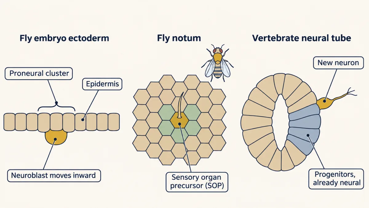
The maps raise three puzzles.
- Who may? Every cell in a cluster could become the precursor. What makes them able, and what decides where the clusters form?
- Who does? If the cells start equal, what breaks the tie, and is the tie always broken?
- Does it carry over? In the neural tube, where cells are already neural, what do the same parts do, and does the proneural factor also decide what kind of neuron forms?
The one idea to carry through this lesson. In the fly, proneural factors say which cells may become neural; Notch says which one does, by comparing each cell with its neighbours. Between two touching cells that start alike, the comparison breaks the tie only if the loop is steep: a small difference must come back round the loop larger than it left.
Step 1: a cluster becomes competent
Read the transcript of “The Casting Call”
Film transcript: The Casting Call
Every cell in a cluster can audition. What gives them that ability? The proneural genes were found in the fly. Two of them, achaete and scute, sit side by side on a chromosome. They are needed for the bristles on the fly's back. Predict. Remove these genes. What happens to the bristles? Then switch them on in extra places.
Remove them, and bristles are missing. Switch them on in extra places, and extra bristles form. A related gene, atonal, was found in 1993. It does the same job for chordotonal organs. These are internal stretch receptors. So different proneural genes start different kinds of sense organ. Force achaete-scute on, and only external sense organs form. Force atonal on, and mostly chordotonal organs form. A year later, atonal was shown to be needed for R8. That is the founding photoreceptor of each eye unit. Without atonal, no R8 forms. The other seven photoreceptors, which R8 recruits, do not form either. All these genes encode the same kind of protein: a basic helix-loop-helix (bHLH) transcription factor.
The helix-loop-helix part lets two proteins pair. The basic part grips DNA at a short sequence called an E-box. A proneural protein works with a partner. In the fly, that partner is Daughterless. In vertebrates, the partners are called E proteins. Vertebrates have relatives of both fly families. Ascl1 is related to achaete-scute. The Neurogenins, and a gene called Atoh1, are related to atonal. So what decides where a cluster forms? In the embryo, clusters still form in the right places without Notch. Regulatory DNA around the achaete-scute genes sets the map. That map was worked out mostly on the notum and wing. There, Notch also helps shape the broader proneural stripes.
That answers the first puzzle. Proneural factors make a cluster able to become neural. Regulatory DNA places it. Proneural factors say who may. Notch will say who does.
From the film
- Proneural genes such as achaete and scute make cells in a cluster able to become neural: remove them and bristles are missing, and switch them on in extra places and extra bristles form.
- Different proneural genes tend to start different kinds of sense organ: achaete-scute mostly external ones, atonal mostly chordotonal organs.
- Proneural proteins are basic helix-loop-helix transcription factors that work as a pair with a partner, Daughterless in the fly and E proteins in vertebrates.
- In the embryo regulatory DNA places the clusters, so proneural factors say who may become neural and Notch says who does.
The proneural genes were found in the fly. achaete and scute, neighbours in a stretch of chromosome called the achaete-scute complex, are needed for bristles: remove them and bristles are missing; switch them on in extra places and extra bristles form. atonal, reported in 1993 by Andrew Jarman, Yves Grau, Lily Jan and Yuh Nung Jan, does the same job for chordotonal organs, internal stretch receptors. In 1994 Jarman, Ellsworth Grell, Larry Ackerman and the Jans showed it is needed for R8, the founding photoreceptor of each unit of the eye.
All of these genes encode basic helix-loop-helix (bHLH) transcription factors. The helix-loop-helix part lets two proteins pair; the basic part grips DNA at a short sequence called an E-box. A proneural protein works as a pair with a partner, Daughterless in the fly and E proteins in vertebrates. Vertebrates have relatives of both fly families: Ascl1 (once called Mash1) is related to achaete-scute, and the Neurogenins and Atoh1 to atonal.
A proneural gene first comes on in a cluster of cells. Think of it as a casting call: everyone in the cluster gets to audition. In 1993 Mar Ruiz-Gómez and Alain Ghysen followed achaete in the ectoderm of staged fly embryos. It came on in clusters of a few cells; later one cell in each cluster held much more achaete RNA and became the neural progenitor. They called the first state competent and the second determined.
Is Notch part of placing the cluster, or only of picking within it? Ruiz-Gómez and Ghysen looked at embryos lacking Notch.
Predict. In an embryo with no Notch, what happens to the achaete clusters? Do they form in the wrong places, fail to form, or something else?
Check your prediction
The clusters still formed in their normal places, but high achaete expression was no longer restricted to one cell in each cluster. So Notch does not place the clusters; it narrows them. Placing the cluster and picking within it are separate events, and where the clusters form is set by regulatory DNA around the achaete-scute genes.
That answers the first puzzle.
Step 2: one cell is picked
Read the transcript of “The Loop, One Arrow at a Time”
Film transcript: The Loop, One Arrow at a Time
Every cell in the cluster now has proneural factor. So what picks one? The cells signal to each other through Notch. The signal says, not you. Delta is a protein in the membrane of one cell. It binds Notch on the cell it touches, and pulls. Two cuts then free the inner part of Notch. That part enters the nucleus. There, with a DNA-binding protein called CSL, it turns on target genes. In a proneural cluster, the targets that matter are the Enhancer of split, genes. They make repressors that switch proneural genes off. Proneural factors do two things in return. They raise their own expression. And, in the standard model, they raise Delta. Now follow two neighbours, A and B, one step at a time.
Step one. A has a little more Delta. B gets more Notch activity, so B makes more repressor. Step two. B is repressed. It has less proneural factor, so it makes less Delta. Step three. The signal coming back to A is weaker. A makes less repressor, and more proneural factor. So A makes more Delta. The returning signal pushes A's Delta up again, in the direction of its original lead. Whether the lead ends up bigger depends on how steep the loop is. That comes next. Each cell inhibits its neighbour. So A inhibits the cell that inhibits A. Two inhibitions in a row act as positive feedback. This is a switch-making arrangement. If the loop is steep enough, noise gives one cell a lead, and the loop magnifies it.
In this ideal pair, which cell wins is chance. Predict. Neither cell has any Notch. What happens to the pair? Neither cell can be told, not you. Both keep proneural genes on, and both become neural. Too many neural cells. Fly geneticists call this the neurogenic phenotype, here in miniature. Now the reverse. Switch Notch on in both cells, whatever Delta they receive. Repressor stays high in both. Neither becomes neural, and the cluster gives no precursor. A double negative between neighbours acts as positive feedback. A small lead can grow.
From the film
- Delta on one cell activates Notch on its neighbour, which turns on Enhancer of split repressors that switch off proneural genes.
- Each cell inhibits its neighbour, so A inhibits the cell that inhibits A; two inhibitions in a row act as positive feedback, and a small lead can grow.
- In an idealised pair, with no Notch in either cell both become neural, and with Notch switched on in both neither does.
What picks one cell from the cluster? The cells signal to each other through Notch, and the signal says “not you”.
You met Notch in the Notch signaling lesson. Delta, a membrane protein on one cell, binds Notch on the cell it touches and pulls. Two cuts then free the inner part of Notch, which enters the nucleus and, with the DNA-binding protein CSL (called Suppressor of Hairless in the fly), turns on target genes.
In a proneural cluster the targets that matter are the Enhancer of split, or E(spl), genes. They encode bHLH repressors that switch proneural genes off. Proneural factors do two things in return: they raise their own expression (self-activation), and, in the standard model, they raise Delta.
Now follow two neighbours, A and B, one hand-off at a time.
- A to B. A has a little more Delta. B’s Notch is pulled harder, so B makes more E(spl).
- Inside B. E(spl) lowers B’s proneural factor, so B makes less Delta.
- B to A. A now receives less Delta. A’s Notch is less active, so A makes less E(spl), keeps more proneural factor and makes more Delta.
The signal that returns to A pushes A’s Delta up again, in the same direction as its original lead. Each cell inhibits its neighbour, so A inhibits the cell that inhibits A. Two inhibitions in a row act as positive feedback, the switch-making arrangement from signaling circuits. The signs say which way the lead moves; whether it comes back larger is a separate question.
Predict. Two neighbouring cells in a fly proneural cluster start with equal Delta, and both express the proneural genes. What happens in each case?
| The pair | Outcome? |
|---|---|
| Normal feedback | ? |
| Cell A has half the normal amount of Notch | ? |
| Neither cell has Notch | ? |
| Both cells have Notch switched on, whatever Delta they receive | ? |
Check your prediction
| The pair | Outcome |
|---|---|
| Normal feedback | Noise gives one cell slightly more Delta or slightly less Notch activity. The loop magnifies that difference until one cell becomes neural and the other becomes epidermis. In this idealised pair, which cell wins is a matter of chance. |
| Cell A has half the normal Notch | A is less sensitive to its neighbour’s Delta, so it is inhibited less, keeps more proneural activity and makes more Delta. A becomes neural, and B becomes epidermis. |
| Neither cell has Notch | Neither cell can receive inhibition. Both keep their proneural genes on and both become neural: too much neural tissue, the fly’s neurogenic phenotype (named below), in miniature. |
| Both cells have Notch always on | E(spl) stays high in both, so proneural genes are repressed in both and neither becomes neural, whatever Delta does. The cluster gives no precursor. |
The second row is the logic of the Heitzler and Simpson mosaics, below. On the real notum and wing, precursors arise at reproducible spots in each cluster, not by chance, because proneural levels are already uneven before lateral inhibition acts; the loop sharpens that bias.
When does the tie break? The loop must be steep
Read the transcript of “When Does the Tie Break?”
Film transcript: When Does the Tie Break?
Two inhibitions make positive feedback. But is positive feedback always enough to split two equal cells? Predict. Give cell A 1 percent more Delta than B. Does the difference always come back larger? No. The difference must come back round the loop larger than it left. Follow it round. B's Notch activity rises, and B's Delta falls. Then A's Notch activity falls, and A's Delta rises. Each step passes the change on, multiplied by a factor. The steeper the step, the bigger the factor. Multiply the factors all the way round. That product is the round-trip factor. Take Collier and colleagues' 1996 model. With steep responses, the 1 percent comes back as nearly 6 The difference grows, and the pair splits.
With shallow responses, the same 1 percent comes back as about 0.14 percent. The difference fades. The two cells stay alike, however long you wait. These numbers come from a model. They are illustrative, not measurements. On a steep loop, an even pair is like a pencil balanced on its point. The slightest tip grows. There are two stable outcomes: A neural, or B neural. A system like this is called bistable. It has two stable outcomes, and the even state between them is unstable. On a shallow loop, the pair is a marble in a bowl. Push it, and it rolls back to the middle. Does the size of the first difference matter much? In this example, a start ten times smaller delays the split by about a third.
So on a steep loop, ordinary noise can start the split. Near the threshold, though, the wait grows long. Now put the cell in a sheet. It hears the average of its six neighbours, so one neighbour's change is diluted. In the model, a pair splits when the round-trip factor is above 1. An ideal hexagonal sheet needs it above 4. When such a sheet splits from a nearly even start, about one cell in three typically wins. That is a spaced pattern, not a guaranteed winner per cluster. Real tissues add help: a head start, a longer reach, or timing.
The notum has a head start. Its proneural levels are already uneven, so precursors arise at reproducible spots. Try the grid in the lesson. Its feedback scale is its own, not the model's. Turn the strength up to about 4 or more and step time forward, and a spaced pattern appears. Between two equal cells, the tie breaks only on a steep loop. A small difference must come back larger than it left.
From the film
- Between two equal cells the tie breaks only if the loop is steep, so that a small difference comes back round the loop larger than it left.
- On a steep loop the pair is bistable, with two stable outcomes and an unstable even state; on a shallow loop the pair settles back to even.
- In a model sheet, touching-neighbour inhibition spaces winners, with about one cell in three winning, and does not by itself guarantee one winner per cluster.
The first row of that table hides a condition. Positive feedback is not, by itself, enough to split two equal cells. What matters is how steep each response is. A response is steep when a small change in input gives a large change in output: how sharply a cell’s Notch activity rises with its neighbour’s Delta, and how sharply its own Delta falls as its Notch activity rises.
Predict. Give cell A 1 percent more Delta than cell B, and let the signal go once round the loop. Does the difference always come back larger?
Check your prediction
No. Each step passes the change on, multiplied by a factor set by its steepness, so multiply the factors round the loop. In the worked example of Collier and colleagues’ 1996 model of lateral inhibition, a 1 percent rise in A’s Delta raises B’s Notch activity by about 1.3 percent. Each 1 percent rise in Notch activity lowers that cell’s Delta by about 1.85 percent, so B’s Delta falls by about 2.4 percent. The same two steps back to A multiply by about 2.4 again (2.39 squared is 5.73), so the 1 percent comes back as about 5.7 percent. The product for the whole trip, here about 5.7, is the round-trip factor. Above 1, the difference grows and the pair splits.
With shallow responses in the same model, the 1 percent comes back as about 0.14 percent. The difference fades, and the two cells stay alike however long you wait. These are illustrative numbers for a small change near the even state, not measurements and not a step in time.
That is the test in the guiding idea. On a steep loop, a perfectly even pair is like a pencil balanced on its point. It could in principle stay in the middle, but the slightest tip grows, and there are two stable outcomes, A neural and B not, or the reverse. A system with two stable outcomes and an unstable even state between them is bistable. On a shallow loop the pair is a marble in a bowl: push it and it rolls back to the middle.
Two more things follow from the same model.
- On a steep loop, the size of the starting difference matters only a little. In the worked example a start ten times smaller delays the split by about a third. So ordinary noise can start the split, though the wait grows long as the loop nears its threshold, and a tissue has only so much time.
- Geometry matters. A cell in a sheet hears the average of its six neighbours, so one neighbour’s change is diluted. In the model a pair splits when the round-trip factor is above 1, but an ideal sheet of hexagonal cells needs it above 4 (twice the gain at each hand-off). When such a sheet splits from a nearly even start, about one cell in three typically wins; other starts, edges and settings change the count.
So on an extended sheet the loop spaces winners apart; it does not by itself guarantee one winner per cluster. Whether a cluster gives exactly one depends on its size and shape: a small enough cluster can give one winner by itself, as the pair does. Real tissues add help: a head start, such as the uneven proneural levels on the notum, a longer reach, or timing.
The grid below is a simpler sketch of the same loop on a sheet of touching cells. At its starting setting nothing happens. Turn the feedback strength up to 4 or more and a spaced pattern appears, with roughly a third of the cells, or a little more, picked. The grid has its own settings, so its thresholds are not the model’s.
Every cell can become the neural precursor; the feedback makes a small difference grow until some cells are picked and their neighbours are held back, but only if the response is steep enough. Time, strength and the size of the grid are relative units: this is a sketch of the logic, not a prediction for a real tissue. The share of cells picked, and the strength at which patterning appears, are set by this sketch's parameters, not by the lesson.
The evidence: neighbours compare
Read the transcript of “Neighbours Compare”
Film transcript: Neighbours Compare
The model says cells compare their signals. How do we know real cells do? By the late 1980s, one thing was clear. Embryos lacking Notch or Delta make too many neural cells. What was not clear was how two neighbours, both carrying the receptor and the ligand, make the choice. Start on the embryo map. Fly embryos lacking Notch or Delta make far too many neuroblasts. They also make too little epidermis on their underside. Genes like these are called neurogenic. Without the receptor, no cell can be told, not you. Now the notum. In 1991, Pascal Heitzler and Pat Simpson made fly mosaics. Patches of mutant cells, or cells with a different level of Notch activity, lay beside normal cells.
Then they asked which cells at the patch borders became precursors. Cells mutant for Notch became neural on their own. Predict. What about cells mutant for Delta, beside normal cells? They did not become neural on their own. They still had Notch, so normal neighbours could still say, not you. Losing the receptor and losing the ligand are not the same. Predict again. A normal cell sits beside cells with less Notch activity than its own. What does it become?
It became epidermis. Beside cells with more Notch activity than its own, the same cell became a neural precursor. In these mosaics, a cell's fate depended on its Notch activity relative to the cells beside it. Take the pair from chapter three, and give cell A half the normal Notch. hears its neighbour less, keeps more proneural factor, and wins. That is the logic of the mosaics, in miniature. Heitzler and Simpson argued that the cells compete, and that Notch receives an inhibitory signal from the precursors. That picture of lateral inhibition with feedback was later written as a mathematical model. That answers the second puzzle, with one qualification.
In a sheet, the loop alone gives spaced winners. Neighbours compare their Notch signals, and the comparison helps decide which cell wins.
From the film
- Fly embryos lacking Notch or Delta make far too many neuroblasts and too little epidermis, so these genes are called neurogenic.
- In fly mosaics, cells mutant for Notch became neural on their own but cells mutant for Delta did not, so losing the receptor and losing the ligand are not the same.
- In these mosaics, a cell's fate depended on its Notch activity relative to the cells beside it.
Two kinds of evidence support this picture. First, the phenotype, on the embryo map. Fly embryos lacking Notch or Delta make far too many neuroblasts and too little epidermis on their underside. Such genes are called neurogenic. Without the receptor, no cell can be told “not you”.
Second, the comparison between neighbours, on the notum. In 1991 Pascal Heitzler and Pat Simpson made fly mosaics in which patches of mutant cells, or cells with a different level of Notch activity, lay beside normal cells. Then they asked which cells at the patch borders became precursors. Cells mutant for Notch became neural on their own. Cells mutant for Delta did not: they still have Notch, so their normal neighbours’ Delta can still tell them “not you”. Losing Delta removes only what a cell sends. A normal cell beside cells with less Notch activity than its own became epidermis, and beside cells with more Notch activity it became a neural precursor. In these mosaics, what decided a cell’s fate was how its signaling compared with that of the cells beside it.
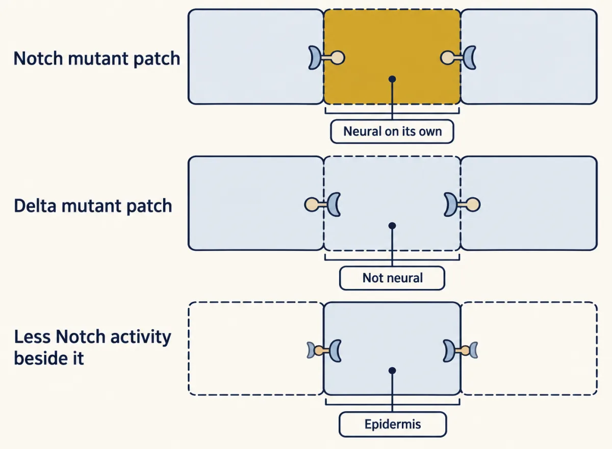
That answers the second puzzle.
Vertebrates: a brake and a clock
Read the transcript of “Vertebrates: a Brake, a Clock and Identity”
Film transcript: Vertebrates: a Brake, a Clock and Identity
In the vertebrate neural tube, the cells are already neural. So what do the same parts do there? Here, proneural factors push a progenitor to become a neuron. Notch holds its neighbours back as progenitors. The vertebrate relatives of Enhancer of split are the Hes genes. Hes inhibits proneural genes. Progenitors make Notch, its ligand Dll1, the repressors Hes1 and Hes5, and proneural factors. A cell that starts to differentiate raises Dll1. That keeps the progenitors around it as progenitors. Neurons first appear spaced apart. New ones arise in the gaps, and cells leave the pool over days. So lateral inhibition here is a brake on how fast progenitors are used up.
A cell held back stays a dividing progenitor, and can compete again later. Is Notch in the stem cell or the non-stem cell? Both cells have the receptor. The cell whose Notch is more strongly activated stays a progenitor. The cell that keeps sending more Delta differentiates. Mice lacking Hes1 make neurons too early, and have severe neural tube defects. Remove Hes5 as well, and progenitors are lost still faster. a surprise about timing. In cultured cells, Hes1 rises and falls about every two hours. Hes1 represses its own gene. Its RNA and protein are short-lived, and making new protein takes time. In mouse neural progenitors, Hes1 oscillates too.
Neurog2 and Dll1 cycle in the opposite phase. In cells becoming neurons, Hes1 falls, and Neurog2 stays high. So a brief pulse of Delta is not a commitment. Predict. In cultured progenitors, use light to hold Ascl1 steady, or make it oscillate. Which become neurons?
Steady Ascl1 pushed cells to become neurons. Oscillating Ascl1 kept them dividing as progenitors. Last question. Does a proneural factor also decide what kind of neuron forms? In the mouse forebrain, Neurog2 is high in dorsal progenitors. Ascl1 is high in ventral ones. Researchers swapped them, each into the other's place.
Does the factor carry the identity, or the territory? Neither, fully. Ascl1 pushed dorsal cells partly ventral. In the ventral forebrain, Neurog2 restored neurons, but did not push them dorsal. The swaps were not mirror images. In vertebrates, the loop is a brake and a clock. The factor carries part of the identity; the tissue and the cell's history shape the rest.
From the film
- In the vertebrate neural tube, Notch-Hes lateral inhibition works as a brake on how fast progenitors are used up, because a cell held back stays a dividing progenitor.
- Hes1 oscillates in cultured cells and in mouse neural progenitors; in culture, steady Ascl1 pushed cells toward neurons while oscillating Ascl1 kept them dividing.
- The Ascl1 and Neurog2 swaps in mouse forebrain were not mirror images, so a proneural factor carries part of a neuron's identity and the tissue and the cell's history shape the rest.
In vertebrates the neural tube is already neural. Proneural factors here do not make cells able to become neural, as in the fly ectoderm. They push a progenitor to become a neuron, and Notch holds its neighbours back as progenitors.
The parts are the same. The vertebrate relatives of E(spl) are the Hes genes, and Hes inhibits proneural genes. Progenitors express Notch, its ligand Dll1 (a vertebrate Delta), the repressors Hes1 and Hes5, and proneural factors such as Ascl1 and Neurog2. A cell that starts to differentiate raises Dll1 and activates Notch in the progenitors around it, which keeps them as progenitors. The neural tube does not settle into the fly’s fixed pattern. Neurons first appear spaced apart, new ones arise in the gaps, and cells leave the progenitor pool over days. Lateral inhibition here works as a brake on how fast progenitors are used up. Why a brake here and a fixed switch in the fly? One difference is that a cell held back here stays a dividing progenitor and can compete again later, while most of the fly’s held-back cells become epidermis.
This answers a question students often bring: is Notch in the stem cell or the non-stem cell? Both cells have the receptor. The cell whose Notch is more strongly activated stays a progenitor; the cell that keeps sending more Delta, because its proneural factor stays on, goes on to differentiate. Progenitors also send Delta in pulses as they oscillate (below), so a high level at one moment is not a commitment. How a dividing progenitor can give its two daughters different amounts of Notch activity, through proteins such as Numb and other mechanisms, is the subject of asymmetric division. The cells themselves are described in radial glia as stem cells, and the later role of Notch in making glia in where glia come from.
The mouse genetics fits the brake. In 1995 Ryoichiro Kageyama’s laboratory, with François Guillemot and others, reported mice lacking Hes1. Their neural tubes expressed more Ascl1, made neurons too early, and had severe neural tube defects. Later mutants lacking Hes5 as well lost progenitors still faster.
Then came a surprise about timing. In 2002 Hiromi Hirata and colleagues found that in several kinds of cultured cells Hes1 rises and falls with a period of about two hours. This happens because Hes1 protein represses its own gene, its messenger RNA and protein are short-lived, and making new protein takes time. In 2008 Hiromi Shimojo, Toshiyuki Ohtsuka and Kageyama imaged reporters in mouse neural progenitors. Hes1 oscillates there too, with a period of about two to three hours, and Neurog2 and Dll1 oscillate in the opposite phase. In cells becoming neurons, Hes1 falls and Neurog2 stays high.
So a progenitor can carry a proneural factor that comes and goes, or one that stays on. Does the difference matter?
Predict. Use light to switch an Ascl1 gene on and off in cultured mouse neural progenitors. In one set of cells, hold Ascl1 steady. In the other, make it oscillate. Which cells keep dividing as progenitors, and which become neurons?
Check your prediction
In 2013 Imayoshi and colleagues in Kageyama’s laboratory did this with optogenetics in culture. Steady Ascl1 pushed cells toward becoming neurons. Oscillating Ascl1 kept them dividing as progenitors. The same factor gave opposite results depending on its timing.
One reading is that oscillating Dll1 lets progenitors keep signaling to each other without any of them committing, and a steady high level of a proneural factor is what pushes a cell to differentiate.
Do proneural genes carry identity? The Ascl1 and Neurog2 swap
Proneural factors start neurogenesis. Do they also say what kind of neuron to make? The mouse forebrain offered a test. Neurog2 is high in dorsal progenitors, which make the excitatory neurons of the cortex. Ascl1 is high in ventral progenitors of the ganglionic eminences (swellings of the ventral forebrain), which make inhibitory neurons, although some Ascl1 is present dorsally too.
A coding-region swap puts one factor’s protein-coding part at the other’s gene, under the other’s regulatory DNA. Before the result, set out what each answer would predict.
| If identity sits in… | Ascl1 made in dorsal cells | Neurog2 made in ventral cells |
|---|---|---|
| the factor | dorsal cells turn ventral | ventral cells turn dorsal |
| the territory | dorsal cells stay dorsal | ventral cells stay ventral |
Predict. Which row did the mouse follow?
Check your prediction
Neither row, fully. In 2000 Carol Fode, Guillemot and colleagues found that removing Neurog2 let Ascl1 rise in the dorsal forebrain, and some dorsal cells then switched on a subset of ventral markers such as Dlx1 and GAD67, a change that required Ascl1. Other ventral markers did not appear. Placing the Ascl1 coding region at the Neurog2 locus induced some ventral markers in dorsal cells as well.
In 2002 Carlos Parras, Carol Schuurmans and colleagues also did the reverse, putting Neurog2 in place of Ascl1. In the ventral forebrain, Neurog2 restored neuron production and the ventral markers, yet did not switch on dorsal markers. Elsewhere it restored neurogenesis in the sympathetic ganglia, but not in the ventral spinal cord or the locus coeruleus.
The two swaps were not mirror images. Ascl1 could push dorsal cells partly ventral; Neurog2 did not push ventral cells dorsal.
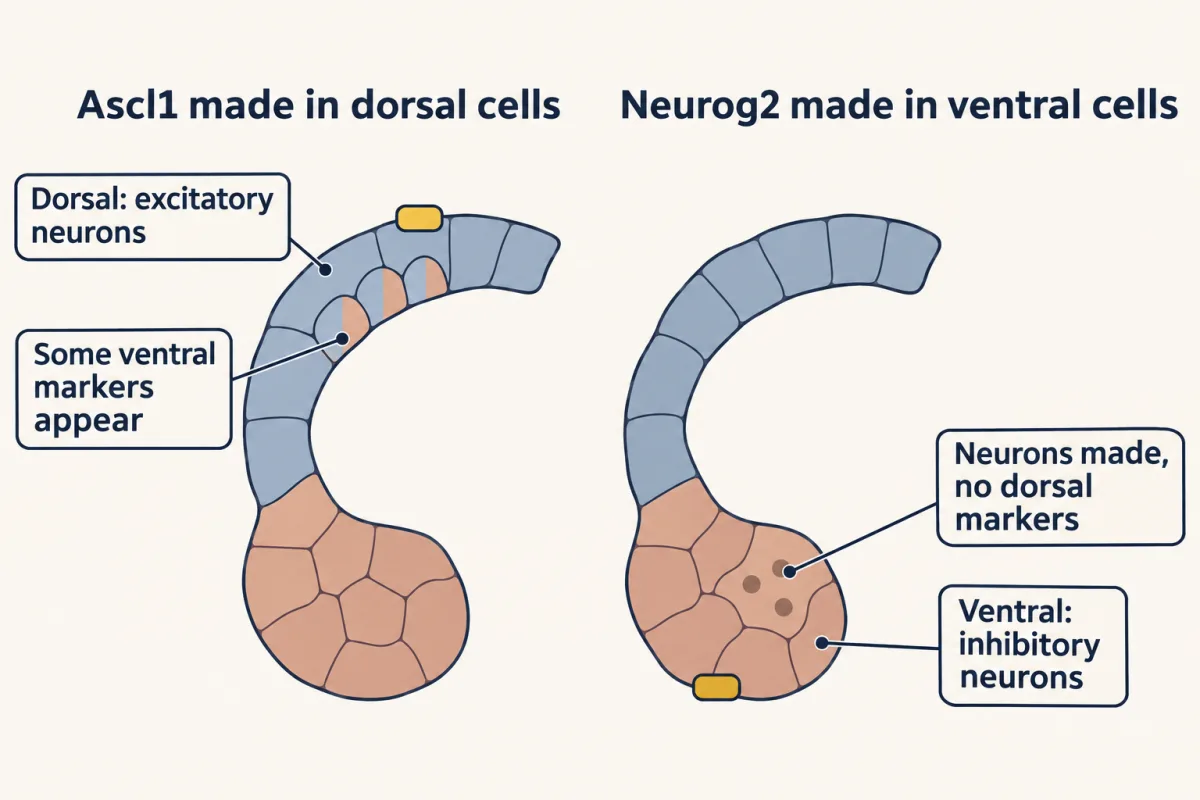
So proneural factors carry partly different identity information, and how much depends on the factor and the tissue. The fly shows both sides. Forcing achaete-scute genes on makes only external sense organs, while forcing atonal on mostly makes chordotonal organs, so the factor itself carries identity. Yet atonal also starts photoreceptors in the eye, so the tissue matters too. A precursor’s identity comes from the proneural factor together with where the cell is and what it has already been told. That answers the third puzzle. When in a lineage those instructions arrive is the subject of birth order and fate.
Putting it together
In the fly, proneural factors say who may and Notch, comparing neighbours on a steep loop, says who does. In the vertebrate neural tube, where cells are already neural, the proneural factor pushes a progenitor toward a neuron and Notch-Hes signalling holds its neighbours back, as a brake that also oscillates.
- Who may? Proneural bHLH factors make a cluster competent. In the embryo, regulatory DNA places the cluster, and Notch-null embryos still form clusters in the right places; on the notum Notch also helps shape the proneural stripes.
- Who does? Delta–Notch–E(spl) feedback between neighbours is a double negative, so it acts as positive feedback, and it splits two equal cells only on a steep loop. In Heitzler and Simpson’s mosaics, fate followed relative Notch activity.
- Does it carry over? In the neural tube the same parts brake how fast progenitors become neurons, and they oscillate; steady proneural expression, not only a high level, goes with commitment. The factor carries part of a neuron’s identity; the tissue and the cell’s history shape the rest.
What the rule covers, and its limits. The steepness test applies to touching cells that all express proneural factor and start alike: the fly embryo’s clusters and the notum’s clusters. In vertebrate progenitors the same parts also form a delayed oscillator, and the lesson does not test a steepness threshold there. On an extended sheet, touching-neighbour inhibition spaces winners rather than guaranteeing one per cluster. A difference imposed from outside, such as uneven proneural levels or a bias inherited through an asymmetric division, can set fates without the tie having to break by itself.
Going deeper
The last cut is regulated proteolysis
The second of the two cuts that free the inner part of Notch is made inside the membrane by γ-secretase. It is a case of regulated proteolysis, the theme of the lesson on ubiquitin and proteolysis. The drug DAPT, one of this lesson’s structures, blocks this cut and so switches Notch off.
Where the cluster map comes from
The map of regulatory sites around the achaete-scute genes that places the clusters was worked out mostly on the notum and wing, by Modolell and colleagues.
Delta level versus Delta activity
“Proneural factors raise Delta” is the standard model, and the lesson uses it. In the fly embryo, though, Delta RNA is broad across the neuroectoderm, and a good part of the difference between cells may lie in Delta’s activity. In the embryo the main protein that makes Delta active as a ligand is Neuralized, an enzyme that tags Delta with ubiquitin and helps the cell take it in. A different E3 ligase with a similar job, Mind bomb, matters more in other fly tissues. A careful reader should not take “raise Delta” to mean only more Delta messenger RNA.
Spacing, and one winner per cluster
The embryo makes one neuroblast per cluster, and sensory organ precursors (SOPs) on the notum are spaced several cells apart, more widely than nearest-neighbour inhibition alone would space them. The longer reach proposed is signaling beyond touching neighbours, through Delta-bearing filopodia, thin cell extensions (de Joussineau and colleagues, 2003; Cohen and colleagues, 2010).
The model behind the numbers
Collier and colleagues wrote the loop for each cell as two equations. Notch activity n moves toward f(d̄), a rising function of the average Delta d̄ of the neighbours, and Delta d moves toward g(n), a falling function of the cell’s own Notch activity. The worked example uses f(x) = xk/(a + xk) and g(x) = 1/(1 + b xh), with a = 0.01 and b = 100; the Hill exponents k and h set how switch-like each response is. Near the even state, a small change passes through f and then g. Because g falls, the product of the two slopes is negative; its size is the gain G for one hand-off between cells, and G² is the round-trip factor. The thresholds in the lesson, round-trip factors of 1 for a pair and 4 for an ideal hexagonal sheet, are G = 1 and G = 2. The steep worked example has k = h = 2 (G about 2.39); the shallow one has k = h = 1 (G about 0.38). A steep exponent alone is not enough: with k = h = 2 but the weaker repression constant b = 10, G is about 0.93, just below 1, and the pair stays even.
Timing, with both rates set to 1 so that one time unit is the mean lifetime of Notch activity: start both cells with Notch activity at its even-state value and cell A’s Delta 1 percent (or 0.1 percent) above B’s; the two Delta levels differ by 0.5 after about 13 (or about 17) time units. A tenfold smaller start adds a fixed time, here about 4 units, and the wait grows without limit as G approaches 1 or the start approaches zero.
Receptor dose: if a cell’s Notch activity is scaled by its share of the normal receptor, a cell with half the Notch of its partner wins from an equal start. A pair in which both cells have half the Notch still splits (G about 1.6), but a pair with a tenth in both stays even (G about 0.07), at the same 1:1 ratio. So the relative signal biases which cell wins, and the absolute signal decides whether the pair splits at all. All of these are illustrative, from published model forms.
Why the Hes1 clock needs a delay
A loop of only two steps, messenger RNA that makes protein and protein that represses the messenger RNA, with each decaying at a steady rate, cannot keep oscillating; it settles to a steady level. Short half-lives, near 20 minutes for both, let Hes1 vanish and the gene restart, but the cycle also needs the delay of transcription and translation, or extra steps in the loop. The models agree with the experiment in which blocking protein synthesis stopped the cycle with the messenger RNA high.
Open questions on identity
How much of fate information sits in the proneural protein and how much in the cell receiving it? Comparisons in programmed stem cells show the two factors open different chromatin, which raises the question of how much of the difference in vivo comes from binding partners, chromatin state and the duration of expression.
The name “neurogenic”
Fly geneticists call Notch, Delta and their partners neurogenic genes, because losing them makes too much neural tissue. The name was used by José Campos-Ortega’s laboratory in the early 1980s for the set, after Donald Poulson’s 1930s descriptions of embryos lacking Notch.
Your notes
Select any text in the lesson to highlight it or add a note to it. Your highlights and notes are saved in this browser and collected in your notebook.
This browser is not letting the site save anything (a private window or strict privacy settings), so highlights and notes will be lost when you leave the page.
How we measure it
Genetic mosaics
An animal is built so that patches of cells carry a different dose or version of a gene than the cells around them. Recording which cells at a patch border take which fate shows whether a gene acts in the cell that sends a signal or the cell that receives it, and whether the comparison between neighbours matters.
In situ hybridization of proneural clusters
A labeled probe that pairs with one messenger RNA shows where a gene is expressed in a fixed embryo. Staging many embryos shows a gene expressed first in a group of cells and later in one, which is how competence and selection were told apart.
Coding-region swap (knock-in)
In mouse embryonic stem cells, the protein-coding part of one gene is replaced with that of another while the original regulatory DNA is kept. Comparing the result with a plain knockout asks whether two related proteins can do each other's job in the same place and time.
Live bioluminescence imaging
A gene's promoter drives a short-lived luciferase whose light is recorded over hours from single cells in culture or in tissue slices. It reveals oscillations that fixed samples cannot, since a snapshot of an oscillating population looks like random variation.
Practice questions on this lesson (3) → MCAT-style, original, each with an explanation
Where this lesson fits
Before thisReview first
- Neural Induction: The Default ModelThis course · Lesson 6
You need the idea that BMP signaling sets neural versus skin fate from there, since proneural genes act in already-neural tissue.
- Ventral to Dorsal: Sonic Hedgehog and BMP in the Neural TubeThis course · Lesson 10
It shows how the neural tube is patterned into progenitor domains, which proneural clusters and Notch then narrow.
- Transcription Factor Codes: Building a Motor NeuronThis course · Lesson 11
It shows how transcription factor codes set neuron identity, which this lesson uses when asking whether proneural factors carry identity.
After thisStudy next
- Asymmetric Division: Numb and the NeuroblastThis course · Lesson 15
It shows how a dividing progenitor gives its daughters different Notch activity, which this lesson only mentions.
- Birth Order and Fate: Temporal Identity and the Inside-Out CortexThis course · Lesson 16
It takes up when in a lineage identity instructions arrive, the question left open after the swap experiments.
- Where Glia Come FromThis course · Lesson 43
It uses Notch-Hes signalling to explain how progenitors switch from making neurons to making glia.
Elsewhere on the siteFilms, simulations and method films in other courses
- The Receptor Is the MessageFilm · Cell Signaling
This film shows the Notch receptor as the message and how contact between two cells decides which becomes the nerve cell.
Suggested by an AI model from the lessons themselves, then checked. How we use AI
Finish line
Test yourself
9 questions
Practice and MCAT-style, with explained answers.
Next · Lesson 15
Asymmetric Division: Numb and the Neuroblast
A stem cell has to keep making new cells and also keep itself. How does one cell divide into two sisters that are different from the moment they are born?
Continue →← Lesson 13 · The Ventricular Zone: Radial Glia as Stem Cells