Developmental Neuroscience · Unit 3: Coordinates: patterning the tube
Transcription Factor Codes: Building a Motor Neuron
Rewritten on : new text, figures, flashcards and film. Your notes and highlights stay, but the text around them may have moved.
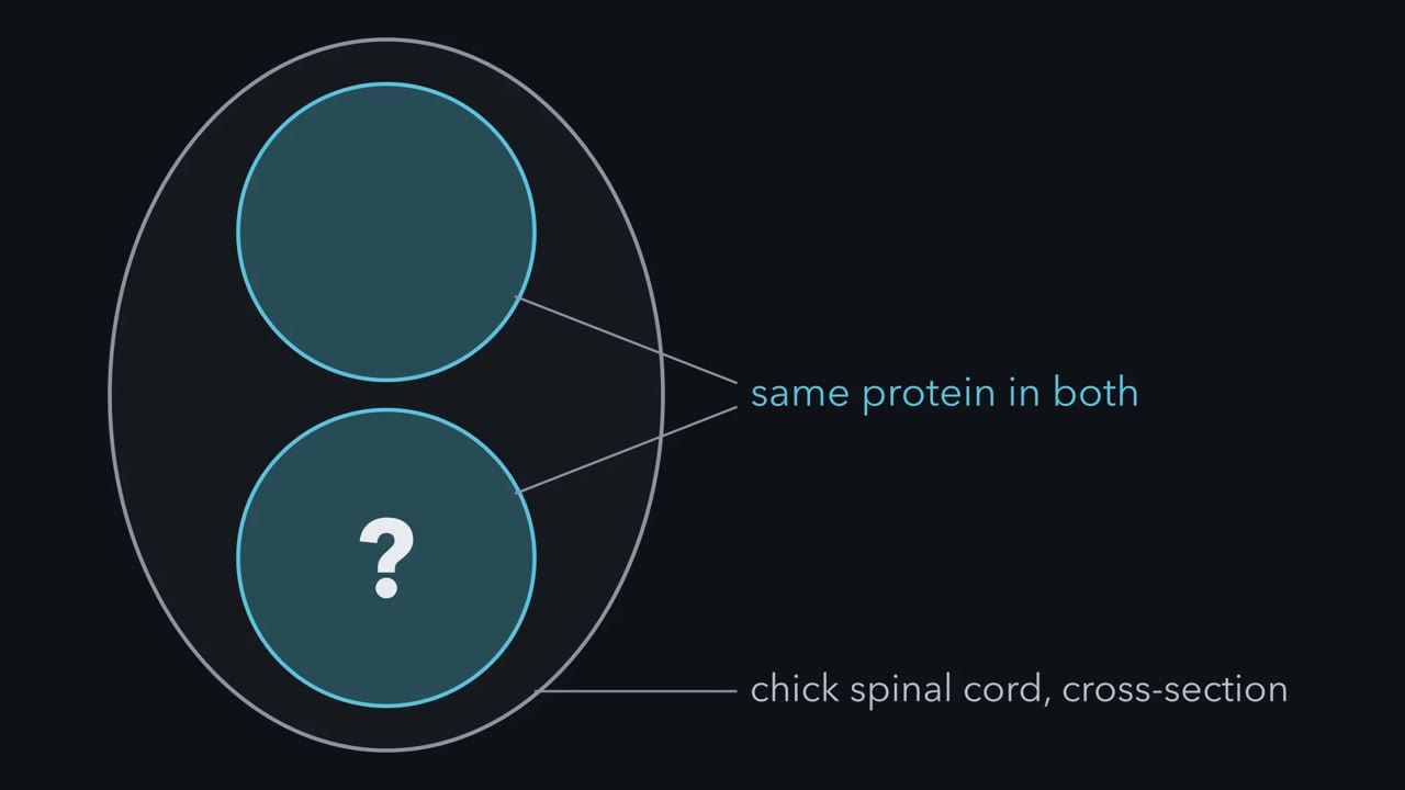
Put Lhx3 into chick spinal cord cells that make no Isl1 and they turn on interneuron genes. Add Isl1 and the same protein turns on motor-neuron genes. If no single gene says motor neuron, how does a set of transcription factors make one, and choose the muscle its axon will reach?
Pause when the reading reaches a film, and offer to play it. Saved on this device.
Watch the film now, or keep listening
Builds on:Ventral to Dorsal: Sonic Hedgehog and BMP in the Neural TubeHead to Tail: Hox Genes, Retinoic Acid and RhombomeresTools for Following Cells and GenesWhy these?
 ▶ Watch One Protein, Two Cell Types · 2:36
Read the transcript of “One Protein, Two Cell Types”
Film transcript: One Protein, Two Cell Types
Two neighbouring cells in a chick spinal cord make the same protein. One becomes a motor neuron, the other an interneuron. The protein is Lhx3. Both newly born motor neurons and V2 interneurons make it. A second protein, Isl1, is made in the motor neurons, not the V2 interneurons. Both are LIM homeodomain proteins. The homeodomain binds DNA. Two LIM domains are handles that other proteins grip. A third player, the cofactor NLI, binds no DNA. It pairs with itself and grips LIM domains. Predict. Put Lhx3 alone into chick cells without Isl1. Then put in both. Which genes come up?
Alone, it turns on V2 interneuron genes, such as Chx10. With Isl1, it turns on motor-neuron genes, such as Mnx1. V2 genes are largely held down. Why? Without Isl1, NLI grips Lhx3 directly. Pairs of these units bind V2 genes.
With Isl1 present, Isl1 grips NLI itself. Then it holds Lhx3 on a short segment. Pairs of this three part complex promote motor neuron genes. NLI is in both. What differs is the bridge, Isl1. Does any Isl1 at all give a clean switch? Try a simple binding model. With equal amounts, about a quarter of the Lhx3 still sits in the interneuron complex. Less Isl1 leaves more. In this model, binding alone gives a mixture, not a switch. The model does not decide a cell's fate. In real cells, a downstream regulator helps lock the choice in. Here is the one idea to carry. No single factor says motor neuron. Here, identity depends on which complexes form.
That depends on which partners are present, and in what ratio. Takeaway: one protein, held by different partners, builds two different cells.
From the film
- No single factor says motor neuron: here, identity depends on which complexes form, and that depends on which partners are present and in what ratio.
- In chick cells, Lhx3 without Isl1 turns on V2 interneuron genes such as Chx10, and Lhx3 with Isl1 turns on motor neuron genes such as Mnx1.
- The cofactor NLI is in both complexes, and what differs is whether Isl1 bridges NLI to Lhx3.
- In a simple binding model, binding alone gives a mixture rather than a clean switch, so real cells need more to lock the choice in.
Put the protein Lhx3 into cells of a chick spinal cord that make no Isl1, and they turn on interneuron genes. Put Lhx3 in together with Isl1, and the same protein turns on motor-neuron genes instead.
No gene in a motor-neuron progenitor says “motor neuron”. The cell expresses a combination of transcription factors, none of which is specific to motor neurons on its own. The code does two jobs. First it makes a cell a motor neuron. Then, along the cord and in the limb, it helps decide which muscle that neuron will reach, though how much of that choice the code sets on its own is still open. Run the same steps in a dish and you can make motor neurons.
One protein, two cell types
Meet the factors. Lhx3 and Isl1 are LIM-homeodomain proteins. Each has a homeodomain, which binds DNA, and two LIM domains, which bind other proteins, not DNA. Think of the LIM domains as handles: whoever holds Lhx3’s handles decides what Lhx3 does. The third player, the LIM cofactor NLI (also called Ldb1), has no DNA-binding domain. It grips LIM domains, and it pairs with itself, so the complexes it builds come in pairs.
Lhx3 is made in two neighbouring cell types: newly born motor neurons, and the V2 interneurons just dorsal to them, local-circuit neurons of the ventral cord. Isl1 is made in the motor neurons, not in V2 interneurons.
Predict. You electroporate the chick neural tube: inject a plasmid into it and drive the plasmid into the cells on one side with a brief electric pulse, so the other side is the control. In condition A, Lhx3 goes into cells that make no Isl1. In condition B, Lhx3 and Isl1 go into the same cells. Which genes come up in each: motor-neuron genes such as Mnx1, V2 interneuron genes such as Chx10, or neither?
Check your prediction
A gives V2 interneuron genes. B gives motor-neuron genes, and V2 genes are largely suppressed (the Design problem below gives the catch). The same protein, Lhx3, is in both. Thaler and colleagues in Pfaff’s laboratory explained why in 2002.
Picture Isl1 as a bridge. It holds NLI with its own LIM domains and holds Lhx3 with a short tail, so Lhx3 hangs off Isl1 instead of off NLI.
- V2 interneuron. NLI grips Lhx3’s LIM domains directly, and that complex triggers V2 differentiation. Two NLI·Lhx3 units, joined through NLI, bind V2 genes.
- Motor neuron. Isl1 grips NLI with its own LIM domains, taking the place Lhx3 would use. Lhx3 then binds a short, high-affinity segment in Isl1’s C-terminal region. Two NLI·Isl1·Lhx3 units, joined through NLI, promote motor-neuron genes.
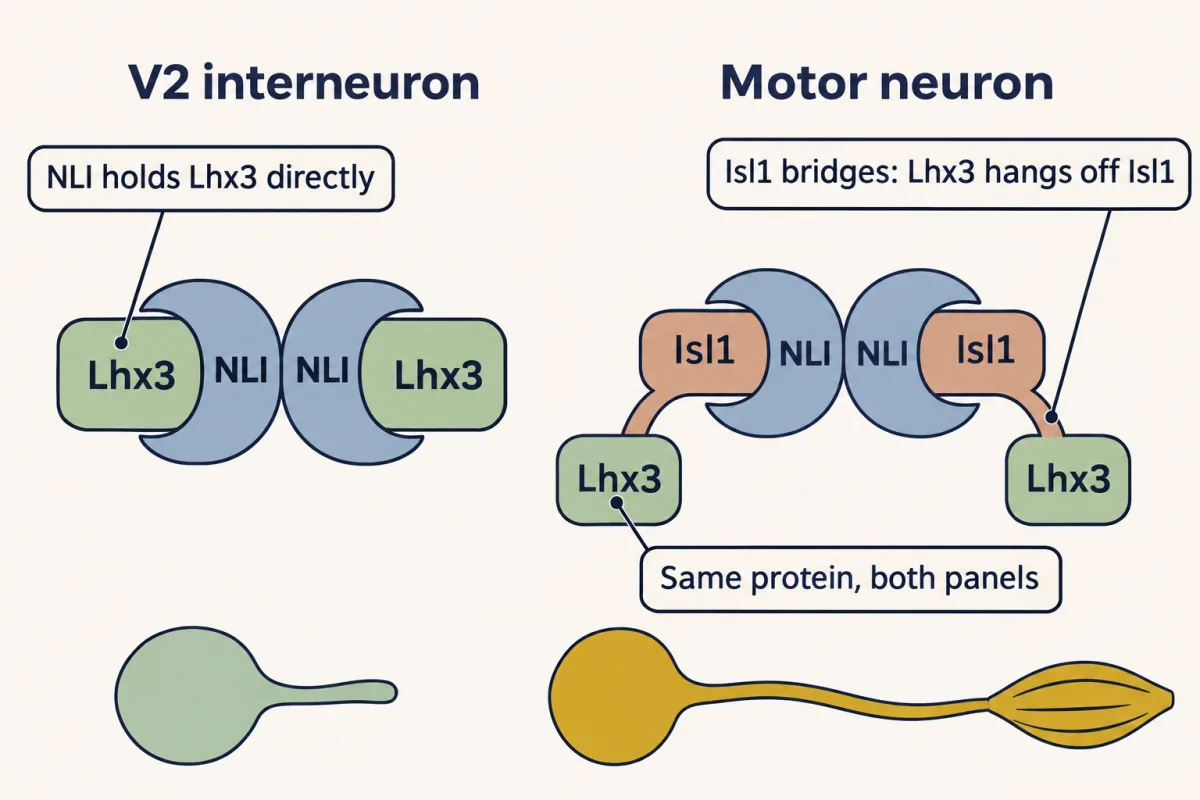
NLI is in both complexes. What differs is whether Isl1 bridges NLI to Lhx3. The first two structures in this lesson show the same surface of Lhx3 held once by the Isl1 segment and once by NLI.
The one idea to carry through this lesson. No single factor says “motor neuron”. In this example, what the cell becomes depends on which complexes its transcription factors form, and that depends on which partners are present and in what ratio. Later layers (Hox proteins with FoxP1, then Lim1 against Isl1) refine which kind of motor neuron it becomes. Their timing overlaps: Hox patterning starts in progenitors.
Scope. This covers motor neurons of the chick and mouse spinal cord and their V2 neighbours; many cranial motor neurons of the hindbrain use other factors, such as Phox2 proteins. It is about specifying a cell’s class and subtype. Keeping the identity is a separate job (Mnx1, below), and so is keeping the cell alive. And presence may not be the whole story: the binding model below, and the excess-Lhx3 caveat in the Design problem, suggest that how much Isl1 there is relative to Lhx3 matters too.
Why the ratio matters
The short answer: even with equal amounts of Isl1 and Lhx3, binding alone leaves some Lhx3 in the V2 complex. A simple model shows it.
Picture an ideal binding model, not a measurement. NLI holds Lhx3 or Isl1, not both, and Lhx3 binds NLI or Isl1, not both; Isl1 can hold NLI and Lhx3 at once, on separate sites. Every pair binds equally tightly, and Lhx3 and NLI are each present at ten times the concentration at which half would be bound (ten times the dissociation constant). The model ignores NLI pairing, DNA and other partners, so it counts single units, not the paired complexes on DNA. Hold Lhx3 and NLI fixed, and vary Isl1.
| Isl1 relative to Lhx3 | Lhx3 in V2 units (NLI·Lhx3) | Lhx3 in motor-neuron units (NLI·Isl1·Lhx3) |
|---|---|---|
| none | 73% | 0% |
| half as much | 45% | 23% |
| equal | 24% | 37% |
| twice as much | 6% | 37% |
| four times as much | 1% | 23% |
The rest of the Lhx3 is free, or held by Isl1 without NLI. The exact numbers depend on these choices (at a hundred times the dissociation constant, equal amounts leave 16 percent in V2 units rather than 24), but the pattern holds. Read the table in three steps.
- A mixture, not a switch. At equal amounts, about a quarter of the Lhx3 is still in the V2 complex. Of the Lhx3 in either complex, the motor-neuron complex holds about 61 percent. If Isl1 binds NLI ten times more tightly than Lhx3 does, that share rises to about 84 percent: better, but still a mixture.
- Too little Isl1 leaves V2 complexes. That is why the Design problem below warns that two separate plasmids do not fix the ratio. A fused Isl1-Lhx3 protein is a cleaner experiment: it fixes the ratio and tethers Lhx3 to its own Isl1 segment, which this simple model does not include (see Going deeper).
- Too much Isl1 lowers the motor-neuron complex too, because spare Isl1 pairs with Lhx3 or with NLI on its own. This last point comes from the model, not from a reported experiment.
So in this model binding alone gives a mixture. The model says nothing about cell fate; real cells add DNA, other partners and downstream regulators. The mouse deletions below show one such regulator, Mnx1, which helps hold the motor-neuron choice in place.
Before the code: a stripe of progenitors
The motor neurons in that Lhx3 choice come from one progenitor domain, and their V2 neighbours from the domain next to it. Both start with position across the neural tube.
Sonic hedgehog from the notochord and then the floor plate sets up a gradient across the neural tube, opposed by bone morphogenetic proteins (BMPs) from above, and progenitors read their position in it; the Hedgehog lesson covers how that signal is received. The read-out is a set of stacked domains, each with its own transcription-factor profile. One of them, called pMN (p for progenitor, MN for motor neuron), makes motor neurons; the V2 interneurons come from p2, just dorsal to it.
A pMN progenitor is still dividing. It carries the bHLH repressor Olig2 along with Nkx6.1 and Pax6, and Olig2 is its tag. Novitch, Chen and Jessell reported in 2001 that Olig2 marks these progenitors and works with Neurogenin2, a factor that makes progenitors leave the cell cycle and become neurons. Together they tie motor-neuron character to the step of becoming a neuron. So the cell becomes a neuron and a motor neuron at the same time. Mice lacking both Olig1 and Olig2 largely lose their motor neurons (the necessity table below has the details). The same pMN domain later turns to making oligodendrocytes, which the glia lesson takes up.
Around its final division, as it becomes postmitotic (no longer dividing), a motor neuron turns on a new set: Isl1 or its relative Isl2, Lhx3, and Mnx1, the gene also called Hb9. These are the markers you would stain for to find a newly differentiated motor neuron in a section; Lhx3 is transient, and later only the medial motor column keeps it.
Enough, needed, and kept
Read the transcript of “Enough, Needed, and Kept”
Film transcript: Enough, Needed, and Kept
If a protein is enough to start making a motor neuron, does every motor neuron need it? A factor is sufficient if adding it produces the effect, in the cells being tested. It is necessary there if the effect does not happen without it. Electroporation in chick tests sufficiency. Gene deletion in mouse tests necessity. In chick, Mnx1, or its relative MNR2, is enough to trigger motor-neuron differentiation. It also represses the neighbouring V2 programme. Predict. Delete Mnx1 in a mouse. Are motor neurons made? Does anything go wrong?
They are made, on schedule and in normal numbers. But for a while they show V2 interneuron features. Their columns and axon paths are disordered. So Mnx1 is not needed to make a motor neuron. It is needed to keep one. A motor neuron has to keep suppressing the interneuron programme. That is the lock.
Back up to the progenitors. Delete both Olig1 and Olig2, and motor neurons are largely lost. Lineage tracing suggested the progenitors make V2 interneurons, then astrocytes, instead. Delete Isl1, and motor neurons are not generated at all. Now a puzzle. Delete Lhx3 and Lhx4. Motor neurons are still made. What changes is their subclass, and which way their axons leave the tube.
Yet Lhx3 sits in the motor-neuron complex. How can both be true? The tests ask different questions. In chick, Isl1 plus Lhx3 is enough to start the programme. In mouse, motor neurons still form without Lhx3 and Lhx4, but of a different kind. Sufficient is not the same as necessary.
Takeaway: enough, needed, and kept are different jobs. Each test answers only one.
From the film
- A factor is sufficient if adding it produces the effect in the cells tested, and necessary if the effect does not happen without it.
- In mouse, deleting Mnx1 still allows motor neurons to form, but they transiently show V2 interneuron features, so Mnx1 is needed to keep a motor neuron rather than to make one.
- Motor neurons still form without Lhx3 and Lhx4, but their subclass and axon exit change, so enough is not the same as needed.
Two words first. A factor, or a combination, is sufficient if adding it produces the effect in the cells and stage tested, using the rest of their machinery (the Lhx3 test above used the cells’ own NLI). It is necessary there if the effect does not happen without it. Each of two techniques tests one of these.
- Electroporation in chick tests sufficiency. Expressing a factor ectopically, in cells that would not normally make it, shows what it can impose.
- Gene deletion in mouse tests necessity. Delete the gene and see what fails to form.
The two ask different questions, and they can give different answers. Note which animal and which manipulation each claim rests on.
Mnx1, or its chick relative MNR2, electroporated into the cord is sufficient to trigger motor-neuron differentiation and to repress the neighbouring V2 programme.
Predict. Now delete Mnx1 in a mouse. Mnx1 is enough to start the programme in chick, but is it needed? Are motor neurons made without it? If they are, what would you look for to see whether anything went wrong?
Check your prediction
They are made, on schedule and in normal numbers. But they transiently take on molecular features of V2 interneurons, such as Chx10, and their columns and axon projections are disordered. Two laboratories reported this in the same issue of Neuron in 1999. Mnx1 is not needed to make a motor neuron. It is needed to keep one: a motor neuron has to keep suppressing the interneuron programme.
Mnx1 is one of the regulators that hold the choice in place. Being needed to make a cell and being needed to keep it are different jobs. The mouse deletions sort the factors into those jobs.
| Mouse lacking | Motor neurons made? | What changes | What it tells you |
|---|---|---|---|
| Olig1 and Olig2 | largely no | oligodendrocyte differentiation is abolished; lineage tracing suggested the pMN cells make V2 interneurons and then astrocytes | the progenitor layer decides what the domain makes |
| Isl1 | no | much of the rest of the tube differentiates normally; a class of Engrailed1 interneurons also fails | needed to make a motor neuron |
| Mnx1 (Hb9) | yes, on schedule and in normal numbers | cells transiently show V2 features; columns and axons disordered | needed to keep the identity, not to make it |
| Lhx3 and Lhx4 | yes | which subclass they become, and which way their axons leave the tube | not needed to make a motor neuron, though enough with Isl1 in chick |
The last row looks like a contradiction: Lhx3 is in the motor-neuron complex, yet motor neurons are made without it. It is not one, because the two tests ask different questions. In chick, Isl1 plus Lhx3 is enough to start the motor-neuron programme. In mouse, motor neurons still form without Lhx3 and Lhx4, but they become a different kind of motor neuron, with a different axon exit. A factor can be sufficient in one test without being necessary in another. These results do not show what lets the cells become motor neurons without Lhx3 and Lhx4.
Which motor neuron: Hox proteins and FoxP1 along the cord
Read the transcript of “Which Motor Neuron, and a Recipe”
Film transcript: Which Motor Neuron, and a Recipe
The same code built a motor neuron in your neck, and one in your lower back. What tells them apart? Position along the cord, written in Hox proteins. Motor neurons sort into columns. The medial column drives back muscles. The lateral column, at limb levels, drives limb muscles. The preganglionic column projects to autonomic ganglia. Hox proteins act inside each cell, as repressors and activators. Hoxc6, at forelimb levels, and Hoxc9, at thoracic levels, repress each other. Predict. In chick, put Hoxc9 into forelimb-level progenitors, and so into the motor neurons they make. What column do they become?
Their lateral column identity is suppressed. They take on a preganglionic identity, from a thoracic column. Timing matters. Hoxc9 given only after the last division suppresses the lateral column, but does not make preganglionic cells.
Hox proteins work with an accessory factor, FoxP1. It gates the limb and autonomic columns. Predict. Delete Foxp1 in a mouse. Are limb-level motor neurons still made? Yes. Motor neurons are still made. What fails is the assignment of which one each is. They resemble the thoracic hypaxial column. In the limb, their axons appear to choose paths and muscles at random.
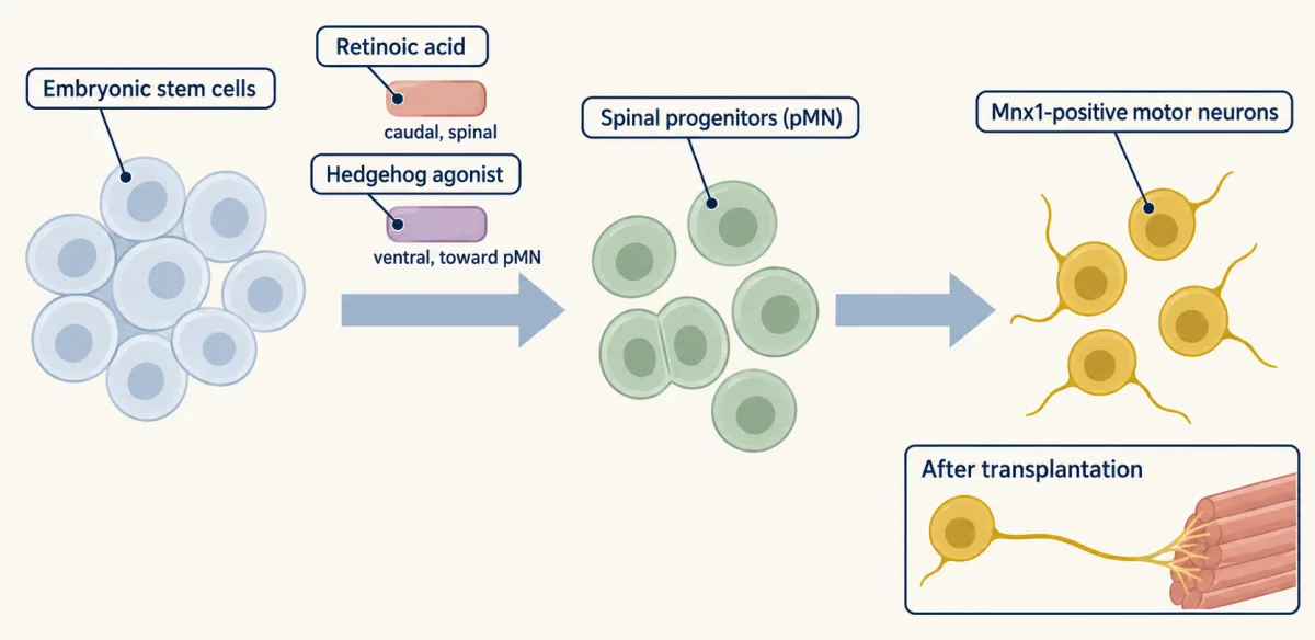
Inside the limb column comes one more choice. Lateral cells make Lim1, and their axons go dorsal. Medial cells keep Isl1, and their axons go ventral. Without Lim1, lateral axons lose their dorsal preference. Eph receptors and ephrins help carry out this choice, alongside other guidance systems. Run these layers forward, and you have a recipe. Retinoic acid gives mouse stem cells a spinal character. A Hedgehog agonist pushes them toward the motor neuron domain. Then motor neurons appear. Put into an embryo, they reach muscle. But this recipe gives mostly medial column cells. One named pool is still hard to make. Takeaway: overlapping layers set a motor neuron's class, its column, and its limb choice. Run them in order, and you have a recipe.
From the film
- Position along the cord is written in Hox proteins, and in chick, Hoxc9 given early in the brachial cord suppresses limb column identity and gives a preganglionic identity.
- In mouse, motor neurons still form without Foxp1, but the limb and autonomic columns are not assigned correctly and axons appear to choose paths at random.
- Within the limb column, lateral cells make Lim1 and their axons take a dorsal path, while medial cells keep Isl1 and their axons go ventral.
- Retinoic acid and a Hedgehog agonist can turn mouse stem cells into motor neurons, but the basic protocol gives mostly medial column cells.
A motor neuron in your neck and one in your lower back were built by the same code, yet one drives a neck muscle and the other a muscle of the leg. The difference is rostrocaudal, along the length of the body, and it is written in Hox proteins, the same family you met setting rhombomere identity in the hindbrain.
First, the groups being sorted. A motor column is defined by where the cell bodies sit and where the axons go. Among the muscle-innervating columns, a pool is the set of neurons that innervates one muscle, such as the biceps.
| Column | Where along the cord | Target | Markers in this lesson |
|---|---|---|---|
| Medial motor column (MMC) | every level | axial muscles of the back | keeps Lhx3; no FoxP1 |
| Lateral motor column (LMC) | limb levels | limb muscles | FoxP1 high; lateral cells Lim1, medial cells Isl1 |
| Preganglionic column (PGC) | mainly thoracic | autonomic ganglia | FoxP1 low |
| Hypaxial motor column (HMC) | thoracic | body-wall (hypaxial) muscles | no FoxP1 |
In mammals a phrenic motor column also innervates the diaphragm. Do not expect a tidy count of pools; the number depends on the species and on which muscles you count.
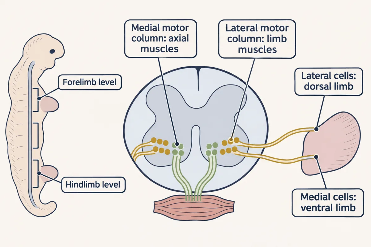
How do Hox proteins sort cells into these columns? Dasen, Liu and Jessell reported in 2003 that sequential phases of Hox-c activity control columnar fate in motor neurons. Graded fibroblast growth factor signalling establishes Hox expression in progenitors, which is translated into a Hox pattern in motor neurons. There, Hox proteins act cell autonomously, inside each cell, as repressors and activators that sort cells into columns. One pair shows the logic. Hoxc6 is made at brachial (forelimb) levels and Hoxc9 at thoracic levels, and each represses the other.
Predict. In chick, you electroporate Hoxc9 early into the brachial neural tube, so that both the progenitors and the motor neurons they make carry it. These motor neurons would normally form the LMC. What should happen to their column identity?
Check your prediction
LMC identity is suppressed, and the cells take on a preganglionic identity (in chick, the column of Terni), a thoracic column. The thoracic Hox protein represses the brachial one and imposes its own level’s column. Timing matters: Hoxc9 given only to motor neurons after their last division suppresses LMC identity but does not impose the preganglionic one, which needs Hoxc9 already in the progenitors. Cross-repression keeps the identities of neighbouring levels apart.
A 2005 paper from the same laboratory described the network in more detail: one set of Hox interactions fixes position along the cord, and another generates diversity among pools within a single segment, with the pattern of connections to target muscles as the read-out.
Hox proteins need help to do this in some columns. FoxP1 is the gate for the Hox-dependent columns: the limb column (LMC) and the autonomic column (PGC). Dasen and colleagues reported in 2008 that FoxP1 acts as a Hox accessory factor, a partner the Hox proteins need to produce their column output. It is expressed in the Hox-sensitive columns, and acting in a dose-dependent way, high in the LMC and low in the PGC. Rousso and colleagues in Novitch’s laboratory showed in the same year that Foxp1 defines columnar identity and sets the pattern of LIM homeodomain proteins.
Predict. Delete Foxp1 in a mouse. Are motor neurons still made at limb level? If so, what do they become, and where do their axons go in the limb?
Check your prediction
Motor neurons are still made; what fails is the assignment of which one each is. The output of the motor-neuron Hox network is abolished in the limb and autonomic columns. Prospective limb and autonomic neurons instead take on a molecular identity like that of the thoracic hypaxial motor column, which Dasen and colleagues read as the ancestral motor neuron state. In the limb, motor axons appear to select trajectories and muscle targets at random.
FoxP1 sorts cells that are already motor neurons. It does not make them.
Which half of the limb: the LIM code sets the first axon choice
The lateral motor column splits again, and the split is a clear case of a transcription factor code handing off to axon guidance. Lateral LMC neurons express Lim1 (Lhx1); medial ones retain Isl1.
Part of the split is set inside the column. Sockanathan and Jessell reported in 1998 that a retinoid made by early-born LMC neurons helps later-born neurons become lateral LMC, so some of the information comes from the motor neurons themselves. For how retinoic acid is received, see nuclear receptors.
Predict. Lateral LMC axons normally take a dorsal path in the limb, and medial LMC axons a ventral one. Kania, Johnson and Jessell removed Lim1 in mouse. What do the lateral LMC axons do without it?
Check your prediction
They lose their dorsal preference and select paths at random. Lim1 in the lateral set ensures that their axons take a dorsal trajectory in the limb (Kania, Johnson and Jessell, 2000). The limb has its say too: Lmx1b in dorsal limb mesenchyme controls both trajectories from outside, and without mesenchymal Lmx1b the choices of both divisions are disturbed.
Much of the downstream machinery is a set of Eph receptors and ephrins, working alongside other guidance systems, which guidance cues and pioneers and topographic maps take up. Once the axons reach muscle, survival during the period of developmental cell death depends partly on support from the target, along with other cells such as glia, and nerve growth factor (NGF) is not the general survival signal for motor neurons; see the neurotrophic hypothesis.
The order is worth holding onto. Position in the tube sets the progenitor domain. The domain sets the postmitotic code. Position along the cord sets the column. The column sets which receptors the axon carries. Only then does the axon meet the cues that steer it. The order is a simplification: the stages overlap (Hox patterning starts in progenitors), an axon meets several cues at once, and signals from the periphery can feed back on a motor neuron’s gene expression.
Making motor neurons in a dish
Run the layers forward and you have a protocol. Wichterle and colleagues reported in 2002 that mouse embryonic stem cells, given the signals that pattern the embryonic cord, became spinal progenitors and then motor neurons along a route that recapitulated the embryonic one. The cells populated an embryonic spinal cord after transplantation, extended axons and formed synapses with target muscles.
Two added molecules do most of the patterning work, roughly one for each axis. Retinoic acid pushes the cells to a caudal, spinal character rather than a forebrain one. A Hedgehog agonist, either Shh protein or a small molecule such as purmorphamine or SAG, ventralises them toward pMN. Then you wait, and Mnx1-positive neurons appear. Retinoic acid is not a dial for axial level, though. On its own it carries cells only as far as hindbrain and cervical Hox profiles, and more caudal identity needs earlier signals (fibroblast growth factor, Wnt and growth differentiation factor) before the retinoid is added. So this basic protocol gives mostly medial motor column cells, the one column in the table above that needs no FoxP1.
Later work added control over subtype: manipulating Foxp1 can bias stem-cell-derived motor neurons toward limb-innervating lateral motor column (LMC) identity (Adams and colleagues, 2015; details in Going deeper). That is the Hox-and-FoxP1 layer, applied on purpose.
What the dish still does badly is the fine grain. You can get motor neurons, and you can bias them toward a column. Getting one named pool, the cells that innervate one muscle, is another matter, which is a fair measure of how much of the code is still unwritten.
Putting it together
The guiding idea, once more in short: in this example, identity depends on which complexes form and in what ratio, and later, overlapping layers refine which kind of motor neuron the cell becomes. Here is the evidence, layer by layer.
| Experiment | Animal | Observed result | What it supports | What it does not establish |
|---|---|---|---|---|
| Lhx3 alone, or Lhx3 with Isl1, electroporated | chick | V2 genes alone; motor-neuron genes with Isl1 | the partner decides Lhx3’s output | that Lhx3 is needed in mouse |
| Mnx1 or MNR2 electroporated | chick | motor-neuron differentiation; V2 programme repressed | Mnx1 is enough to start the programme | that it is needed |
| Olig1 and Olig2 deleted | mouse | motor neurons largely lost; no oligodendrocytes | the progenitor layer comes first | the separate roles of Olig1 and Olig2 |
| Isl1 deleted | mouse | no motor neurons | Isl1 is needed to make one | how Isl1 acts |
| Mnx1 deleted | mouse | made in normal numbers; transient V2 features | Mnx1 keeps the identity | — |
| Lhx3 and Lhx4 deleted | mouse | made; subclass and axon exit change | enough is not the same as needed | — |
| Hoxc9 in brachial progenitors and motor neurons | chick | LMC suppressed; preganglionic identity | Hox proteins assign the column | how pools within a column are set |
| Foxp1 deleted | mouse | made; hypaxial-like; axons choose at random | FoxP1 gates Hox output in the LMC and PGC | — |
| Lim1 deleted | mouse | lateral LMC axons lose the dorsal preference | Lim1 sets the first limb choice | the guidance receptors it controls |
| Retinoic acid and a Hedgehog agonist | mouse stem cells | motor neurons that reach muscle after transplantation | the pathway, run in order, is a recipe | caudal levels, or one named pool |
Design problem
You can electroporate chick dorsal spinal cord, where Lhx3 is absent and Isl1 is confined to a small intermediate population of interneurons, and you can also look at an Isl1 knockout mouse. Predict the marker result for each of three conditions: (a) Lhx3 alone; (b) Isl1 plus Lhx3; (c) the ventral spinal cord of the Isl1 knockout mouse, where motor neurons would normally form.
Saved in this browser only.
One way to do it
(a) Lhx3 alone leaves Lhx3 free to bind NLI, so you expect ectopic V2 interneuron genes such as Chx10, and no motor-neuron genes. (b) Isl1 plus Lhx3 allows the Isl1-Lhx3 complex to form, so you expect ectopic motor-neuron genes such as Mnx1. Interneuron genes should fall, but two separate constructs do not fix the ratio, and excess Lhx3 can still raise V2 genes. (c) Without Isl1 the complex cannot form, and motor neurons are not generated. One prediction ties all three together: the cell responds to the combination, not to either factor alone.
Going deeper
A fused Isl1-Lhx3 protein
A single fused Isl1-Lhx3 protein holds the two factors at a fixed one-to-one ratio in one molecule, with Lhx3 tethered to its Isl1 segment. Expect the same outcome as Isl1 plus Lhx3 given as separate constructs, and more reliably, since the ratio cannot drift. Lee and colleagues reported in 2012 that an Isl1-Lhx3 fusion induces motor-neuron genes while suppressing interneuron programmes, in the cord and in stem cells.
The progenitor code in more detail
Mizuguchi and colleagues, misexpressing Olig2 and Neurogenin2 in chick, reached a conclusion similar to Novitch, Chen and Jessell’s in the same year, 2001.
Who reported the Olig mutants
Two groups reported Olig loss-of-function mice in 2002; the double-mutant lineage tracing in the lesson is from Zhou and Anderson. Keep the scope in mind: this is the mouse spinal cord, and Olig2 is the one required for motor-neuron and oligodendrocyte specification in the cord, while Olig1 contributes to oligodendrocyte development and maturation, especially in the brain.
Programming limb motor neurons
Adams and colleagues in 2015 programmed limb-innervating lateral motor column (LMC) neurons from mouse and human embryonic stem cells by manipulating Foxp1. The mouse-derived cells preferentially projected toward limb muscle in explant and transplant assays; the human result was at the marker level.
Is the code big enough?
Capacity is probably not the hard part. In principle, four LIM genes, each on or off, give 16 combinations, and six on-off factors would give 64, enough to label even 50 pools. Which combinations cells actually use, and how they are read into pool identity, is open, as is how much of pool identity is transcriptional at all.
Your notes
Select any text in the lesson to highlight it or add a note to it. Your highlights and notes are saved in this browser and collected in your notebook.
This browser is not letting the site save anything (a private window or strict privacy settings), so highlights and notes will be lost when you leave the page.
How we measure it
In ovo electroporation in chick
A plasmid is injected into the lumen of the neural tube of a chick embryo and a brief electric pulse drives it into the cells on one side. The other side is the control in the same embryo, so you can ask whether a factor is sufficient to impose a fate.
Targeted gene deletion in mouse
A gene is disrupted in the germ line, and the embryo is stained for markers of each cell class. This asks whether the factor is necessary, which is a different question from the electroporation experiment and often has a different answer.
Antibody staining of transverse sections
Antibodies against transcription factors label nuclei in a section through the cord, and several can be read in one section in different colours. Combinatorial codes were found this way, because the information is in which markers coincide in one cell.
Directed differentiation of stem cells
Embryonic stem cells are treated with the signals that pattern the embryo, in the order the embryo uses them, and the cells are then scored for markers or transplanted. It tests whether a proposed pathway is sufficient outside the embryo.
Practice questions on this lesson (3) → MCAT-style, original, each with an explanation
Where this lesson fits
Before thisReview first
- Ventral to Dorsal: Sonic Hedgehog and BMP in the Neural TubeThis course · Lesson 10
The Hedgehog and BMP gradient that sets the pMN and p2 progenitor domains is used here without being re-taught.
- Head to Tail: Hox Genes, Retinoic Acid and RhombomeresThis course · Lesson 8
You need Hox genes writing position along the axis from there to read the column code along the cord.
- Tools for Following Cells and GenesThis course · Lesson 4
The lesson reads chick electroporation and mouse knockout results as sufficiency and necessity tests, which depend on these tools.
After thisStudy next
- Pioneers, Guideposts and the Logic of Guidance CuesThis course · Lesson 22
Axon guidance cues act on the Lim1 and Isl1 limb code, steering lateral and medial axons.
- Too Many Neurons: Naturally Occurring Cell DeathThis course · Lesson 28
Motor neurons made by this code are overproduced, and about half die while competing for muscle.
- Where Glia Come FromThis course · Lesson 43
The same Olig2 pMN domain that makes motor neurons later switches to making oligodendrocytes.
Elsewhere on the siteFilms, simulations and method films in other courses
- Engineered mutants: site-directed, dominant-negative and constitutively activeMethod film
The fused Isl1-Lhx3 protein shows how engineered protein changes test which complex a cell needs.
Suggested by an AI model from the lessons themselves, then checked. How we use AI
Finish line
Test yourself
8 questions
Practice and MCAT-style, with explained answers.
Next · Lesson 12
Mapping the Cortex: Areas and Arealization
Add one secreted protein to the back of a mouse embryo's cortex and a second, partial whisker map can appear there.
Continue →← Lesson 10 · Ventral to Dorsal: Sonic Hedgehog and BMP in the Neural Tube