Biochemistry · Lipid Metabolism
Fatty Acid Degradation: The β-Oxidation Spiral
Rewritten on : new text and flashcards. Your notes and highlights stay, but the text around them may have moved.
Fat comes apart two carbons at a time, and that is how a fasting body keeps its tissues running when glucose is scarce.
Pause when the reading reaches a film, and offer to play it. Saved on this device.
Watch the film now, or keep listening
Builds on:The Citric Acid Cycle: Harvesting ElectronsOxidative Phosphorylation: The Electron-Transport ChainSignal-Transduction Pathways: How Cells Compute With Molecular SwitchesWhy these?
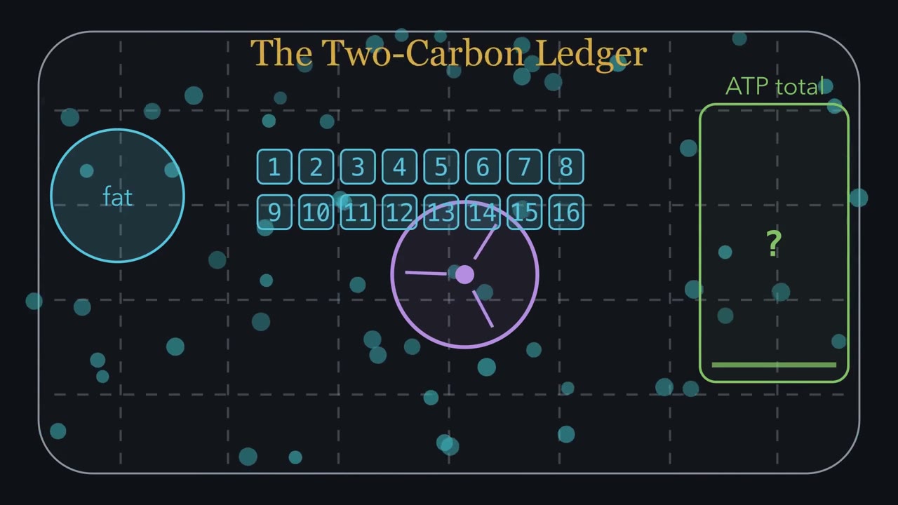 ▶ Watch The Two-Carbon Ledger · 3:17
Read the transcript of “The Two-Carbon Ledger”
Film transcript: The Two-Carbon Ledger
How can one fatty acid become roughly 106 ATP? Watch a 16-carbon chain enter this mitochondrial workshop, then count what leaves. In its simplest form, beta β-oxidation handles a saturated, even-numbered chain. Fat stores many electrons on carbon–hydrogen bonds. The workshop harvests them while removing two carbons per turn. First, however, the chain must reach the machinery. During fasting or exercise, fat droplets release fatty acids into blood. Albumin carries them to energy-using tissues. Activation attaches coenzyme A and spends two ATP equivalents.
Long-chain cargo then crosses the inner membrane through the carnitine shuttle. Inside the matrix, count from the coenzyme A end. Carbon 2 is α, and carbon 3 is β. A plain chain offers no cutting handle. Three moves rebuild the beta β-carbon, preparing the bond beside it. First oxidation makes a double bond and reduces FAD to FADH₂. Hydration adds water, placing an OH on the beta β-carbon. Second oxidation makes a ketone and reduces NAD⁺ plus to NADH. Now thiolase can cut. Thiolase adds fresh coenzyme A and releases one two-carbon acetyl-CoA. The remaining chain is two carbons shorter, so it returns to the first step. Beta β-oxidation converts chain length into a ledger of turns, carrier pairs, and acetyl coenzyme A. Beta -oxidation converts chain length into a ledger of turns, carrier pairs, and acetyl coenzyme A.
Predict the palmitate ledger. A 16-carbon chain becomes how many 2-carbon acetyl-CoA units, after how many turns? Place your answer beside the empty counters. Eight acetyl-CoA units leave, but only seven turns run. Seven cuts make eight pieces, and the last turn releases two acetyl-CoA. Therefore, 7 FADH₂ and 7 NADH also enter the ledger. Each carrier pair yields about 4 ATP through oxidative phosphorylation. Each acetyl-CoA yields about 10 through the citric acid cycle and oxidative phosphorylation. Seven pairs give 28; eight acetyl-CoA give 80. Subtract the 2-ATP activation toll. The result is roughly 106 ATP. That count applies to a saturated, even chain burned completely to carbon dioxide.
Double bonds and odd chains change the accounting. The reusable rule remains. Count the turns, the acetyl-CoA, and the activation toll.
From the film
- For a saturated, even chain, beta-oxidation removes two carbons per turn.
- Activation costs two ATP equivalents before beta-oxidation begins.
- Long-chain fatty acids use the carnitine shuttle to enter the mitochondrial matrix.
- Palmitate yields 8 acetyl-CoA, 7 FADH₂, 7 NADH, and roughly 106 net ATP.
Angus Barbieri went 382 days without food, living on his own body fat.
In 1965, at 27 and 456 pounds, he stopped eating under medical supervision. He lived on water, tea and coffee, with daily vitamins and, for stretches, potassium and sodium supplements, and finished at 180 pounds. Nobody measured his brain’s fuel, but by Cahill’s fasting data his blood ketones would have stayed high and his brain would have run largely on ketone bodies made from his own fat, with the glucose it still needed coming from gluconeogenesis. How did stored fat become fuel his tissues, and later his brain, could use?
Why fat is the reserve: the electrons
Cells make most of their ATP by taking electrons from fuel (oxidizing it) and passing them down the electron transport chain, so a fuel is worth roughly the electrons it can give up. Nearly every carbon in a fatty acid is a CH₂ group, as electron-rich as a carbon inside a chain can be. A typical glucose carbon is H–C–OH: it already carries an oxygen, so glucose is partly oxidized before the cell starts.
Per carbon, fat gives more ATP than glucose (the ledger below counts how much). Per gram the gap is wider: a gram of fat stores more than twice the energy of a gram of dry carbohydrate, because glucose’s oxygen atoms add weight without adding fuel. Storage widens it again: glycogen is kept with about twice its weight in water, fat nearly dry, so gram for gram of stored depot fat holds roughly six times as much. The cost of that density is access: fat is hidden in droplets as triacylglycerol, fatty acids do not dissolve in water, and each one must be released, ferried through blood and carried into a mitochondrion before a bond is burned.
The one idea to carry through this lesson. The film above shows how a saturated, even-numbered fatty acid becomes an energy ledger: fat’s fuel is the electrons on its CH₂ carbons, one repeated cycle spends them two carbons per turn, and the yield is a count of turns and acetyl-CoA, minus a 2-ATP activation toll. Below, we build the chemistry and exceptions behind that count.
The count holds for a saturated, even-numbered chain burned all the way to CO₂. The rest of the chapter is logistics, three stages as in the film (release, activation and import through a gate, then the cycle), and the edges of the rule: double bonds, odd chains, and a fasting liver that makes more acetyl-CoA than it can burn.
Stage 1: Mobilization, unlocking the droplet
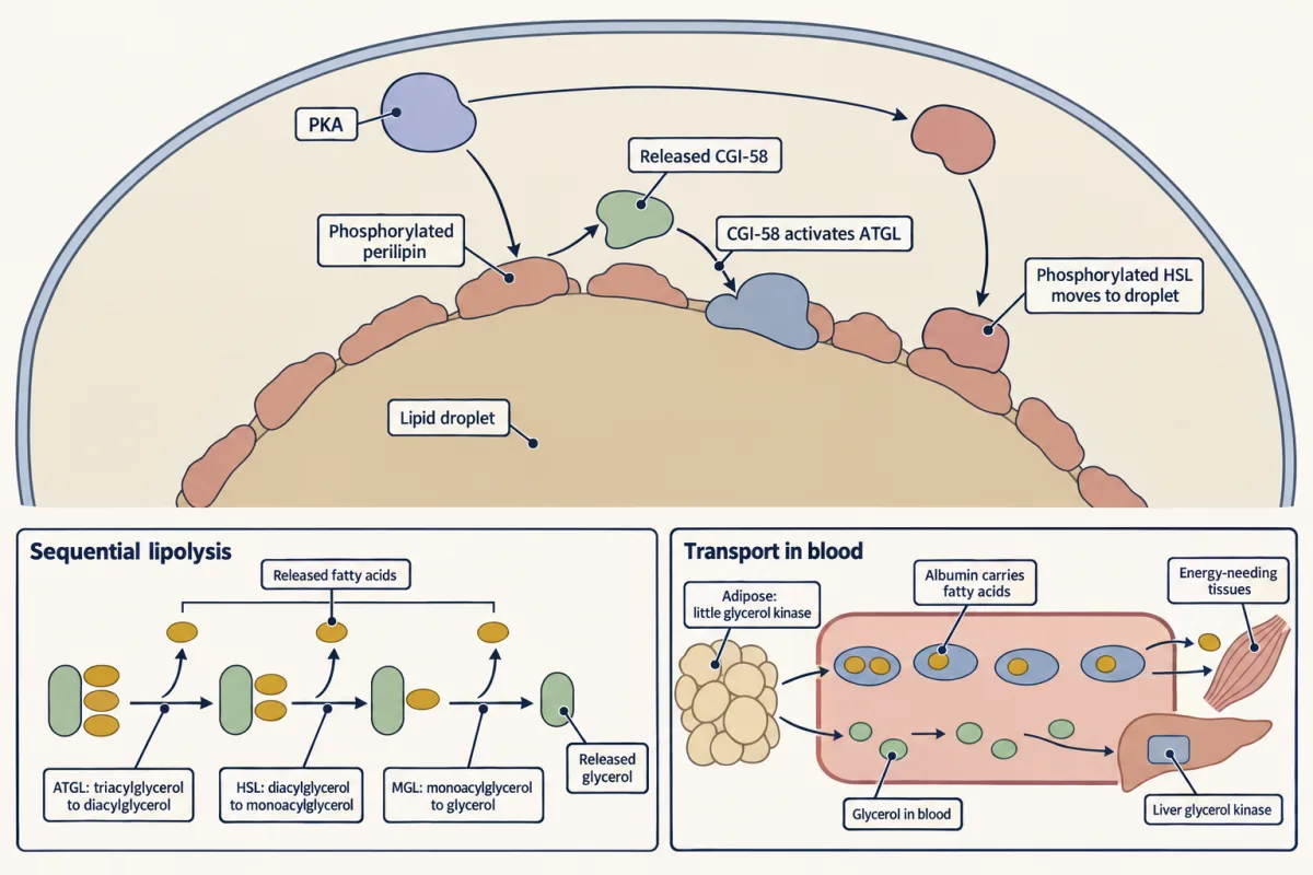
Triacylglycerol is glycerol holding three fatty acids by ester bonds, and about 95% of its energy sits in the chains. When you fast or exercise, epinephrine and norepinephrine bind β-adrenergic receptors (7-transmembrane receptors) on the fat cell, raise cAMP and activate protein kinase A (PKA), while falling insulin takes the brake off. It is the same cAMP → PKA signal that breaks down glycogen, so both storage depots empty together. Textbooks also list glucagon here; know it for exams, but its direct effect is mainly in rodent fat cells and small in human ones, where the catecholamines do most of the work.
PKA does two things: it loosens the protein coat on the fat droplet, which frees an activator for the first lipase, and it switches on a second lipase that then moves onto the droplet. Three lipases then remove the three fatty acids one at a time. These released free (nonesterified) fatty acids, not the fat you eat or the fat you store, are what travels in blood, bound to serum albumin. The glycerol is water-soluble and goes to the liver, where glycerol kinase phosphorylates it so it can enter glycolysis or gluconeogenesis.
Stage 2: Activation and the carnitine gate
First the fatty acid is activated: acyl-CoA synthetase couples it to coenzyme A. The reaction splits ATP to AMP plus pyrophosphate (PPᵢ), not to ADP, and hydrolysis of the PPᵢ makes the step irreversible.
Predict. How many high-energy phosphate bonds does activation spend?
Check your prediction
Two. ATP → AMP + PPᵢ breaks one, and PPᵢ → 2 Pᵢ breaks the second, so activation costs 2 ATP, not 1. Charge the toll against the yield at the end.
Acyl-CoA cannot cross the inner mitochondrial membrane, so it hands its cargo to carnitine (Latin carnis, flesh). Carnitine palmitoyltransferase I (CPT-I), in the outer mitochondrial membrane, swaps CoA for carnitine. A translocase ferries the acylcarnitine across the inner membrane, and CPT-II inside regenerates acyl-CoA.
CPT-I is the main control point for burning long-chain fatty acids. It is inhibited by malonyl-CoA, the first committed intermediate of fatty acid synthesis.
Predict. A drug strongly activates acetyl-CoA carboxylase, the enzyme that makes malonyl-CoA. What happens to fat burning in the mitochondria?
Check your prediction
It falls. More malonyl-CoA shuts CPT-I, so long chains cannot get in. That is the gate’s logic: while the cell is building fat, malonyl-CoA is high and the cell does not waste energy burning what it is busy making.
Stage 3: Beta-oxidation, one cycle run again and again
The film above numbers the chain from the CoA end and turns its β-carbon into a cutting handle. In palmitoyl-CoA, coenzyme A is attached to carbon 1 through a thioester, a sulfur-linked ester; carbon 2 is the α-carbon and carbon 3 the β-carbon. A plain bond between CH₂ groups gives an enzyme nothing to grab, so the cycle first rebuilds the β-carbon into a ketone (C=O).
Chemically, the new ketone gives CoA–S–C(=O)–CH₂–C(=O)–R: carbons 1, 2 and 3 in order, with R the rest of the chain. Carbon 2 then sits between two carbonyl groups, the thioester on one side and the new ketone on the other. Thiolase’s cysteine attacks the ketone carbon, the bond between carbons 2 and 3 breaks, and the thioester steadies the negative charge left on carbon 2, so the two-carbon piece leaves as acetyl-CoA.
Predict. Turning a CH₂ into a C=O means taking electrons away and adding an oxygen. Using an electron carrier, then water, then an electron carrier again, what three moves would do it?
Check your prediction
Oxidize, hydrate, oxidize: a double bond, then an OH on carbon 3, then a ketone. You have seen this sequence before: the citric acid cycle uses it to turn succinate into fumarate, then malate, then oxaloacetate.
The four reactions, with their carriers:
- Acyl-CoA dehydrogenase removes two hydrogens from carbons 2 and 3 and makes a trans double bond. The product is a trans-Δ²-enoyl-CoA; Δ² means the double bond starts at carbon 2. The electrons go to the enzyme’s FAD, making FADH₂.
- Enoyl-CoA hydratase adds water across the double bond, putting an OH on the β-carbon. No carrier is used.
- 3-Hydroxyacyl-CoA dehydrogenase oxidizes that OH to a ketone. These electrons go to NAD⁺, making NADH.
- β-Ketothiolase uses a fresh CoA to make that cut between carbons 2 and 3, releasing acetyl-CoA. The rest of the chain, now two carbons shorter, carries the fresh CoA straight back to step 1.
Why FAD first and NAD⁺ second? Making a carbon–carbon double bond out of two CH₂ groups releases too little energy to push electrons onto NAD⁺, so the enzyme hands them to its own tightly bound FAD, just as succinate dehydrogenase does in the citric acid cycle. Oxidizing an alcohol to a ketone is a closer match for NAD⁺, and the cut that follows pulls that step forward.
The film’s ledger shows these products stacking up. Use the cycle below to follow their chemistry turn by turn.
regulated step − used up · + produced
Activation, done once before the spiral begins. The fatty acid is adenylated to an acyl-adenylate, then CoA displaces AMP. ATP is split to AMP plus pyrophosphate, and hydrolysis of that pyrophosphate to 2 Pᵢ pulls the reaction forward and makes it irreversible. The cost is 2 ATP equivalents, not 1, because ATP went to AMP rather than ADP — the most-missed line in the ledger.
Acyl-CoA is too charged to cross the inner membrane, so CPT-I, in the outer mitochondrial membrane, swaps CoA for carnitine, a translocase carries acylcarnitine across, and CPT-II regenerates acyl-CoA inside. CPT-I is the main control point for long chains: malonyl-CoA, the first committed intermediate of fatty acid synthesis, inhibits it, so the cell avoids building and burning fat at the same time.
Removing a hydrogen from C2 and one from C3 installs a trans double bond between them, handing two electrons to FAD to make FADH₂. Chain-length isozymes (VLCAD, MCAD, SCAD; LCAD matters mainly in rodents) split the labor across long-to-short substrates.
Clinical. MCAD deficiency (roughly 1 in 10,000 to 20,000 births) stalls medium chains here, and with them every long chain on its way down, causing hypoketotic hypoglycemia under fasting; some deaths were once misread as SIDS; it is now usually caught on the newborn heel-prick. In Jamaican vomiting sickness from unripe ackee, a metabolite of hypoglycin A inhibits this same step.
Water adds across the new double bond with strict stereochemistry, placing an OH on the β-carbon (the L-3-hydroxy product). No cofactor; this just sets up the next oxidation.
The β-carbon hydroxyl is oxidized to a ketone, reducing NAD⁺ to NADH. The carbonyl now polarizes the C2–C3 bond, priming it for cleavage. The β-carbon is now a ketone, the handle thiolase needs.
An active-site cysteine attacks the β-keto carbon, releasing the two carbons at the CoA end as acetyl-CoA and holding the rest of the chain on the enzyme; a second CoA then takes that chain off as an acyl-CoA shortened by two carbons that re-enters at the dehydrogenase. Each acetyl-CoA can feed the citric acid cycle; each FADH₂ and NADH feeds the electron-transport chain.
Chain (palmitoyl-CoA): C16 · turn 0 of 7
The ledger: counting packets
Predict. Palmitate has 16 carbons. How many turns of the cycle does it take to cut it all into acetyl-CoA, and how many acetyl-CoA come out?
Check your prediction
Seven turns, eight acetyl-CoA. Each turn makes one cut, and a ruler needs seven cuts to become eight pieces. The last turn splits a four-carbon chain into two acetyl-CoA at once. Counting eight turns is the most common mistake here.
At this course’s rates, through the electron transport chain each NADH is worth about 2.5 ATP and each FADH₂ about 1.5. Each acetyl-CoA burned in the citric acid cycle is worth about 10 ATP: it makes 3 NADH, 1 FADH₂ and 1 GTP, and 3 × 2.5 + 1.5 + 1 = 10.
In words Net ATP equals the number of turns times four, for each turn's FADH2 and NADH, plus ten for each acetyl-CoA, minus two for activation.
- turns“turns”
- turns of the β-oxidation cycle: half the number of carbons, minus one
- acetyl-CoA“acetyl CoA”
- two-carbon units released: half the number of carbons
- 1.5 + 2.5“one point five plus two point five”
- ATP from one FADH₂ plus one NADHATP
- 10“ten”
- ATP from one acetyl-CoA through the citric acid cycle and the electron transport chainATP
- 2“two”
- the activation toll: ATP split to AMP, which spends two high-energy bondsATP
Sanity check The ATP values per carrier are rounded averages. Older textbooks that count 3 per NADH and 2 per FADH₂ land near 129 for palmitate. Learn the counting, not the number. For a saturated, even chain of n carbons the sum always comes to 7n − 6 (for palmitate, 7 × 16 − 6 = 106).
Try one number
Palmitate, 16 carbons: 7 turns and 8 acetyl-CoA.
From the turns: 7 FADH₂ × 1.5 = 10.5 ATP, and 7 NADH × 2.5 = 17.5 ATP. Together, 28.
From the acetyl-CoA: 8 × 10 = 80 ATP.
Gross: 28 + 80 = 108 ATP. Subtract the activation toll: 108 − 2 = 106 ATP.
Need a refresher? units and prefixes
- Spiral turns
- 7
- Acetyl-CoA
- 8
- FADH₂
- 7
- NADH
- 7
- Activation
- −2 ATP ATP → AMP
- Net ATP
- 106
turns × 4 + acetyl-CoA × 10 − 2 = 7 × 4 + 8 × 10 − 2 = 106
C16 palmitate runs 7 turns of the spiral, giving 8 acetyl-CoA, 7 FADH₂, and 7 NADH: about 106 net ATP, 3.3 times what one glucose yields. Most of it (80 ATP) comes later, when the acetyl-CoA burns in the citric acid cycle; the spiral's own carriers add 28, minus 2 for activation. Per carbon, fat still wins (6.6 vs 5.3) because its carbons start out more reduced.
Modern P/O ratios (2.5 ATP/NADH, 1.5/FADH₂, ~10/acetyl-CoA). Older textbooks counting 3 and 2 give higher totals (palmitate ≈ 129). Even chains only here; odd chains leave a propionyl-CoA.
Compare sugar: glucose, fully oxidized, gives about 32 ATP from 6 carbons, about 5.3 per carbon. Palmitate gives 106 from 16, about 6.6 per carbon: about a quarter more, and one palmitate is worth a little more than three glucose. The per-carbon gain comes from the extra electrons; for fat’s role as the reserve, the per-gram gap and dry storage above matter even more.
MCAD deficiency: a block in the middle stalls the spiral
Predict. Step 1 comes in chain-length versions, and most dietary fat is long-chain. Why would losing only the medium-chain enzyme, MCAD, stop a child from burning fat during a fast?
Check your prediction
Every long chain becomes a medium chain on its way down the spiral, so a block at medium length stalls much of fat burning partway through. Too little acetyl-CoA reaches the liver’s ketone pathway, so ketones stay low, and tissues burn glucose instead while the liver loses the energy it needs to make more. Blood sugar falls without ketones appearing: hypoketotic hypoglycemia.
MCAD deficiency affects roughly 1 in 10,000 to 20,000 births and is most common in people of northern European ancestry. A stomach bug or a skipped breakfast can set off the crash. Some deaths from it were once written off as SIDS. Now a newborn heel prick, read by tandem mass spectrometry, catches most cases, though a few babies crash before the result is back. One enzyme, one heel prick, thousands of lives. The same step is the target in Jamaican vomiting sickness, where a metabolite of hypoglycin A from unripe ackee fruit poisons several of the acyl-CoA dehydrogenases.
Special cases that bend the rule
Two kinds of real fat break the rule’s assumptions of an identical packet per turn and a chain that ends in acetyl-CoA.
- Unsaturated chains. After a few cycles, a natural cis double bond lands in a position (cis-Δ³, starting at carbon 3) the standard enzymes cannot process. cis-Δ³-enoyl-CoA isomerase moves it to the trans-Δ² position and the cycle resumes. The double bond is already there, so that turn skips the FAD step: stearate (C18, saturated) nets 120 ATP, oleate (C18, one cis bond at an odd-numbered position) 118.5. Some polyunsaturated chains need a second helper, 2,4-dienoyl-CoA reductase, which spends NADPH.
- Odd chains. The last thiolase cut leaves a three-carbon propionyl-CoA instead of an acetyl-CoA. A biotin enzyme converts it to methylmalonyl-CoA, an epimerase flips its arrangement at one carbon, and a vitamin B12 enzyme rearranges it into succinyl-CoA, a citric acid cycle intermediate. A B12 deficiency stalls this step and spills methylmalonic acid into urine.
Why even-chain fat gives no net glucose
The odd-chain route matters because of a hard rule: mammals cannot make net glucose from even-chain fatty acids by the usual route. Each turn of the citric acid cycle brings in two carbons as acetyl-CoA and loses two as CO₂, so no net oxaloacetate is left over to seed gluconeogenesis. Succinyl-CoA is a net addition of a four-carbon intermediate, so that surplus can be drawn off as oxaloacetate and become glucose. Apart from a small trickle through acetone (some is metabolized back to pyruvate), only the odd-chain tail and the glycerol backbone make net sugar.
Ketone bodies: the liver’s fasting export
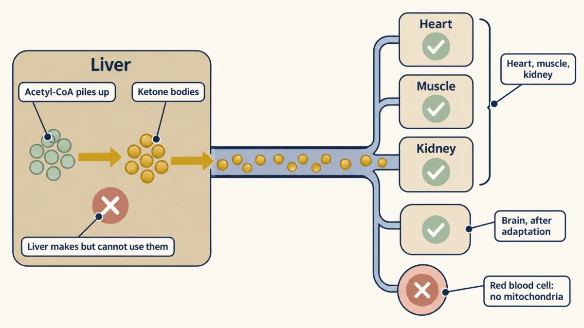
When glucose is scarce, low insulin floods the liver with fatty acids and opens carnitine palmitoyltransferase I (CPT-I). The liver’s β-oxidation then makes acetyl-CoA faster than its citric acid cycle can burn it. Fasting signals also raise the amount of HMGCS2 (HMG-CoA synthase), the regulated enzyme that commits acetyl-CoA to ketone bodies, and oxaloacetate drawn off for gluconeogenesis adds to this.
The liver repackages the surplus. Thiolase, HMGCS2 and 3-hydroxy-3-methylglutaryl coenzyme A (HMG-CoA) lyase build acetoacetate, which is reduced to D-3-hydroxybutyrate (the dominant form in blood) or decarboxylates spontaneously to acetone. Acetone is exhaled as the fruity “acetone breath” that can get diabetic ketoacidosis mistaken for drunkenness.
The liver keeps only the carrier part of each packet, because the acetyl-CoA leaves in the ketones instead of entering its own citric acid cycle. If it exports palmitate’s eight acetyl-CoA as four acetoacetate, it keeps 7 × 4 − 2 = 26 ATP, about a quarter of the 106. The tissues that burn the ketones collect most of the rest. Each acetoacetate gives them two acetyl-CoA, worth 20, minus one GTP, because reloading it onto CoA spends a succinyl-CoA: 4 × 19 = 76. Exported as D-3-hydroxybutyrate instead, each ketone carries one of the liver’s NADH with it, so the liver keeps about 16 and the tissues get about 86. Liver plus tissues total 102, not 106; the missing 4 are the four GTP the tissues give up to reload the ketones onto CoA.
The liver cannot use its own ketones. It lacks thiophorase (succinyl-CoA:3-ketoacid CoA transferase), the enzyme that reloads acetoacetate onto CoA so it can be split back into two acetyl-CoA. Red blood cells cannot use ketones either, having no mitochondria. Heart, muscle, kidney and, after adaptation, the brain run on them happily. George Cahill’s starvation studies measured the payoff: by day 40 of a fast, protein breakdown falls from roughly 75 to 20 grams a day as the brain shifts onto ketones, sparing muscle. The same ketotic state is the basis of the ketogenic diet, which reduces seizures in many children with drug-resistant epilepsy; how it does so is still not settled. Russell Wilder introduced the diet for epilepsy in 1921, and the Charlie Foundation revived it in 1994.
The citric acid cycle chapter follows the acetyl-CoA the rest of the way to CO₂, and oxidative phosphorylation shows where the FADH₂ and NADH you tallied cash out. The next chapter, fatty acid synthesis, runs the same two-carbon logic in the building direction, starting from the malonyl-CoA that keeps the carnitine gate shut.
Going deeper
The main line above does not depend on these details.
A second route to lipolysis
The catecholamine–cAMP signal is not the only trigger: heart-derived natriuretic peptides also start lipolysis, through cGMP rather than cAMP.
Inside the droplet: perilipin, CGI-58 and three lipases
Besides perilipin, whose release of CGI-58 switches on adipose triglyceride lipase (ATGL), protein kinase A (PKA) phosphorylates hormone-sensitive lipase (HSL), which moves onto the droplet. In order, ATGL takes triacylglycerol to diacylglycerol, HSL takes that to monoacylglycerol, and monoacylglycerol lipase removes the last fatty acid. When CGI-58 is mutated and ATGL cannot be switched on, fat accumulates in tissues that should not hold it: Chanarin-Dorfman syndrome.
Albumin’s spare capacity
The fatty acids leave the fat cell on serum albumin, which has about seven fatty acid binding sites but normally carries only one or two.
Who does step 1 at each chain length
The chain-length versions of step 1 are VLCAD, MCAD and SCAD, plus LCAD, which matters more in rodents than in people. Mind the naming trap. VLCAD acts on roughly C12 to C20 chains, palmitate included, and sits on the inner mitochondrial membrane; for those long chains, steps 2 to 4 are done by one membrane-bound complex, the mitochondrial trifunctional protein. Shorter chains use separate soluble enzymes in the matrix. “Very-long-chain fatty acids” (about C22 and longer) are first shortened in peroxisomes, where step 1 hands its electrons to O₂, making H₂O₂, so less ATP is made.
Where step 1’s electrons go
Acyl-CoA dehydrogenase’s bound FADH₂ passes its electrons to ubiquinone through electron-transferring flavoprotein (ETF) and ETF:ubiquinone oxidoreductase, skipping complex I, which is why each is worth about 1.5 ATP, like the FADH₂ of succinate dehydrogenase.
Chains that skip the shuttle
The carnitine gate is a long-chain checkpoint. Short- and medium-chain fatty acids mostly cross into the matrix as free acids without the carnitine shuttle, most clearly in liver, and are activated to acyl-CoA inside.
CPT1A in Arctic populations
The CPT1A P479L variant, common in Inuit and other Arctic populations, lowers carnitine palmitoyltransferase I (CPT-I) activity and makes it less sensitive to malonyl-CoA, which shows how much the import step matters. It carries genetic signs of past selection, though what it was selected for is still debated, and it has been linked to fasting hypoglycemia and higher infant death rates, without proof that it causes those deaths.
Your notes
Select any text in the lesson to highlight it or add a note to it. Your highlights and notes are saved in this browser and collected in your notebook.
This browser is not letting the site save anything (a private window or strict privacy settings), so highlights and notes will be lost when you leave the page.
How we measure it
Energy-ledger accounting (the activation toll)
The yield from a fatty acid comes from a step-by-step tally: one acetyl-CoA, one FADH2, and one NADH per cycle, run (n/2 − 1) cycles for an even chain, add the extra acetyl-CoA the last cleavage releases, burn the acetyl-CoA through the TCA cycle and the carriers through the ETC, then subtract 2 ATP for activation (ATP goes to AMP plus 2 Pi, which costs two high-energy bonds). For palmitate that lands near 106 net ATP, and the activation toll is the single most-missed line in the books.
Stable-isotope tracing of flux
Rudolf Schoenheimer fed animals fatty acids and amino acids tagged with deuterium and 15N and followed where the labels went. The tracers showed that fat stores are continuously torn down by lipolysis and rebuilt by re-esterification — the dynamic-state view that turns metabolism from a static map into measurable flux.
Newborn heel-prick screening (tandem mass spectrometry)
A drop of blood from a newborn's heel is analyzed by tandem MS for acylcarnitine signatures. A medium-chain acylcarnitine spike flags MCAD deficiency, usually before the first fasting crisis, turning a disorder whose deaths were sometimes misread as SIDS into something caught and managed. One enzyme, one heel prick, thousands of lives.
Practice questions on this lesson (7) → MCAT-style, original, each with an explanation
Where this lesson fits
Before thisReview first
- The Citric Acid Cycle: Harvesting ElectronsThis course · Lesson 19
You need to know how acetyl-CoA is burned for NADH, FADH2 and GTP to count palmitate's ATP.
- Oxidative Phosphorylation: The Electron-Transport ChainThis course · Lesson 20
The 2.5 and 1.5 ATP per NADH and FADH2 used in the ledger come from there.
- Signal-Transduction Pathways: How Cells Compute With Molecular SwitchesThis course · Lesson 13
The epinephrine, cAMP and PKA relay that starts fat mobilization is built there.
After thisStudy next
- Fatty Acid Synthesis: Building the Chain Two Carbons at a TimeThis course · Lesson 26
It builds fat in the other direction, starting from the malonyl-CoA that shuts the carnitine gate here.
- Integration of Metabolism: How the Body Switches FuelsThis course · Lesson 30
It shows how fasting hormones switch fat burning and ketone making up or down alongside other pathways.
- Protein Turnover and Amino Acid Catabolism: Tag, Recognize, Destroy, RecycleThis course · Lesson 28
Fasting fat burning and ketones spare the muscle protein whose breakdown this lesson tracks.
Suggested by an AI model from the lessons themselves, then checked. How we use AI
Finish line
Test yourself
9 questions
Practice and MCAT-style, with explained answers.
Next · Lesson 26
Fatty Acid Synthesis: Building the Chain Two Carbons at a Time
Cytoplasm, ACP, NADPH: three choices that keep building fat from colliding with burning it.
Continue →Also in the arcade for this lesson: Wire the Cell: Fasting liver makes ketones
← Lesson 24 · The Pentose Phosphate Pathway: NADPH and Five-Carbon Sugars