Sensory Systems · Unit 7: Smell and taste
From Glomeruli to Piriform Cortex
Rewritten on : new text, flashcards and film. Your notes and highlights stay, but the text around them may have moved.
The bulb hands piriform cortex a tidy map of receptor identity, and the cortex scatters it. How can a scattered pattern still carry which odours are alike, and come to mean something?
Pause when the reading reaches a film, and offer to play it. Saved on this device.
Watch the film now, or keep listening
Builds on:One Receptor per Neuron: How Olfactory Neurons ChooseSensory Neurons by Their Genes: the Atlas EraWhy these?
 ▶ Watch What the Bulb Sends · 3:21
Read the transcript of “What the Bulb Sends”
Film transcript: What the Bulb Sends
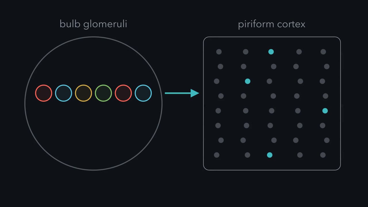
Your nose hands the brain an orderly map, and the cortex scatters it. So what does the cortex keep? A smell starts with molecules binding receptors in the nose. Each receptor neuron makes one type of receptor. Neurons of one type send their axons to the same small knot of synapses in the bulb. That knot is a glomerulus. In mice, each receptor type usually has two glomeruli in each bulb. An odour binds some receptor types and not others. So it activates a set of glomeruli. Here is a question. Raise the concentration of the same odour. Do the same glomeruli light, or more of them?
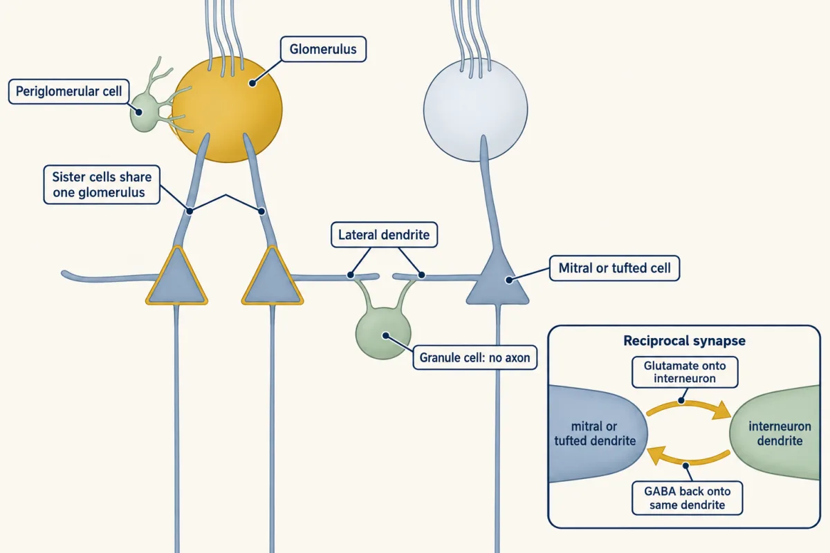
Usually more. Higher concentrations often recruit extra glomeruli, though this can level off. So the set is characteristic at a given concentration. The layout is a map of receptor identity. Where glomerulus sits tells you which receptor feeds it. The layout is only coarsely related to chemistry. So it is not a fine chemical map. Mitral and tufted cells carry the pattern out. Each sends one main dendrite into one glomerulus. Cells that send their dendrites into the same glomerulus are called sister cells. They share one input. Inhibitory interneurons sit around them. Periglomerular cells work in the glomerular layer. Granule cells have no axons.
Mitral and tufted axons run on to piriform cortex, the main olfactory area of the cortex. There is no relay in the thalamus. There the tidy map seems to vanish. One odour switches on neurons scattered across the cortex. That raises four puzzles. Does the output of one glomerulus stay together in the cortex? If it spreads out, how can the cortex still carry which odours are chemically alike? Can a scattered pattern acquire learned meaning? And does an odour's pattern stay put from week to week? One idea answers the second puzzle. Where a piriform neuron sits tells you little about which odours excite it. In our simple model, its glomerular inputs and its threshold decide its response.
Real tuning also depends on input strengths, timing and the cortex's own network. Each neuron gets input from several scattered glomeruli. It fires only when enough are active together. So the more glomeruli two odours share, the more neurons they share, wherever those neurons sit. With sparse responses, random wiring like this keeps the order of similarity on average. But related odours come out less alike than in the bulb. Anything tighter is not explained by this model. The cortex's own network and experience are the candidates. So the bulb sends a map of which receptors are active. The rest of this story is what the cortex keeps of it.
From the film
- The olfactory bulb sends piriform cortex a map of which receptor types are active, and in the cortex one odour switches on scattered neurons.
- An odour is characterized by the set of glomeruli it activates at a given concentration, and higher concentrations often recruit more of them.
- A simple model explains why a scatter can keep which odours are alike, and the cortex's own network and experience are candidates for anything tighter.
In the olfactory bulb, smell arrives as a map: receptor neurons of one type converge on a glomerulus, so each odour lights up a characteristic pattern of glomeruli at a given concentration. Piriform cortex does not keep that map. An odour activates neurons scattered across the cortex, yet chemically similar odours still evoke similar patterns there. The cortex’s own network then reshapes those patterns, so related odours cluster more strongly than in the bulb’s input, and a pattern can acquire learned meaning. Reviews of piriform cortex describe odour quality as coded sparsely and in a distributed way, and put learning and memory at the centre of how odours are perceived (Gottfried, 2010; Wilson and Sullivan, 2011).
Smell and taste ended with receptor neurons converging on glomeruli and the bulb projecting to piriform cortex. This lesson follows that projection into the cortex, nearly all of it in mice. The question at each stage is what the circuit keeps of the receptor map, what it adds, and what it gives up. How the receptor choice and the axon wiring arise is in olfactory receptor choice, partners, layers and self-avoidance and topographic maps.
What the bulb sends, and the puzzle
Each olfactory receptor neuron makes one type of receptor, and neurons of one type send their axons to the same small knot of synapses in the bulb, a glomerulus (in mice, usually two per bulb for each receptor type). An odour binds some receptor types and not others, so it activates a set of glomeruli. That set is characteristic of the odour at a given concentration. Raising the concentration often recruits extra, less sensitive glomeruli, though recruitment can level off.
The bulb’s layout is therefore a map of receptor identity: where a glomerulus sits tells you which receptor feeds it. The layout is only coarsely related to chemistry, so it is not a fine chemical map.
Mitral and tufted cells are the principal output neurons of the mouse main olfactory bulb. Each sends a primary dendrite into one glomerulus and an axon toward the cortex. Cells that send their dendrites into the same glomerulus are sister cells: they share one input. Around and beneath them sit interneurons: periglomerular cells, GABAergic cells that work in the glomerular layer, and granule cells, which lack axons and contact the lateral dendrites of mitral and tufted cells.
Mitral and tufted axons reach piriform cortex, the main olfactory area of the cortex, without a thalamic relay. There the tidy map seems to vanish: an odour switches on neurons scattered across the cortex. That raises four puzzles.
- Scatter. Does the output of one glomerulus stay together in piriform cortex, or spread out?
- Similarity. If it spreads out, how can the cortex still carry which odours are chemically alike?
- Meaning. Can a scattered pattern acquire learned meaning, and what would that show?
- Stability. Does an odour’s pattern in the cortex stay put from week to week?
The first puzzle is anatomy, taken next. The second needs one term and one idea: a piriform neuron’s glomerular inputs are the glomeruli whose mitral and tufted axons reach it. It also receives input from other cortical neurons.
The one idea to carry through this lesson. Where a piriform neuron sits tells you little about which odours excite it. In the simple feed-forward model used here, its glomerular inputs and its threshold decide its response: each neuron samples several glomeruli scattered across the bulb and fires only when enough of them are active together. So the more glomeruli two odours share, the more neurons they share, wherever those neurons sit. With sparse responses, this random wiring keeps the order of similarity on average, but related odours come out less alike than in the bulb. Real responses also depend on synaptic strengths, timing and inhibition. Anything tighter, such as related odours clustering more strongly, completion from partial input or learned meaning, is not explained by this model; the cortex’s own network and experience are the candidates tested below.
Following one glomerulus
Read the transcript of “Following One Glomerulus”
Film transcript: Following One Glomerulus
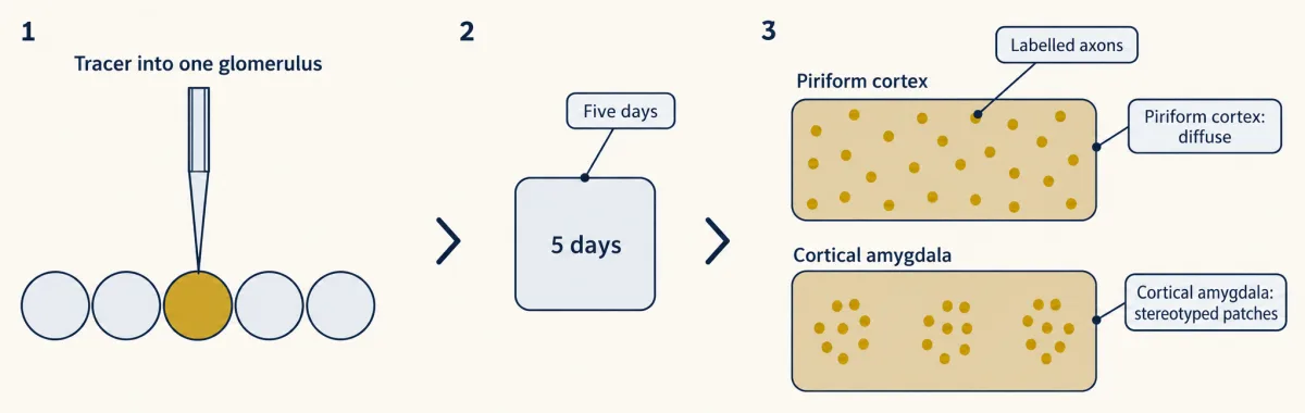
If the cortex kept the bulb's map, where would one glomerulus send its output? A topographic map keeps neighbours together. Then one glomerulus should land in one patch of cortex. Sosulski and colleagues asked whether the wiring does that. They put a tracer into a single glomerulus of an adult mouse. Five days later, they imaged where the labelled axons went. They looked in two targets: piriform cortex and, for comparison, the cortical amygdala. Pause and predict. In piriform cortex, will the axons gather in a patch or spread out? Must both targets agree?
In piriform cortex the axons spread diffusely, with no apparent spatial preference. In the cortical amygdala, the same glomerulus gave stereotyped patches. One tracer in one glomerulus showed two different rules in two different targets. It would have been easy to conclude that the bulb projects at random everywhere. The cortical amygdala shows it does not. So the scatter is not a property of the whole bulb. It was seen in piriform cortex, not in the amygdala. A tracer image shows where axons go. It does not show which synapses each neuron makes. Recordings of responses tell the same story. Stettler and Axel imaged odour responses in piriform cortex of living mice.
An odour activated a dispersed ensemble. Its neurons were scattered across the area, not gathered in a patch. A neuron's receptive field is the set of odours that excite it. A single piriform neuron could respond to chemically dissimilar odours. Its receptive field was discontinuous in chemical space. This does not say piriform carries no chemical information. It says the arrangement is not a topographic map. Diffuse is also not the same as random. Later projection mapping found spatial structure in how bulb neurons route their axons. It was not a map of odour chemistry. So the scatter is real, and it belongs to piriform cortex. Next comes the harder question: how can a scatter keep chemistry?
From the film
- In mice, one glomerulus sent axons diffusely across piriform cortex but to stereotyped patches in the cortical amygdala.
- In piriform cortex an odour activates a dispersed ensemble, and a single neuron can respond to chemically dissimilar odours.
- The scatter belongs to piriform cortex, not to the whole bulb, and diffuse is not the same as random.
If piriform cortex kept the bulb’s map, the output of one glomerulus should land in one patch of cortex, the way a topographic map keeps neighbours together. Sosulski and colleagues (2011) labelled the output axons of a single glomerulus in adult mice with a tracer and imaged where they went, in piriform cortex and, for comparison, in a second target, the cortical amygdala.
Predict. If the cortex kept the map, what would the labelled axons look like in piriform cortex? What would it mean if the two targets looked different?
Check your prediction
A kept map would show up as a patch of labelled axons. If the bulb sprayed its axons at random everywhere, both targets would look diffuse. If the two targets differ, the rule belongs to the target.
What they found. In piriform cortex the axons projected diffusely with no apparent spatial preference. In the cortical amygdala the same glomerulus gave spatially stereotyped patches. So dispersal belongs to one target, not to the whole bulb.
Recordings of responses tell the same story. Stettler and Axel (2009) imaged odour responses in mouse piriform cortex in vivo and found that an odour activated a dispersed ensemble, neurons scattered across the area and not gathered in a patch. A single neuron could respond to chemically dissimilar odours: its receptive field, the set of odours that excite it, was discontinuous in chemical space. That does not mean piriform carries no chemical information, only that its arrangement is not a topographic map.
Diffuse is also not the same as random: later projection mapping found spatial structure in how bulb neurons route their axons, though not a map of odour chemistry (Chen and colleagues, 2022). That settles the first puzzle. The second is harder.
How a scatter keeps similarity
Read the transcript of “How a Scatter Keeps Similarity”
Film transcript: How a Scatter Keeps Similarity
If one glomerulus spreads its output all over the cortex, is the chemistry lost? First, what does similar mean for a whole population of neurons? Record many piriform neurons while a mouse smells odour A. Write down each neuron's response. This one fired. That one did not. A third fired a little. That list is A's population pattern. Do the same for odour B. Two odours are represented similarly if many of the same neurons respond to both. Real analyses correlate the responses of many neurons. That is a related measure, but not the same number. Now the wiring. Tracing found that each piriform neuron gets input from several glomeruli spread across the bulb.
Driving one glomerulus did not make the cells fire. In this simple model, a neuron fires only when enough inputs are active together. Nothing about the neuron's position enters the model. Here is a toy. Each neuron listens to two glomeruli and fires only if both are active. Wiring is random. Odour A activates ten glomeruli. A related ester, B, also activates ten, and shares six with A. An unrelated amine, C, shares none. Any pair of A's glomeruli can feed a neuron. Those neurons respond to A. The pairs are counted on screen. Which of them also respond to B? Only neurons whose two inputs both sit among the six shared glomeruli. Pause and predict.
The odours share sixty percent of their glomeruli. What share of A's neurons also respond to B? About one third, on average. The odours share most of their glomeruli, but only a third of their responsive neurons. And for C, no A neuron responds at all, because no glomeruli are shared. Two results follow. B is still far more like A than C is. The order of similarity survives. But the overlap has shrunk. Both of a neuron's inputs must be shared, so six tenths times five ninths gives one third.
Where the neurons sit never entered the calculation. A larger toy, with a thousand glomeruli and twenty inputs per neuron, gives the same two results. The order is kept. Related odours become less alike, not more. So in this model, where a neuron sits does not matter. Which glomeruli feed it does.
So here is a test for real data. If piriform worked like this model, related odours should be less alike there, not more. In this model, random wiring keeps the expected order of likeness, but makes related odours less alike. Anything tighter needs more than this model.
From the film
- In a simple model, a piriform neuron fires only when enough of its scattered glomerular inputs are active together, and where it sits does not matter.
- Random wiring of this kind keeps the expected order of odour similarity.
- The same wiring makes related odours less alike than in the bulb, so anything tighter needs more than this model.
If one glomerulus spreads its axons all over piriform cortex, is the chemistry lost? First, what does “similar” mean for a whole population?
A population pattern. Record from many piriform neurons while a mouse smells odour A, and write down each neuron’s response: this one fired, that one did not, a third fired a little. That list is A’s population pattern. Do the same for odour B. Two odours are represented similarly if their lists overlap, that is if many of the same neurons respond to both. Real analyses correlate the responses of many neurons, a related measure but not the same number.
How a piriform neuron is wired. Tracing back from single piriform neurons found that each receives input from mitral cells of several glomeruli spread across the bulb (Miyamichi and colleagues, 2011). In recordings from anterior piriform cortex, driving one glomerulus with light did not make the cells fire, while combinations of co-active glomeruli did (Davison and Ehlers, 2011). The simple model used here keeps just those two facts: a neuron fires only when enough of its inputs are active together, because one input alone does not bring it to threshold (see action potential).
Worked problem: overlap after a scatter. Take a toy neuron that listens to two glomeruli and fires only if both are active. Wiring is random, so every pair of glomeruli is equally likely to feed a neuron. Odour A activates 10 glomeruli. A related ester, B, also activates 10 and shares 6 of them with A. An unrelated amine, C, shares none (esters and amines are two different chemical families).
Step 1: which neurons respond to A? Those whose two inputs both fall among A’s 10 glomeruli. There are 10 × 9 / 2 = 45 such input pairs.
Step 2: which of those also respond to B? Those whose two inputs both fall among the 6 shared glomeruli: 6 × 5 / 2 = 15 pairs.
Step 3: overlap. 15 / 45 = 1/3. On average, in a large randomly wired population, a third of the neurons that respond to A also respond to B. The odours share 60 percent of their glomeruli but only about 33 percent of their responsive neurons.
Step 4: the unrelated odour. No glomeruli are shared, so no A-responsive neuron can also respond to C. The overlap is 0.
Check: B is still far more like A than C is, so the order survives. The overlap has shrunk because both of a neuron’s inputs must be shared: 6/10 × 5/9 = 1/3, close to squaring the shared fraction (0.6² = 0.36).
Where the neurons sit never entered the calculation. A larger toy with 1,000 glomeruli gives the same two results: the order of similarity is kept, and related odours become less alike, not more, as long as responses stay sparse (see Going deeper).
Predict. Pashkovski and colleagues (2020) imaged two things in mice breathing at a controlled sniff rate, with the same panel of odours for the comparison: the bulb’s axon boutons inside piriform cortex (the terminals of mitral and tufted cells, so the cortex’s input), and piriform neurons in layers 2 and 3, where its principal cells sit (its output). Which comparisons would test whether the scatter keeps chemistry, and adds anything? If piriform worked like the toy, how should the similarity of related odours compare between boutons and cortical neurons?
Check your prediction
Three comparisons do it. Is there a spatial map? Ask whether neurons near each other share responses more often than distant ones. If they do not, there is no spatial map. Is there structure anyway? Ask whether chemically related odours give more similar population patterns than unrelated ones. Has the cortex added something? Compare the boutons’ similarity structure with the cortex’s on the same panel.
By the toy, piriform should largely keep the bulb’s order of similarity, with related odours less alike than in the boutons, not more.
What they found. The first half held: in both the boutons and the piriform neurons, chemically related odours gave more similar patterns than unrelated ones, so the scattered cortex kept the chemistry. The second half did not: piriform neurons clustered related odours more tightly than their own input did. To picture it in made-up numbers, related pairs might correlate at about 0.4 in boutons and 0.7 in cortex. The authors also passed their bouton responses through a published random feed-forward model: it largely kept the relationships but made the patterns less alike, as the toy predicts, and the real cortex did not match it. Something in the cortex added structure that this random model did not give.
This is a different kind of map. A spatial map says that similar odours activate nearby places. A representational geometry says that similar odours evoke similar population patterns, wherever the cells sit. Piriform cortex has the second without the first.
What the cortex’s network adds
Read the transcript of “What the Cortex Adds”
Film transcript: What the Cortex Adds
Does piriform cortex just scatter its input, or does it add something of its own? Pashkovski and colleagues imaged two things in mice, with the same panel of odours for the comparison. First, bulb axon boutons inside piriform cortex. These are the terminals of mitral and tufted cells, the cortex's input. Second, piriform neurons in layer 2/3, the cortex's output. The mice breathed at a controlled sniff rate. Pause and predict. If piriform followed our simple random model, how should related odours compare between boutons and cortex?
By that model, the cortex should largely keep the bulb's relationships. Related odours should be less alike than in the input, not more. The first half held. In boutons and in cortex, related odours gave more similar patterns than unrelated ones. The scattered cortex kept the chemistry. The second half did not hold. Piriform neurons clustered related odours more tightly than their own input did. Made-up numbers on screen show the direction. The cortex value sits above the input value. The authors also fed their bouton data through a random-wiring model. It kept the order, but made the patterns less alike. So something in the cortex added structure that the random model did not give.
This is a different kind of map. A spatial map puts similar odours in nearby places. A representational geometry gives similar odours similar population patterns, wherever the cells sit. Piriform cortex has the second without the first. What could add the structure? Piriform pyramidal cells, its main excitatory neurons, are linked to each other by association fibres. These are recurrent connections within the cortex. Reviews treat them as a place where experience is stored. Strengthening synapses among neurons that are active together is thought to bind a scattered ensemble into a familiar odour. Then the whole ensemble could respond even when part of the input is missing.
This is pattern completion. Pashkovski's team tested the network directly. A genetic tool blocked transmitter release from excitatory piriform neurons. Their bulb input still arrived, so they still responded. But they excited their neighbours, including inhibitory cells, much less. Pause and predict. With the network weakened, what should happen to the cortex's odour relationships?
Its odour relationships became more like the bulb's. Responses did not simply weaken. Tuning broadened and correlations rose overall, yet the pattern moved toward the bulb's. One caveat. Piriform also sends feedback to the bulb, and connects to other olfactory areas. So piriform network activity is needed for the change. It is not shown that local connections alone produce it. The cortex keeps the bulb's chemistry, then reshapes it with its own network.
From the film
- In mice, related odours gave more similar patterns than unrelated ones in both bulb boutons and piriform cortex.
- Piriform neurons clustered related odours more tightly than their input did, so the cortex adds structure that the random model does not give.
- Blocking transmitter release from excitatory piriform neurons moved the cortex's odour relationships toward the bulb's, but this does not show that local connections alone produce the change.
What could add the structure? Piriform pyramidal cells, its main excitatory neurons, are linked to each other by association fibres, recurrent connections within the cortex, and reviews of the field treat those connections as a place where experience is stored. Strengthening the synapses among neurons that are active together is thought to bind a scattered ensemble into a familiar odour object. That would help an animal tell a familiar odour from similar ones, and let the whole ensemble respond even when part of the input is missing, a process called pattern completion (Wilson and Sullivan, 2011). The same network is a candidate for the extra clustering.
Pashkovski and colleagues tested the network directly. A genetically targeted silencing tool blocked transmitter release from excitatory piriform neurons, attenuating the cortex’s associational network. Their input from the bulb still arrives, so they still respond to odours and can still be imaged, but they excite their neighbours, including inhibitory cells, much less.
Predict. If piriform’s own network reshapes the odour relationships, what should silencing do to the cortical similarity structure?
Check your prediction
The relationships should move back toward the input’s, closer to the structure in the boutons. That need not mean every correlation falls.
What they found. Responses did not simply weaken: tuning broadened, more neurons responded and odour correlations rose overall, as expected if recurrent excitation normally recruits inhibition. Even so, which odours resembled which moved toward the boutons’ pattern. Piriform also sends feedback to the bulb and connects to other olfactory areas, so this shows that piriform network activity is needed for the change, not that local connections alone produce it. Passive odour experience could also reshape the cortical relationships.
| Recording | Odour relationships |
|---|---|
| Bulb boutons in piriform cortex (the input) | Related odours more alike than unrelated ones; tracks chemistry |
| Random feed-forward model fed the bouton responses | Relationships largely kept; related patterns less alike |
| Piriform neurons | Related odours clustered more strongly; some pairs regrouped |
| Piriform neurons, excitatory output blocked | Broader, more correlated responses; relationships closer to the boutons’ |
What stays unproven. A fixed panel does not sample all odours, fluorescence is not a full record of spikes, and similar neural patterns do not show that the mouse perceives the odours as similar; that needs a behavioural test.
Teaching an arbitrary ensemble
Read the transcript of “Teaching an Arbitrary Ensemble, and a Code That Drifts”
Film transcript: Teaching an Arbitrary Ensemble, and a Code That Drifts
If an odour is an ensemble of neurons, what happens if you make one without any odour? Choi and colleagues expressed channelrhodopsin in subsets of piriform neurons in awake mice. Channelrhodopsin is a light-gated channel. Light drives those neurons, whatever the nose is doing. In one group, the light was paired with a reward. In another, with a shock. Pause and predict. What will each group learn? Would that show the mice smelled something?
Paired with water, the light came to trigger licking. Paired with shock, it came to trigger flight. Small ensembles were enough, though they gave a weaker response than larger ones. Mice could also learn to tell different activated groups of neurons apart. So activity in these neurons is sufficient as a learned cue. Paired with an outcome, it was enough to learn an association. It does not show that the mice smelled anything. The authors withheld that claim. Nor does it test where the association is stored, in piriform or in the areas it feeds. Now the last puzzle. Does an odour's pattern stay put? Schoonover and colleagues recorded single piriform neurons over days to weeks.
They used a decoder. It is a program trained to guess which odour was given, from the recorded activity. Its score is compared with chance, the score of random guessing. Pause and predict. Train the decoder on the first day. How well will it do a month later?
It approached chance. The odour responses had drifted. Daily exposure to an odour slowed the drift. Drift does not mean the animal's smell changed. A decoder is a tool the experimenter holds. A downstream circuit may read a moving population consistently. If decoders retrained each day still work, the information is there, but the mapping changed. That reading needs checks that the same cells were tracked. So an odour does not always light the same set of cortical neurons, at least not in these recordings. What stays stable on top of a moving code is still an open question. A scattered pattern can be taught meaning, and the pattern itself moves over weeks.
From the film
- Activating small ensembles of piriform neurons with light, paired with reward or shock, was enough for mice to learn an association, which does not show that they smelled anything.
- In recordings over days to weeks, a decoder trained on the first day approached chance later, so odour responses drifted.
- Drift does not mean the animal's smell changed, because a downstream circuit may read a moving population consistently.
If an odour is an ensemble, what happens if you create one without an odour? Choi and colleagues (2011) expressed channelrhodopsin, a light-gated channel, in subsets of layer 2/3 piriform neurons in awake mice and activated them with light. Light drives those neurons whatever the nose is doing. In one group the activation was paired with a reward, and in another with a shock.
Predict. What should each group learn? Would learning show that the mice smelled something?
Check your prediction
If an arbitrary pattern can acquire meaning, the reward group should learn an appetitive response to the light and the shock group an aversive one. Learning would show that the pattern became a usable signal, not that it felt like an odour.
What they found. Pairing the activation with a water reward taught the mice to lick when the light came on, and pairing it with a shock taught them to flee: opposite learned behaviours from the same kind of pattern. Small ensembles were enough to drive a learned response, though a weaker one than larger ensembles, and mice could learn to discriminate different activated subpopulations. (For optogenetic tools, see hacking the cell; for learned associations, synaptic plasticity and memory consolidation.)
So activity in these neurons, even an arbitrary pattern, is sufficient as a learned cue: paired with an outcome, it was enough for the mouse to learn an association. It does not show that piriform is necessary, that is required, for natural odour learning, nor where the association is stored, in piriform’s own synapses or in the areas that piriform feeds. The authors withheld the claim that their artificial network reproduced natural odour processing, and the ensemble sizes tested are not the size of a natural odour representation.
A code that drifts
The distributed code is also not fixed. Schoonover and colleagues (2021) recorded single units in mouse piriform cortex over days to weeks. To ask whether the responses stayed the same, they used a decoder (a classifier): a program trained to guess which odour was given from the recorded activity. Its score is compared with chance, the score of random guessing.
Odour-evoked responses drifted: a classifier trained on the first recording day approached chance by 32 days, a measured end point rather than a time constant. Daily exposure to an odour slowed the drift; fear conditioning did not stabilize the responses. This is an active question rather than a settled rule; a 2026 review reads drift on multiple timescales as a sign that piriform cortex may encode temporal context as well as odour identity (Friedrich and Zenke, 2026).
Drift does not mean that an animal’s smell changed. A decoder is a tool the experimenter holds, and a downstream circuit may read a moving population consistently. The finding does change what a cortical ensemble can mean: the idea that an odour always lights the same set of cortical neurons is not what these recordings show. That reading assumes the recording itself stayed stable, which Going deeper shows how to check.
More than a map: inhibition and timing in the bulb
Read the transcript of “More Than a Map: Timing in the Bulb”
Film transcript: More Than a Map: Timing in the Bulb
So far the bulb's output was a set of active glomeruli. Is that all the cortex receives? No. The bulb reshapes the pattern with inhibition, and the pattern carries timing. The key contact is a reciprocal dendrodendritic synapse, one dendrite signalling to another and back. A mitral or tufted dendrite releases glutamate onto the interneuron. The interneuron releases GABA back onto the same dendrite. The loop can inhibit the cell that excited it. That is recurrent inhibition. One interneuron touches many output cells, so it can also inhibit neighbours. That is lateral inhibition. Granule cells are not the only source of this inhibition. This resembles the lateral inhibition of sensory coding.
But the bulb is not a retina. Each mitral cell is influenced by a few scattered glomeruli, not a ring of neighbours around it. Now timing. A sniff is a cycle of breathing in and breathing out. A spike's phase is where in that cycle it falls. A review of bulb coding concluded that the relative timing of activity in the bulb conveys odour information. Whether and how downstream circuits use that timing has been debated. Tufted and mitral cells lock to opposite phases of the sniff. They are parallel channels, not copies. Sister cells make a sharp test. Do cells that share one glomerulus send the same message? Dhawale and colleagues recorded in anaesthetized adult mice.
A light-based method found which recorded cells received input from one parent glomerulus. So sisters were defined by their input, not by how they responded to odours. Pause and predict. Sisters share one input. Will their rates match? Will their timing against the breath match?
Counting mean spikes, sister responses were correlated. But against the breathing cycle, an odour shifted the timing of sisters differently. Rate made them look alike. Timing did not. A shared glomerulus is a shared address, not a shared message. Read this within its limits. The mice were anaesthetized, and mitral and tufted cells were pooled. So some timing differences may reflect cell type or state. So the cortex receives a timed map shaped by inhibition. It turns that into a distributed code that its network reshapes and that drifts.
From the film
- The bulb reshapes its output with recurrent and lateral inhibition through reciprocal dendrodendritic synapses, and granule cells are not the only source.
- The relative timing of activity in the bulb conveys odour information, though how downstream circuits use it has been debated.
- In anaesthetized mice, sister cells shared an input and correlated rates, but an odour shifted their timing against the breath differently, so a shared glomerulus is a shared address, not a shared message.
So far the bulb’s output has been treated as a set of active glomeruli. What the cortex actually receives is richer: the bulb reshapes the pattern with inhibition, and the pattern carries timing.
The key contact is a reciprocal dendrodendritic synapse, one dendrite signalling to another and back. A mitral or tufted dendrite releases glutamate onto the interneuron, and the interneuron releases GABA onto the same dendrite. A reviewer of the rodent literature (Burton, 2017) summarized the consequences: the loop can inhibit the cell that excited it (recurrent inhibition), and because one interneuron touches many output cells, it can inhibit neighbours that share it (lateral inhibition). This resembles the lateral inhibition of sensory coding, built from dendrites, but the bulb is not a retina (see Going deeper).
A sniff is a cycle of inhalation and exhalation, and a spike’s phase is where in that cycle it falls. A review of bulb coding concluded that the relative timing of activity in the bulb conveys odour information, although whether and how downstream circuits use it has been debated (Uchida and colleagues, 2014). The two output types already differ in timing: tufted and mitral cells lock to opposite phases of the sniff cycle (Fukunaga and colleagues, 2012). They are parallel channels, not copies.
Sister cells make a sharp test of timing: do cells that share one glomerulus send the same message? Dhawale and colleagues (2010) recorded in adult anaesthetized mice and used a light-based method to find which recorded mitral and tufted cells received input from one parent glomerulus, so sisters were defined by their input and not by how they responded to odours.
Predict. If the glomerulus fixed the message, what would their mean rates and their spike timing against the breath look like?
Check your prediction
Both would match: similar rates and similar timing, like copies of one input line.
What they found. Counting mean spikes, sister responses were correlated. Looking at spike timing relative to the respiration cycle, odours changed the timing of sisters differently. A shared glomerulus is a shared address, not a shared message.
Read the result within its limits. The units were pooled mitral and tufted cells, which fire at different sniff phases, and the mice were anaesthetized, so some timing differences may reflect cell type or state.
So the cortex receives a timed map shaped by inhibition, and turns it into a distributed code that its own network reshapes and that drifts.
Smell loss in people, and a learned odour map
Two applications sit beside this circuit story. Early in the COVID-19 pandemic, Brann and colleagues (2020) found the ACE2 entry gene in non-neuronal olfactory cells and localized ACE2 protein to sustentacular support cells in mouse dorsal epithelium, not in sensory neurons, which suggested non-neuronal mechanisms. Khan and colleagues (2021) examined adult postmortem tissue from people who had died with COVID-19 and found sustentacular cells to be the main olfactory mucosal target, with no evidence of infection of sensory neurons or the bulb parenchyma in their samples, though they allowed that mechanisms may differ between people. Infection of olfactory neurons and spread along their axons into the brain is therefore not the usual route on current evidence, which suggests neuronal infection is rare.
Poorer smell identification also predicted later disease in cohort studies: Parkinson disease in the Health ABC study (Chen and colleagues, 2017) and Alzheimer dementia in a North Manhattan cohort (Devanand and colleagues, 2015). These are associations, not diagnoses, and smell loss has other causes such as chronic rhinosinusitis.
A different route to odour structure skips the brain. Lee, Mayhew and colleagues (2023) trained a graph neural network on approximately 5,000 labelled molecules and represented every molecule as a point in 256 learned dimensions, a principal odor map. In a test on new molecules rated by a human panel, the frozen model’s description profile was closer to the panel’s average than the median panellist’s was for just over half of the molecules. This is a computational embedding of structure and reported odour, not anatomy of the bulb or cortex.
Ahead: taste coding takes the same question to a sense with few receptors, and multisensory integration and prediction asks how smell combines with other senses.
Going deeper
Preparations behind the main results
Sosulski and colleagues electroporated a dextran tracer into a single glomerulus of an adult mouse, waited five days, and imaged the labelled output axons in fixed, flattened cortex and in excised amygdala.
Pashkovski and colleagues imaged male mice 8 to 16 weeks old. The mice were semi-paralysed and ventilated, in a state the authors called wakefulness on electroencephalogram (EEG) criteria, with an imposed 8 Hz sniff cycle, and cortical neurons were tested with three panels of 22 odours (the bulb-bouton comparison used one of them, called the tiled panel).
Dhawale and colleagues recorded in mice 60 to 400 days old, anaesthetized with ketamine and xylazine.
Diffuse, but not random
Diffuse axons leave open how individual synapses are chosen, because a tracer image is not a connectivity matrix. Barcoded sequencing of the projections of thousands of single bulb neurons (Chen and colleagues, 2022) found that bulb neurons aimed at one anterior-posterior position in piriform also project to matched targets outside it.
A larger toy, and a published model
The worked problem’s rule is a simplification of the tracing and recordings in the main text. In the larger binary toy, our own calculation, each odour activates 100 of 1,000 glomeruli, and each neuron samples 20 different glomeruli at random and fires if at least 5 of them are active. About 4 percent of neurons then respond to an odour. Two odours that share 60 percent of their glomeruli share about 31 percent of their responsive neurons, and two that share none share about 2 percent. The shrinking depends on that sparseness. If the same neurons needed only 2 active inputs, about 61 percent would respond to each odour, and related odours would share about 77 percent of their responders, more than their 60 percent of glomeruli (unrelated ones would still share about 58 percent). The model Pashkovski and colleagues used on their bouton data (from Schaffer and colleagues, 2018) is richer: each model neuron samples many excitatory and inhibitory bulb inputs and gives graded responses. The real cortex kept many of the relationships it predicted, but showed stronger correlation structure and selectively regrouped some pairs, which the model did not capture.
Inhibition in the bulb is not a retina’s
Recordings in rats found that each mitral cell is influenced by a handful of spatially scattered glomeruli, not a centre-surround field of neighbours (Fantana and colleagues, 2008), and glomerular-layer circuits also spread inhibition across the bulb and set its gain (Banerjee and colleagues, 2015). The review of the rodent literature (Burton, 2017) concluded that granule cells alone do not account for all odour-evoked inhibition of mitral and tufted cells.
Two output channels
In mice, tufted cells respond with shorter latency over a wider range of odour concentrations and project mainly to anterior olfactory cortex, while mitral cells project to all olfactory cortical areas (Igarashi and colleagues, 2012).
How small an artificial ensemble can be
In Choi and colleagues’ experiments, ensembles of as few as about 300 channelrhodopsin-expressing neurons could drive a learned response (flight in about 30 percent of aversive-task trials at about 300 cells and about 95 percent above 500). The figure of 300 counts cells that expressed channelrhodopsin, and only about 40 percent of such cells showed an activity marker after light, so the number of neurons actually driven is uncertain.
Worked problem, part a: the light-pulse train
The aversive-conditioning photostimulus used 20 Hz pulses of 25 ms each. Suppose a train lasts 3 s (our assumption) with one pulse starting each period.
Step 1: period. 1/20 s = 50 ms. Step 2: pulses. 20 s⁻¹ x 3 s = 60 pulses. Step 3: off-time per period. 50 ms - 25 ms = 25 ms. Step 4: duty cycle. 25/50 = 0.50. Step 5: light-on time. 60 x 25 ms = 1,500 ms = 1.5 s. Check: 3 s x 0.50 = 1.5 s.
These are stimulus quantities, not a count of 60 spikes.
- Period = 1/f
- Pulses = f × duration
- Off-time per period
- Duty cycle = on ÷ period
- Total light-on time
- Check: duration × duty
In this sketch: square light pulses, one pulse starting each period, stimulus quantities only, no neural response modelled. The drawing is computed from the three settings and is not a recording.
Rule to keep: duty cycle = pulse width ÷ period, and light-on time = pulses × width = duration × duty. These are stimulus quantities, not spike counts.
Sisters: further limits, and the awake bulb
Timing differences between sisters do not tell you which synapses produced them, so the Dhawale experiment does not show that granule-cell inhibition did. In awake mice, sisters of one genetically identified glomerulus, M72, gave temporally diverse excitatory and inhibitory responses to most of its ligands and stereotyped early responses only to its strongest ligand (Arneodo and colleagues, 2018), so sisters can differ in more than timing.
Drift: a changing code or a changing recording
A classifier trained on day 1 piriform responses decodes odours well on day 1 and near chance a month later. Design the experiments that distinguish a changing neural code from a changing recording, and say what the result does not show.
One way to do it
Is the recording stable? Check that the same units are tracked: compare waveform shape, isolation quality and baseline firing across days, and repeat with a subset that is clearly the same cells.
Is the failure about the decoder? Train a new decoder on each day and compare it with the frozen day-1 decoder. If daily decoders work and the frozen one fails, odour information is present every day while the mapping changed. If daily decoders also fail late, the odour information is lost or hidden, so check recording quality, odour delivery and the animal’s state before reading anything into it.
Does behaviour drift too? Test the animal’s discrimination of the same odours on the same days. If behaviour holds steady while the decoder fails, the behaviour tolerates the change, though this does not say which cells or readout keep it stable.
What controls drift? Compare animals with daily exposure, with fear conditioning and with neither, as the paper did, and ask which factor changes the rate.
What stays unproven. A failed decoder alone does not show a changed percept, and a change in code does not show which synapses changed.
Worked problem, part b: the odour map’s prospective test
A human panel characterized 400 new odorants, and 323 were retained for the final evaluation with no retraining.
Step 1: fraction retained. 323/400 = 0.8075, or 80.75 percent. Step 2: how many molecules. The main text’s just over half is 53 percent: 0.53 x 323 = about 171 molecules for which the model’s profile beat the median panellist’s. It is not classification accuracy.
The map did not predict suprathreshold intensity as concentration varied, and the roughly 500,000-molecule picture plots predictions.
Smell and later disease: the numbers
In the Health ABC study of 2,462 community-dwelling people aged 70 to 79 at enrolment, the lowest tertile of a smell identification test had a Parkinson disease hazard ratio of 4.8 against the highest tertile (95% CI 2.0 to 11.2, from only 42 incident cases; Chen and colleagues, 2017).
Your notes
Select any text in the lesson to highlight it or add a note to it. Your highlights and notes are saved in this browser and collected in your notebook.
This browser is not letting the site save anything (a private window or strict privacy settings), so highlights and notes will be lost when you leave the page.
How we measure it
In vivo recording with optical identification of input
Light is used to find which output neurons receive input from one glomerulus, and extracellular electrodes then record their spikes during odour presentation. The method defines sister cells by their input, independently of how they respond to odours.
Optogenetic ensemble activation
A light-gated channel is expressed in a subset of neurons, and light then drives them whatever the nose is doing. It tests whether an activity pattern is sufficient to drive a learned behaviour. It does not show that the same neurons are needed for natural smelling.
Longitudinal recording and cross-day decoding
The same preparation is recorded over days to weeks, and a classifier trained on one day is tested on later days. A fall in accuracy means the mapping from activity to odour changed, and it needs controls to separate a changing code from a changing recording.
Single-cell RNA sequencing
Transcripts are counted in individual cells, which are then grouped into types by what they express. It shows which cells express a gene such as ACE2; it cannot by itself show infection.
Practice questions on this lesson (3) → MCAT-style, original, each with an explanation
Where this lesson fits
Before thisReview first
- One Receptor per Neuron: How Olfactory Neurons ChooseThis course · Lesson 26
It explains how each neuron picks one receptor, so neurons of one type converge on a glomerulus and the bulb map exists.
- Sensory Neurons by Their Genes: the Atlas EraThis course · Lesson 3
It shows how sensory neurons are sorted into types by their genes, which this lesson assumes when it talks about receptor identity.
After thisStudy next
- Combining Senses and Predicting ThemThis course · Lesson 29
It asks how a distributed cortical code like this one combines with other senses and with predictions.
- Taste from Tongue to CortexThis course · Lesson 28
It asks the same question of what a circuit keeps of the receptor map in a sense with few receptor types.
Elsewhere on the siteFilms, simulations and method films in other courses
- A Cell Cannot Name a MoleculeFilm · Neuroscience
It shows that a receptor cell reports only how active it is, which is why odours need many separate lines.
- Patterns, not labelsSimulation · Neuroscience
It lets you see how a pattern across many receptors, not a single label, identifies a smell.
- A Part Recalls the WholeFilm · Neuroscience
It shows how a partial pattern of co-active cells can pull up a whole stored pattern, as in an ensemble.
- Set Up: Smell DetectivesFilm · Biology Together
It shows with a pinched-nose test that smell shapes how food tastes, a simple start to smell.
Suggested by an AI model from the lessons themselves, then checked. How we use AI
Finish line
Test yourself
9 questions
Practice and MCAT-style, with explained answers.
Next · Lesson 28
Taste from Tongue to Cortex
A mouse licks sugar water and keeps licking. Which taste does its brain register, and how much does the mouse want it? Are those one signal or two, and where can they be pulled apart?
Continue →← Lesson 26 · One Receptor per Neuron: How Olfactory Neurons Choose