Cell Signaling · Unit 8: Evolution, disease and tools
Hacking the Cell: Reporters, Actuators and Genetic Tools
How do you watch a signal inside a living cell, and how do you start one in a cell of your choosing?
Pause when the reading reaches a film, and offer to play it. Saved on this device.
Watch the film now, or keep listening
Builds on:Calcium: The Messenger Kept at Arm's LengthSensing the World: Vision, Smell, Nitric Oxide and cGMPProtein Modules: SH2, SH3, PTB, PDZ and PHWhy these?
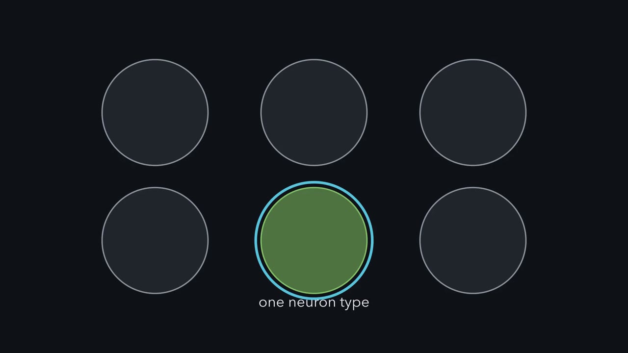 ▶ Watch Tools Behind a Stop, Chosen by Cre · 2:29
Read the transcript of “Tools Behind a Stop, Chosen by Cre”
Film transcript: Tools Behind a Stop, Chosen by Cre
Suppose you want a mouse in which one type of neuron glows when its calcium rises. You also want to switch that same type on with light. Put the gene for green fluorescent protein behind a promoter that every cell reads, and you get a green mouse. But you need the tools in one cell type, and nowhere else. How? Two kinds of tool do the work. A reporter is an engineered protein that changes its light when a signal happens. G -Camp is a calcium reporter, built from green fluorescent protein. An actuator is an engineered protein that changes cell activity on command. Channel rhodopsin 2 is an actuator. Blue light opens its pore, cations flow in, the neuron fires.
G -Camp's blue light opens it too. Now give each gene a stop. Put a stop signal in front of the reporter gene, between two sites called LOX -P. The two sites point the same way. The actuator gene gets its own stop in the same way. With the stop in place, neither gene is read. Kree is an enzyme from a bacterial virus. It cuts and rejoins DNA at LOX -P sites. When two sites point the same way, Kree removes what lies between them. The stop is cut out. The cut is permanent, so that cell and its descendants can read the gene. So who makes Cre? Make it from a promoter active mainly in one cell type. That cell type cuts out the stops, and makes both proteins.
In most other cells the stops stay in place. Check each line with a reporter. To build the mouse, breed three parts together. The first is Kree, placed in a gene that only your neuron type reads. The second is a reporter behind a stop. The third is an actuator behind a stop. Cells that make Kree remove the stops, so mainly they and their descendants make both proteins. Keep this rule. Tools wait behind stops, and Kree removes them. Check the line with a reporter.
Two kinds of engineered protein let you work on a signal inside a living cell: a reporter shows you where and when it happens, and an actuator starts or stops it on command. Genetic methods then put either kind into one cell type in a mouse. The reporters began with a jellyfish: a gene copied out of it and put into gut bacteria in 1992 made the bacteria glow green, with nothing else from the jellyfish. A cell that builds its own fluorescent tag can be watched while it is alive.
The idea: watch a signal, or push one
Two words sort the tools. A reporter is an engineered protein that changes its light when a signal happens, so you can watch it in a living cell. An actuator is an engineered protein that changes a cell’s activity on command, usually with a flash of light: it can start a signal or shut one down. Using light and a gene to control cells is called optogenetics, and the word is often stretched to cover reporters used alongside it.
Think of a voltmeter and a switch. A reporter can show that calcium rises in a neuron each time a mouse turns left. A reporter cannot tell you whether firing that neuron is enough to turn the mouse. For that you have to drive those neurons yourself, with an actuator or an electrode, and watch what the mouse does.
“How does fluorescence work?” A molecule absorbs a photon, which lifts an electron to a higher energy level. A little of that energy is lost as heat before the electron falls back and emits a photon. So the light that comes out has a longer wavelength than the light that went in: blue in, green out.
The GFP gene alone turned out to be enough. Put into E. coli, its coding stretch made the bacteria fluoresce green, which showed that no jellyfish enzyme was needed to finish the protein.
The structure explains why. GFP is a barrel with a helix through its middle. Once the protein folds, three neighboring amino acids on that helix react with one another, lose water, and are oxidized by oxygen from the air. That leaves a rigid ring system that absorbs violet and blue light and emits green. The recipe is the sequence itself, so many kinds of cell with the gene and oxygen make their own fluorescent tag, though the first version folded poorly in warm cells, and another gene joined to GFP gives a protein with the lamp attached.
Building a reporter: a glowing protein joined to a detector
“What would a synaptic protein look like?” Fuse GFP to gephyrin, a scaffold protein found under inhibitory synapses, and a neuron’s dendrites show rows of bright dots. That is the simplest reporter: it tells you where a protein is.
To make the light depend on a signal, join GFP to a detector you have already met.
- A lipid reporter. A PH domain that binds phosphatidylinositol 3,4,5-trisphosphate (PIP3) (see the lesson on lipid anchors and addresses), fused to GFP, moves from the cytosol to the membrane when PI 3-kinase makes PIP3.
- A pH reporter. A GFP that dims in acid, placed inside a synaptic vesicle (pH about 5.5), brightens when the vesicle fuses and the reporter meets fluid at pH 7.4.
- A calcium reporter. In the GCaMP family, GFP’s original ends are joined and new ends are cut beside the light-emitting group. Calmodulin (from the calcium lesson) is attached to one new end and a peptide that it grips to the other. When calcium rises, calmodulin wraps around the peptide and plugs the gap in the barrel, which shuts water out from the light-emitting group, and the GFP brightens.
- A kinase reporter. Put a peptide that the kinase phosphorylates, and a domain that binds the phosphorylated peptide (an SH2 domain, for a tyrosine kinase), between a cyan and a yellow fluorescent protein. Phosphorylation folds the chain. Two fluorescent proteins within a few nanometers pass energy from one to the other without a photon (FRET), so the balance of cyan and yellow light changes as the kinase works.
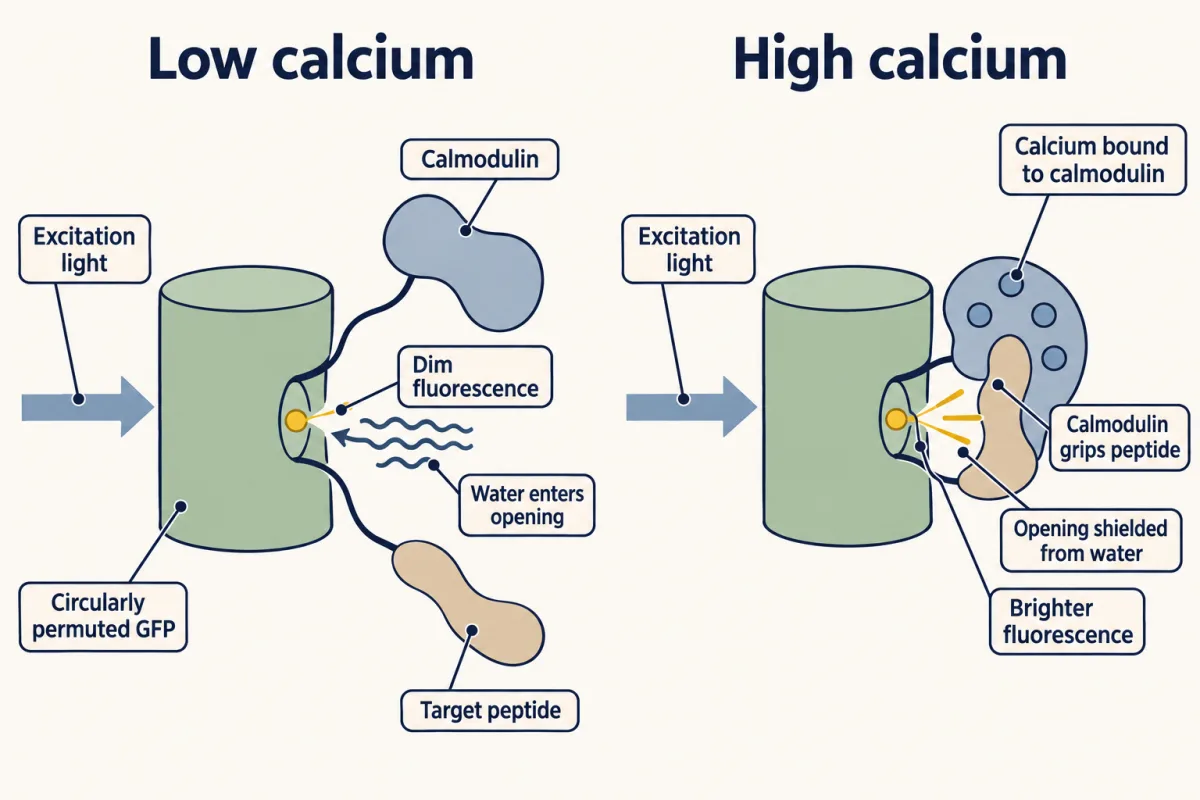
A reporter also takes part in what it reports. A calcium reporter soaks up some of the calcium it measures, and its own binding and shape changes are slow, so a fast train of spikes can blur into one slow rise.
An actuator: light in, current out
Read the transcript of “Light In, Current Out”
Film transcript: Light In, Current Out
In an earlier lesson you met rhodopsin. It has seven helices holding retinal. Retinal changes shape when it absorbs a photon, and that sets off a G protein. Some microbes have opsins on a similar plan that skip the relay. Channelrhodopsin-2, from a green alga, is itself an ion channel. Now shine blue light on channelrhodopsin-2. Blue light changes its retinal. The pore opens within milliseconds. Cations flow into the neuron, with little choice among them. That depolarizes the membrane. At threshold, the neuron fires. Georg Nagel, Peter Hegemann, Ernst Bamberg and colleagues had shown in 2002 and 2003 that channelrhodopsins from the green alga Chlamydomonas are light-gated channels.
The same work showed that channelrhodopsin-2 passes cations, and that it works in mammalian cells. In 2005 Edward Boyden, Feng Zhang, Ernst Bamberg, Georg Nagel and Karl Deisseroth reported on hippocampal neurons in culture. Given the gene for channelrhodopsin-2, the neurons fired action potentials in step with millisecond flashes of blue light. Other laboratories reported similar results at about the same time.
In the lesson on the senses and cGMP you met rhodopsin: seven helices holding retinal, which changes shape when it absorbs a photon and sets off a G protein. Some microbes have opsins on a similar plan that skip the relay. Channelrhodopsin-2, from a green alga, is itself an ion channel. Blue light changes its retinal, the pore opens within milliseconds, and cations flow into the neuron. That depolarizes the membrane, and at threshold the neuron fires.
Channelrhodopsin-2 had already been shown to work in mammalian cells. In 2005 Edward Boyden, Feng Zhang, Ernst Bamberg, Georg Nagel and Karl Deisseroth reported that hippocampal neurons in culture, given the channelrhodopsin-2 gene, fired action potentials in step with millisecond flashes of blue light. Other laboratories reported similar results at about the same time.
Opsins are sorted by what light does through them:
- Channelrhodopsin-2 and its relatives: blue light opens a channel that passes cations with little choice among them. The neuron switches on. Other channelrhodopsins answer to other colors, and some pass anions instead.
- Halorhodopsins: yellow light drives chloride into the cell, which can stop the neuron firing. These are pumps, moving one chloride per photon, so they need bright light, and the chloride they load can weaken the inhibition. Light-gated chloride channels are now used more often for silencing.
- OptoXRs: green light activates a rhodopsin whose inner loops come from another G-protein-coupled receptor, so light starts that receptor’s pathway.
A virus can carry the gene into one brain region, though the adeno-associated virus (AAV) vectors used most have room for only a short promoter, and light arrives through an optical fiber.
A drug can do the job of light. In one chemogenetic approach the cell gets an engineered G-protein-coupled receptor, called a DREADD, that barely answers its natural ligand and answers a synthetic drug instead, so in the main only cells with the receptor react. The drug still needs a control of its own: clozapine-N-oxide, the one used most, turns into clozapine in the body, and clozapine acts on the animal’s own receptors. A channel from the lesson on ion channels works too: put the transient receptor potential vanilloid 1 (TRPV1) gene into one neuron type of a mouse whose own TRPV1 gene has been knocked out, and capsaicin opens a cation channel in those neurons alone (Güler and colleagues, 2012).
How can we make a green animal?
Start with a transgene: DNA built from a promoter (the stretch that sets where and when a gene is read) and a coding sequence. Injected into a fertilized mouse egg, it sometimes joins a chromosome at a random place, and if it reaches the germ line it is inherited from then on. In 1982 Palmiter and colleagues did this with a metallothionein promoter joined to a growth hormone gene, and the mice grew much larger than their littermates. Use a promoter active in every cell and the coding sequence for GFP, and you have a green mouse.
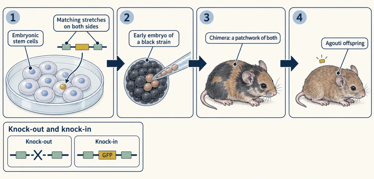
To change one chosen gene you work in embryonic stem cells, cells from an early embryo that can still become any tissue. Give them DNA that matches the target gene on both sides of your change, and in a few cells the matching stretches swap your version into the gene’s own place. Destroying the gene this way is a knock-out; writing something new into it, such as GFP, is a knock-in. The altered cells are injected into an early embryo, the pup is a chimera, and if the stem cells formed its sperm or eggs, its offspring can carry the change. CRISPR now shortens this path, as the paragraph after the Cre and lox text describes.
“How do you get expression in 2 different neuron types?” The tool is Cre and lox. Cre is an enzyme from a bacterial virus that cuts and rejoins DNA at a 34-base-pair site called loxP. Put two loxP sites around part of a gene, pointing the same way, and Cre removes what lies between them. Make Cre from a promoter that is active in one cell type, and the gene is lost there and, in the main, nowhere else. Every Cre line has to be checked with a reporter, because brief expression during development or in the germ line can cut the DNA in other cells too. To switch a gene on, put a stop signal between two loxP sites in front of it. Where Cre is made the stop is cut out, and the cut is permanent, so that cell and its descendants read the gene wherever the reporter’s own promoter is active, which is how cell lineages are traced. With several fluorescent proteins for Cre to pick among at random, neighboring cells take different colors. For the second neuron type, add a second enzyme of the same kind with its own sites (Flp, from yeast): Cre switches on one gene in one type and Flp another gene in the other.
CRISPR shortens the path: an RNA guide brings a DNA-cutting enzyme to a matching sequence in a fertilized egg, so many knock-outs and knock-ins no longer need stem cells or chimeras.
Past the limit of light
A lens cannot focus light to a point. The glow of one molecule spreads into a blur, and two molecules closer than about half a wavelength merge. Ernst Abbe put the limit as a formula in the 1870s:
smallest separable distance ≈ wavelength ÷ (2 × numerical aperture)
Numerical aperture measures how wide a cone of light the lens collects, about 1.4 for a very good lens. For green light the limit is about 200 nanometers; a synaptic vesicle is about 40 across.
One way past it came through Jennifer Lippincott-Schwartz’s laboratory at the NIH. In 2002 her postdoc George Patterson made a GFP that starts dark and switches on after a flash of violet light. In 2005 the physicist Eric Betzig described to her a microscope that needed such a molecule, and she gave him and Harald Hess room in her laboratory. The method is PALM. Switch on a few scattered molecules, so that no two blurs overlap, and find the center of each blur, which can be done far more precisely than the blur is wide. Let those molecules bleach and repeat thousands of times. The stacked positions make a picture with detail down to tens of nanometers. The paper appeared in 2006, as did two related methods, STORM from Xiaowei Zhuang and FPALM from Samuel Hess.
Other methods take other routes: STED (proposed by Stefan Hell in 1994, first shown to work in 1999) switches off fluorescence around the rim of the spot with a ring-shaped second beam, and structured illumination computes finer detail from striped lighting.
Design problem
“How can I make a mouse that does this?” First, how can we make a green animal? Then go one step further: design a mouse in which only one type of neuron glows when its calcium rises, and in which you can switch that same type of neuron on with light. Say which reporter, which actuator, and how you make sure that both are made in that cell type and nowhere else.
Saved in this browser only.
One way to do it
The green animal is a transgene: a promoter active in every cell, joined to the GFP coding sequence and injected into fertilized eggs.
For the second mouse, breed together three parts.
- An address. Cre knocked into a gene that only your neuron type reads.
- A reporter. A GCaMP gene behind a lox-flanked stop.
- An actuator. A channelrhodopsin gene behind its own lox-flanked stop.
Only cells that make Cre remove the stops, so in the main only they and their descendants make both proteins. Check the line with a reporter before trusting it.
One trap: GCaMP is read with blue light and channelrhodopsin-2 is opened by blue light, so looking would also be pushing. Switching to a red-shifted channelrhodopsin does not fix this, because retinal itself absorbs blue light. Change the reporter instead: keep channelrhodopsin-2 and use a red calcium reporter, such as RCaMP or jRGECO, which is read with red light that leaves the channel shut. Then test the pair, and check that your imaging light alone drives no current.
Going deeper
How GFP became a tool
From 1961 Osamu Shimomura and Frank Johnson extracted and purified aequorin, a jellyfish protein that gives off blue light when calcium binds it. Traces of a second protein, green and fluorescent, came along in the extract. In 1971 Morin and Hastings renamed it green fluorescent protein, GFP.
Until 1992 nobody knew whether the GFP gene alone would be enough. The common guess was that a jellyfish enzyme had to finish the protein. That year Douglas Prasher published the cloned gene, and in Martin Chalfie’s laboratory at Columbia the rotation student Ghia Euskirchen put its coding stretch into E. coli. The bacteria fluoresced green.
Once the protein folds, serine 65, tyrosine 66 and glycine 67 on the central helix react with one another, lose water, and are oxidized by oxygen from the air.
Who showed channelrhodopsins are channels
Georg Nagel, Peter Hegemann, Ernst Bamberg and colleagues had shown in 2002 and 2003 that channelrhodopsins from the green alga Chlamydomonas are light-gated channels, and that channelrhodopsin-2 passes cations and works in mammalian cells.
Selecting and breeding targeted mice
Drugs select the rare correct cells (random insertions keep an added herpes thymidine-kinase gene, and ganciclovir kills the cells that keep it). The chosen cells, from a strain with a brown ‘agouti’ coat, are injected into an early embryo of a black strain. The pup is a chimera, a patchwork of both, and if the stem cells formed its sperm or eggs, its agouti offspring can carry the change.
Your notes
Select any text in the lesson to highlight it or add a note to it. Your highlights and notes are saved in this browser and collected in your notebook.
This browser is not letting the site save anything (a private window or strict privacy settings), so highlights and notes will be lost when you leave the page.
How we measure it
Genetically encoded fluorescent reporters
You join the gene for a fluorescent protein to the gene for a detector, such as calmodulin or a PH domain, and the cell makes the reporter itself. A fluorescence microscope then records the reporter's brightness, position or color over time in living cells, which tells you when and where the signal happened.
Optogenetic stimulation
You express a microbial opsin such as channelrhodopsin-2 in chosen cells and deliver light, in a dish from a lamp or laser and in an animal through an optical fiber. Recording the cell's voltage or the animal's behavior during the light tells you whether activity in those cells is enough to cause the effect.
Gene targeting and Cre/lox conditional genetics
You change one chosen gene in embryonic stem cells by homologous recombination, make chimeric mice from those cells, and breed the change into a line. With loxP sites placed around a piece of DNA, Cre recombinase removes that piece in the cells that make Cre and in their descendants, so a gene can be removed or switched on in largely one cell type.
Single-molecule localization microscopy (PALM)
You switch on a few photoactivatable fluorescent proteins at a time, find the center of each separate blur, and repeat for thousands of camera frames. The summed positions give an image with detail of tens of nanometers, well below the diffraction limit of about 200 nanometers.
Practice questions on this lesson (3) → MCAT-style, original, each with an explanation
Where this lesson fits
Before thisReview first
- Calcium: The Messenger Kept at Arm's LengthThis course · Lesson 15
The calcium reporter here is built from calmodulin and its peptide, whose calcium binding you need first.
- Sensing the World: Vision, Smell, Nitric Oxide and cGMPThis course · Lesson 17
Rhodopsin and retinal from there are the starting point for light-gated opsins used as actuators.
- Protein Modules: SH2, SH3, PTB, PDZ and PHThis course · Lesson 9
The kinase reporter uses an SH2 domain to read a phosphorylated peptide, as taught there.
Elsewhere on the siteFilms, simulations and method films in other courses
- Engineered mutants: site-directed, dominant-negative and constitutively activeMethod film
Reporters and actuators are made by changing chosen parts of proteins, which this method film shows.
- Targeted cell ablationMethod film
Genetically targeted toxins are another tool delivered to one chosen cell type by the same Cre-based systems.
- Live-cell time-lapse imagingMethod film
Fluorescent reporters are read out by filming living cells over time.
Suggested by an AI model from the lessons themselves, then checked. How we use AI
Finish line
Test yourself
8 questions
Practice and MCAT-style, with explained answers.
That's the last lesson · Next course
Developmental Neuroscience
How a nervous system builds itself, one local decision at a time.
Start the next course →Before you move on: review the questions you missed , or go back to all lessons in this course.