Cell Biology · Unit 2: Membranes and compartments
Nuclear Architecture and Membraneless Compartments
Rewritten on : new text, flashcards and film. Your notes and highlights stay, but the text around them may have moved.
How does a nucleus keep things apart with no walls around its compartments?
Pause when the reading reaches a film, and offer to play it. Saved on this device.
Watch the film now, or keep listening
Builds on:Organelles as Distinct EnvironmentsExperiments That Distinguish MechanismsThe Nuclear Envelope and Nuclear TransportWhy these?
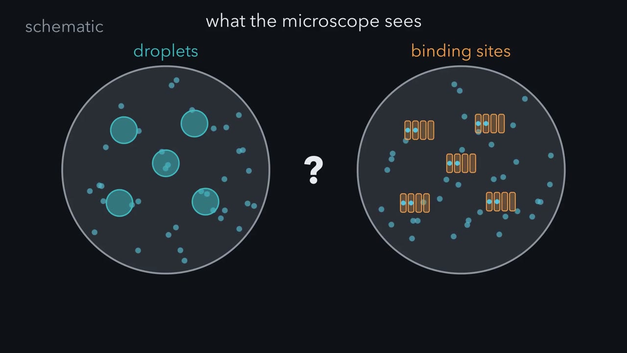 ▶ Watch Bigger or Brighter? · 3:35
Read the transcript of “Bigger or Brighter?”
Film transcript: Bigger or Brighter?
Two nuclei, each dotted with bright spots of a glowing protein. Is a spot a droplet of that protein? Or is it a cluster of binding sites, such as many copies of one DNA sequence? Under a microscope, the two can look alike.
A nucleus has no membrane-bound compartments inside it, yet it holds many bodies like these. The largest, the nucleolus, builds ribosomal subunits. Many of their molecules carry several weak sticky patches, so each can hold many partners at once. That is multivalency. The usual checks cannot tell droplets from sites. Both kinds of spot look round. Droplets fuse, and site clusters can drift together until they look merged. Bleach one with a laser, and it glows again, because molecules that bind and let go refill it too. A bright spot shows only that the protein is concentrated there. So change the amount instead. Give both nuclei one dial: the total protein, averaged over the whole nucleus.
Add a gauge that reads the concentration outside the spots. Now double the protein in both. What will each outside gauge do? The droplet needle holds still. Think of salt water past saturation: added salt stops dissolving and forms a second phase. At equilibrium under fixed conditions, one protein on its own holds the outside at the saturation concentration. The extra builds more of a second, denser phase: the droplets. They take up more room, but glow no brighter.
Bigger, not brighter. On the site side, the needle climbs. Think of the sites as parking spaces, a fixed number, with plenty still free. Some protein is parked and the rest is outside, so the outside rises with the total. The spots fill in and glow brighter, but grow no bigger. Brighter, not bigger.
Keep turning. On the site side, the parking lot fills up. The spots brighten more slowly, and nearly every extra molecule stays outside, so that needle climbs faster. Within this range, the droplets keep taking up more room, and their needle stays put. So here is the test, in its simplest form. Add more protein, and watch where the extra goes. At equilibrium under fixed conditions, one protein forming a phase puts it into more droplet volume, outside held still. A fixed set of sites puts it onto those sites: brighter spots, until they are full. This clean version is for one protein that separates on its own. Real bodies hold many kinds of molecule, some may form on chromatin, and tiny spots blur size into brightness.
So where the extra goes is a clue, not a proof. The text below shows what each complication changes.
From the film
- A bright spot in the nucleus shows only that a protein is concentrated there, not how.
- Both kinds of spot look round and refill after bleaching, and site clusters that drift together can look fused.
- At equilibrium under fixed conditions, one protein forming a phase on its own adds droplet volume while the outside stays at saturation.
- On a fixed array of sites that does not change size, extra protein mainly brightens the spots until the sites are full.
- Where the extra goes is a clue, not a proof: mixtures, chromatin and tiny spots can blur it.
A human nucleus is only several micrometres across and holds roughly two metres of DNA, but it has no membrane-bound compartments inside it, and it is still not a uniform soup.
The short answer to the hook: chromosomes keep to territories, bodies gather molecules by binding them to scaffolds, and some bodies may form by phase separation. Any membraneless body that concentrates proteins and nucleic acids is a biomolecular condensate, whatever made it. The first film’s droplets are one proposed way to make one, liquid-liquid phase separation: above a certain concentration a solution demixes into a dense phase (the droplets) and a dilute phase (here the nucleoplasm, the nuclear interior around them). For proteins it is thought to be driven by multivalency, many weak, reversible binding sites per molecule (repeated domains, disordered regions, RNA; that lesson). The first film’s sites are clustered sites: many copies of a DNA or RNA sequence in one place, bound with no demixing.
The one idea to carry through this lesson. A bright spot shows that something is concentrated, not how. Add more protein and ask where the extra goes. For one protein demixing on its own, at equilibrium under fixed conditions, the extra adds dense-phase volume (bigger drops or more of them) while the inside and outside concentrations stay fixed. On a fixed set of sites that does not grow, the extra raises occupancy: spots brighten until the sites fill, and only then does the outside climb. Each pattern supports its model; neither proves it alone.
The first film showed that comparison; it leaves out what the imaging checks really measure and why the graphs have their shapes. The second film tours the bodies and chromatin a real nucleus holds, then examines liquid-phase tests in a frog nucleolus and in heterochromatin. The last section (Where the test is weaker) takes up the first film’s closing complications.
Why looking harder does not help
Why did the first film’s usual checks pass both kinds of spot? Look at what each one measures. Roundness alone shows neither softness nor mechanism, because clusters below the resolution of the microscope look round too. Real fusion, two bodies merging and relaxing into one round body, is what drops do, but clusters that drift together can look merged unless that relaxation is seen. FRAP, fluorescence recovery after photobleaching (that lesson), measures turnover, which binding and unbinding also give.
Predict. 1,6-Hexanediol, an alcohol thought to weaken hydrophobic contacts, dissolves a nuclear body. What would you want to know before calling the body a liquid?
Check your prediction
What else the alcohol does. Itoh and colleagues found that 5% hexanediol for 5 minutes strongly suppressed chromatin motion in live HeLa cells, and foci of one nuclear body, the Cajal body (below), fell in number in the same treatment. A reagent that changes chromatin this much cannot tell you which mechanism held a body together.
Half-body FRAP has more power: bleach half of a large body and follow both halves and the nucleoplasm beside it. A droplet has a boundary, so molecules are expected to mix inside it faster than they cross its edge. That pattern favours a phase without proving one: the weak interfaces of real condensates may slow crossing only a little, and molecules that rebind sites inside a dense cluster can also mix internally.
Add more protein: the numbers
The first film’s picture was the simple case; here is the fuller one: why each graph has its shape, and where it stops. Total (along the bottom) is averaged over the whole nucleus; outside (up the side) is measured between the spots.
- A phase of one protein. Below the saturation concentration, csat, there are no bodies and outside equals total: a 45° line. Above it the line goes flat: like salt past its solubility, extra protein forms more dense phase, whose own concentration, cdense, also stays fixed. That holds at equilibrium and fixed conditions, until the dense phase fills the nucleus (φ = 1, below).
- Sites. While they are far from full, each molecule has the same chance of being bound at any moment, so a fixed fraction stays outside: a straight line through zero, below the 45° line. Once every parking space is taken, extra protein stays outside and the line bends up. This assumes fixed sites; if added protein brought in new sites or reshaped the chromatin that carries them, the spots could grow too.
A flat outside line alone is weaker evidence than it looks: cooperative binding can flatten it too (Where the test is weaker).
For a phase, the amounts must add up: the protein in the dilute phase plus the protein in the dense phase is all of it.
In words The bodies' share of the nucleus is the excess over saturation, divided by the gap between the dense and dilute concentrations.
- φ“phi”
- the fraction of nuclear volume taken up by the dense phase0 to 1
- ctotal“c sub total”
- the protein's average concentration over the whole nucleusμM
- csat“c sub sat”
- the saturation concentration, where the dilute phase staysμM
- cdense“c sub dense”
- the concentration inside the bodiesμM
Sanity check At ctotal = csat, φ = 0: the coexistence boundary, with a nonzero dense fraction just above it. At ctotal = cdense, φ = 1: the dense phase fills the nucleus. In between, at equilibrium and fixed conditions, csat and cdense stay fixed, so only φ grows.
Where it comes from optional
Step 1: ctotal = csat × (1 − φ) + cdense × φ (the dilute volume times its concentration, plus the dense volume times its).
Step 2: multiply out: ctotal = csat + φ × (cdense − csat).
Step 3: solve for φ.
Need a refresher? subscripts and Greek letters · rates and “per”
Predict. A protein has csat = 1 μM and cdense = 50 μM. Its total rises fourfold, from 2 to 8 μM. Do its bodies double, quadruple or more than quadruple in volume?
Check your prediction
More: sevenfold. φ goes from 1/49 = 2.0% of the nucleus at 2 μM, through 3/49 = 6.1% at 4 μM, to 7/49 = 14.3% at 8 μM. Volume tracks the excess over csat, which goes from 1 to 7, not the total, which goes from 2 to 8. The nucleoplasm stays at 1 μM.
Worked problem (made-up data). Two proteins, each tagged at its own gene, are set to total nuclear concentrations of 2, 4 and 8 μM.
- Protein A: outside 1.0, 1.0 and 1.1 μM, while the total body volume grows at the same brightness. Bigger, not brighter: consistent with the simple phase model.
- Protein B: outside 1.5, 2.8 and 5.9 μM, or 1.5/2 = 0.75, 2.8/4 = 0.70 and 5.9/8 = 0.74 of the total, while its foci brighten in proportion. A constant fraction outside is the straight line through zero: sites far from full.
- Dilute phase
- Dense phase
- Bodies fill (φ)
An idealized model, in this sketch: one dense phase at 50 μM, a saturation concentration of 1 μM, a dilute phase held at that value, and made-up data. The number and places of the dots are arbitrary, only their total area means anything. Binding to clustered sites or bridging chromatin can also make bright spots, and a protein that fails this test may still phase separate as one part of a mixture of several components. A protein that passes has not been shown to need the body for any function. Keep the rule φ = (c_total − c_sat) / (c_dense − c_sat).
Below 1 μM, the outside follows the total and φ stays zero; above 1 μM, watch φ grow while the outside stays at 1 μM.
Those are made-up proteins. The second film picks up here: it tours the bodies and chromatin a real nucleus holds, then examines two systems tested for liquid-like behaviour.
Read the transcript of “Real Bodies, Real Tests”
Film transcript: Real Bodies, Real Tests
Chapter one ended on a clue: add protein and see whether a spot grows bigger or brighter. Now meet the bodies and chromatin a real nucleus holds. Which have been tested as liquids, and what did those tests show? Some order needs no spot at all. After a cell divides, each chromosome partly unpacks but keeps a region of its own: a chromosome territory.
Picture balls of yarn in a box, with loose loops poking across where two meet. A sequencing method called Hi-C maps which stretches of chromatin touch. Its maps reveal two broad compartments. A is open and mostly active. B is closed and mostly quiet, often against a mesh under the nuclear envelope. Standard maps average over millions of cells, not one nucleus. Now the bodies. The nucleolus forms around repeated genes for ribosomal RNA.
Mammalian nucleoli have three parts along an inside-out assembly line. Fibrillar centres hold the genes and copying machinery. New RNA emerges at their borders, where processing begins. The outer granular component assembles ribosomal subunits. Smaller bodies sit around it, each known by what it holds. Speckles hold splicing factors; Cajal bodies, the marker protein coilin; PML bodies, the PML protein. Paraspeckles are built on a long RNA called NEAT1. These bodies are real. They exchange molecules with the nucleoplasm, and several help with a job. But how does each one form? Only a few have had their material properties tested closely. The strongest liquid-phase evidence here comes from the nucleolus in the egg-forming cells of a frog. Its dense fibrillar and granular components behave like two liquids that coexist without mixing.
What should purified nucleolar proteins do when mixed with RNA in a tube? They reproduce the layering: drops with one phase inside the other. This is strong evidence that the frog nucleolus contains coexisting liquid phases. Heterochromatin, densely packed and mostly silent chromatin, adds a third mechanism. A protein can bridge distant stretches until the fibre collapses into a compact ball. Physicists count that as a phase transition of the chromatin. But it makes a ball of chromatin, not a liquid drop of protein. The verdict splits by system. In early fruit fly embryos, foci of HP1a looked liquid-like. In mouse fibroblasts, half-body FRAP found no preferential mixing inside heterochromatin clusters, favouring collapsed chromatin.
Neither study speaks for all heterochromatin. Whether phase separation builds a given body in a given living cell is still contested. The text below shows where the test itself grows weaker.
From the film
- Chromosomes keep to territories, and Hi-C reveals open A and closed B compartments.
- Mammalian nucleoli have three parts that build ribosomal subunits, read from the inside out.
- Speckles, Cajal bodies, PML bodies and paraspeckles are each known by what they hold.
- The frog oocyte nucleolus provides strong evidence for coexisting liquid phases.
- Chromatin bridging is a third mechanism, and for heterochromatin the verdict splits by system.
What the nucleus holds
The second film toured the territories and bodies; here is the detail behind them.
Chromosomes keep to territories, mostly
During mitosis each chromosome is a compact rod. Afterwards it partly unpacks, but it does not spread through the whole nucleus. Labelled with fluorescent DNA probes, a method called chromosome painting, each one fills a region of its own, a chromosome territory. The DNA is packed as chromatin, DNA wound on histone proteins, and how tightly it is packed affects whether its genes are used (that lesson). Picture balls of yarn in a box: each keeps its own space, but where two press together, loose loops of one poke into the other.
Predict. Why can painting a whole nucleus not show whether neighbouring territories mix at their edges?
Check your prediction
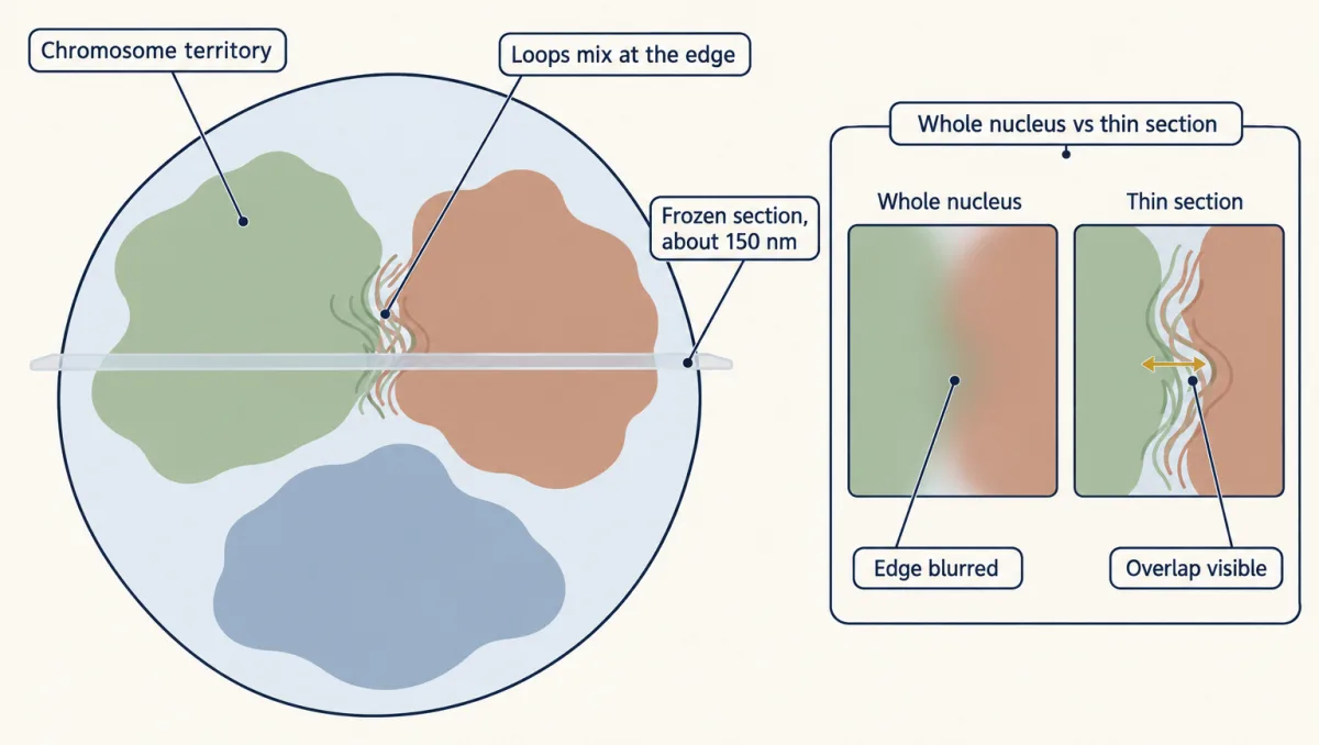
A conventional light microscope blurs everything below a few hundred nanometres (that lesson), and in a whole nucleus light from above and below blurs every edge. In 2006 Miguel Branco and Ana Pombo painted thin frozen sections of fixed human lymphocytes, which removes most of that blur. Neighbouring territories overlapped at their edges, and how much two chromosomes overlapped correlated with how often that pair takes part in translocations (exchanges of pieces between chromosomes) measured in human lymphocytes after ex vivo irradiation. That is a correlation, not proof that overlap causes any given translocation.
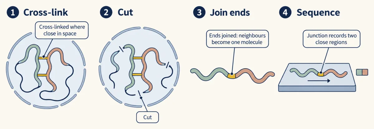
Hi-C, a sequencing method, gives a second view: chromatin is cross-linked in the cell, cut and re-joined, so segments lying close in space end up in one DNA molecule, and sequencing millions of these junctions maps which parts of the genome touch. The 2009 maps of human cells, at a resolution of one megabase, showed that open and closed chromatin segregate into two genome-wide compartments, A (open, gene-rich, mostly active) and B (closed, mostly quiet). Closed chromatin often lies against the nuclear lamina, the protein mesh under the nuclear envelope (that lesson). The maps average over millions of cells: they report how often two regions are near each other, not where they sit in one nucleus. Later single-cell versions map one nucleus at a time.
Bodies without membranes
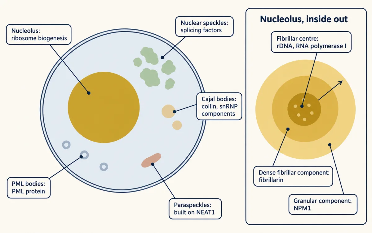
A nuclear body is a region of the nucleoplasm where particular molecules are concentrated, with no membrane around it. The largest, the nucleolus, carries out ribosome biogenesis. It forms around the repeated genes for ribosomal RNA (the rDNA) and, in mammalian cells, has three parts, like an assembly line read from the inside out. The fibrillar centre holds the rDNA and the machinery of RNA polymerase I, the enzyme that transcribes it; active transcription occurs at or near its border with the dense fibrillar component. In that component, which is rich in fibrillarin, the newly made ribosomal RNA precursor (pre-rRNA) begins to be processed. The outer granular component, rich in nucleophosmin (NPM1), is where ribosomal subunits are assembled before export.
The smaller bodies, with one key experiment each:
| Body | What it holds | One key experiment |
|---|---|---|
| Nuclear speckles | Pre-mRNA splicing factors, which exchange with the nucleoplasm (splicing) | Mouse embryonic stem cells: genes near speckles showed more co-transcriptional splicing (splicing while the RNA is still being made) than genes farther away. Human HEK293T cells: tethering a reporter pre-mRNA to speckles increased its splicing |
| Cajal bodies | Coilin, their marker, and components of the small nuclear ribonucleoproteins (snRNPs) that splice pre-mRNA | Engineered mammalian cells: tethering Cajal-body components to chromatin started apparently functional Cajal bodies from scratch |
| PML bodies | The PML protein that builds them, SUMO (a small protein tag) and partner proteins | Cultured mammalian cells and mice: oxidation drove PML to multimerize and nucleate the bodies, which then enhanced stress-induced SUMO modification of partners inside |
| Paraspeckles | Their scaffold, the long noncoding RNA NEAT1, and its bound proteins | Mice lacking Neat1 have no paraspeckles yet were reported viable and fertile in the laboratory; later work found defects in pregnancy and lactation |
These facts are well supported: the bodies exist, they exchange molecules with the nucleoplasm, and several of them help with a job. How each forms is the second film’s question, and few have had their material properties tested closely.
Real bodies: one strong case, one split verdict
The strongest liquid-phase evidence here comes from the nucleolus in oocytes of the frog Xenopus laevis. There the fibrillarin-rich and NPM1-rich regions behave as two liquids that coexist without mixing, and purified nucleolar proteins mixed with RNA reproduce the layering (see Marina Feric’s card). This is strong evidence that these frog nucleoli contain coexisting liquid phases, not that every nucleolus is a single liquid phase.
Heterochromatin, densely packed, mostly silent chromatin, adds a third mechanism. A protein can bridge chromatin, cross-linking distant segments so the fibre collapses into a compact globule. Physicists count this collapse as a phase transition of the chromatin polymer, but the result is a globule of chromatin, not a liquid drop of the protein.
Predict. A protein makes a focus by bridging one stretch of chromatin. If you double the protein, must the focus act like the first film’s sites?
Check your prediction
Not necessarily. If the chromatin stretch, its compaction and its sites are fixed and nearly full, extra protein mostly raises occupancy, like sites. But more bridges can pull in and compact more chromatin, changing the size of the focus. Bridging is its own mechanism, which the comparison does not sort automatically.
The verdict on heterochromatin splits by system. Purified Drosophila HP1a demixed in the test tube, and HP1a foci in early Drosophila embryos looked liquid-like, with mature domains proposed to contain liquid and stable parts. In mouse fibroblasts, by contrast, chromocentres, dense clusters of heterochromatin, behaved like collapsed chromatin globules rather than HP1 liquid pools, and half-body FRAP found no preferential mixing inside them. Test-tube numbers for mouse HP1α do not settle it either (Going deeper). Neither study speaks for all heterochromatin.
Where the test is weaker
The first film ended on three complications. Here is what each changes, after a look-alike neither chapter showed.
A flat outside line can mislead. If many molecules must bind together before a site holds any (cooperative binding), the sites stay nearly empty at low concentration and then fill over a narrow range, like a threshold, so the outside stays nearly flat while they fill.
Predict. A protein’s outside concentration creeps only from 1.6 to 2.2 μM while its total triples, from 3 to 9 μM. Is it a phase?
Check your prediction
Not yet. A made-up model of cooperative sites gives just these numbers; its outside climbs (to 4 μM at a total of 14 μM) only once the sites are full. Ask where the extra went: more body volume at the same brightness supports a phase, brighter foci of the same size support sites.
Mixtures. A protein that fails the test may still phase separate as one part of a mixture of several components, which need not buffer each one. In human cells, the dilute-phase concentration of the nucleolar protein NPM1 rose with its total expression, which was explained by its interactions with other nucleolar components (Riback and colleagues, 2020).
Seeds. In engineered Corelet experiments in living cells, local capture of a protein on a small engineered seed triggered phase separation below the global concentration needed for it. If chromatin acts as a seed in the same way, a body could form on it where the average level looks too low.
Tiny spots. Below the blur of the microscope, a few hundred nanometres, size and brightness cannot be told apart.
Function. A protein that passes has not been shown to need the body for any function. Speckle tethering shows that location matters, not that phase separation is the reason, and that mice without paraspeckles survive in the laboratory does not mean the bodies do nothing.
What is contested, then, is whether phase separation is the mechanism for a given body in a given living cell, and whether its material state matters for its job.
Design problem
A laboratory reports that protein X forms round nuclear foci that fuse, recover after photobleaching and dissolve in 1,6-hexanediol. It concludes that X drives a phase separation needed for transcription. What is wrong with the argument, and what would you do instead?
Saved in this browser only.
One way to do it
None of the three observations is diagnostic: roundness, apparent fusion and FRAP recovery also fit binding to clustered sites or chromatin bridging, and hexanediol also suppresses chromatin motion.
Instead, tag X at its own gene (its endogenous locus) and check that the tag does not change its amount or behaviour. Raise and lower its level, measure concentrations inside and outside the foci, and ask where the extra goes: into more focus volume at a steady outside concentration, or into brighter foci of fixed size (support for a model, not proof). Use half-body FRAP to ask whether X mixes inside a focus faster than it crosses the edge. Make a mutant that keeps DNA binding but loses its multivalent region, and see whether foci and transcription change together. Finally, test transcription separately from assembly, with a control that moves the same genes to or from the foci by another route, much as Bhat and colleagues tethered a reporter pre-mRNA to speckles to test splicing. Only then can you ask whether the foci themselves, rather than protein X alone, are needed.
Going deeper
More on territories, compartments and bodies
Branco and Pombo (2006) activated their human lymphocytes with phytohemagglutinin before fixing and sectioning them. Deeper Hi-C maps in 2014, at kilobase resolution, also found smaller contact domains and thousands of chromatin loops whose anchors usually bind the protein CTCF. Immunofluorescence usually shows about 20 to 50 nuclear speckles per nucleus in mammalian cells. PML bodies take their name from promyelocytic leukaemia, in which a fusion of PML with a retinoic acid receptor is associated with disrupted bodies.
The HP1α turbidity numbers
Purified mouse HP1α mixed with DNA turned cloudy (turbid), a sign that droplets may be forming, reaching half-maximal turbidity at 45 μM untagged and 23 μM with a GFP tag. These are in vitro values: a number that a tag alone can roughly halve, measured in a tube, cannot be read as the protein’s saturation concentration in a nucleus.
Your notes
Select any text in the lesson to highlight it or add a note to it. Your highlights and notes are saved in this browser and collected in your notebook.
This browser is not letting the site save anything (a private window or strict privacy settings), so highlights and notes will be lost when you leave the page.
How we measure it
Cryo-FISH with chromosome paints
Fixed cells are cut into thin frozen sections, about 150 nm in the Branco and Pombo work, and whole chromosomes are labelled with fluorescent DNA probes. The thin section removes most out-of-focus light, so overlap between two territories can be measured rather than blurred.
Hi-C
Chromatin is cross-linked in the cell, cut, and re-joined so that segments close in space are ligated together, then the junctions are sequenced. The result is a genome-wide map of contact frequencies averaged over many cells, from which open and closed compartments can be read.
Half-body FRAP
Fluorescence is bleached in one half of a nuclear body and recovery is followed in both halves and in the surrounding nucleoplasm. Whether molecules refill the bleached half mainly from the other half or from outside tells internal mixing apart from exchange across the edge.
Concentration titration with endogenous tagging
The protein is tagged at its own genomic locus, then its level is raised or lowered, for example by inducible expression or induced degradation. Measuring concentrations inside and outside the bodies at each level tests whether there is a threshold and a buffered dilute phase, and whether the extra protein makes the bodies bigger or only brighter.
Practice questions on this lesson (3) → MCAT-style, original, each with an explanation
Where this lesson fits
Before thisReview first
- Organelles as Distinct EnvironmentsThis course · Lesson 6
It shows how membrane-bound compartments hold distinct contents, the contrast this lesson draws for compartments with no membrane.
- Experiments That Distinguish MechanismsThis course · Lesson 3
You need its logic of tests that tell mechanisms apart to judge what round shape, FRAP and hexanediol can show.
- The Nuclear Envelope and Nuclear TransportThis course · Lesson 8
It describes the nuclear envelope and the nucleoplasm that this lesson's bodies and territories sit inside.
After thisStudy next
- Mitosis and Chromosome SegregationThis course · Lesson 19
It follows how chromosomes that sit in territories in interphase condense and are segregated in mitosis.
- Cell Identity, Cell State and DiseaseThis course · Lesson 25
It takes up how chromatin and regulation set cell identity, building on compartments and nuclear organization.
- Designing a Cell Biology ExperimentThis course · Lesson 26
It uses the same skill of designing a test that separates mechanisms, as in this lesson's design problem.
Suggested by an AI model from the lessons themselves, then checked. How we use AI
Finish line
Test yourself
8 questions
Practice and MCAT-style, with explained answers.
Next · Lesson 11
Vesicle Budding, Targeting and Fusion
A yeast mutant keeps making secretory enzymes at 37 °C but cannot release them. Which step of delivery broke, and how could you tell?
Continue →