Genetics and Molecular Biology · Unit 4: From gene to protein, and its control
RNA Processing: Caps, Tails and Splicing
Rewritten on : new text, flashcards and film. Your notes and highlights stay, but the text around them may have moved.
Why is a message copied from more than 5,000 nucleotides of DNA only 737 nucleotides long?
Pause when the reading reaches a film, and offer to play it. Saved on this device.
Watch the film now, or keep listening
Builds on:Transcription: From DNA to RNADNA Is the Genetic MaterialWhy these?
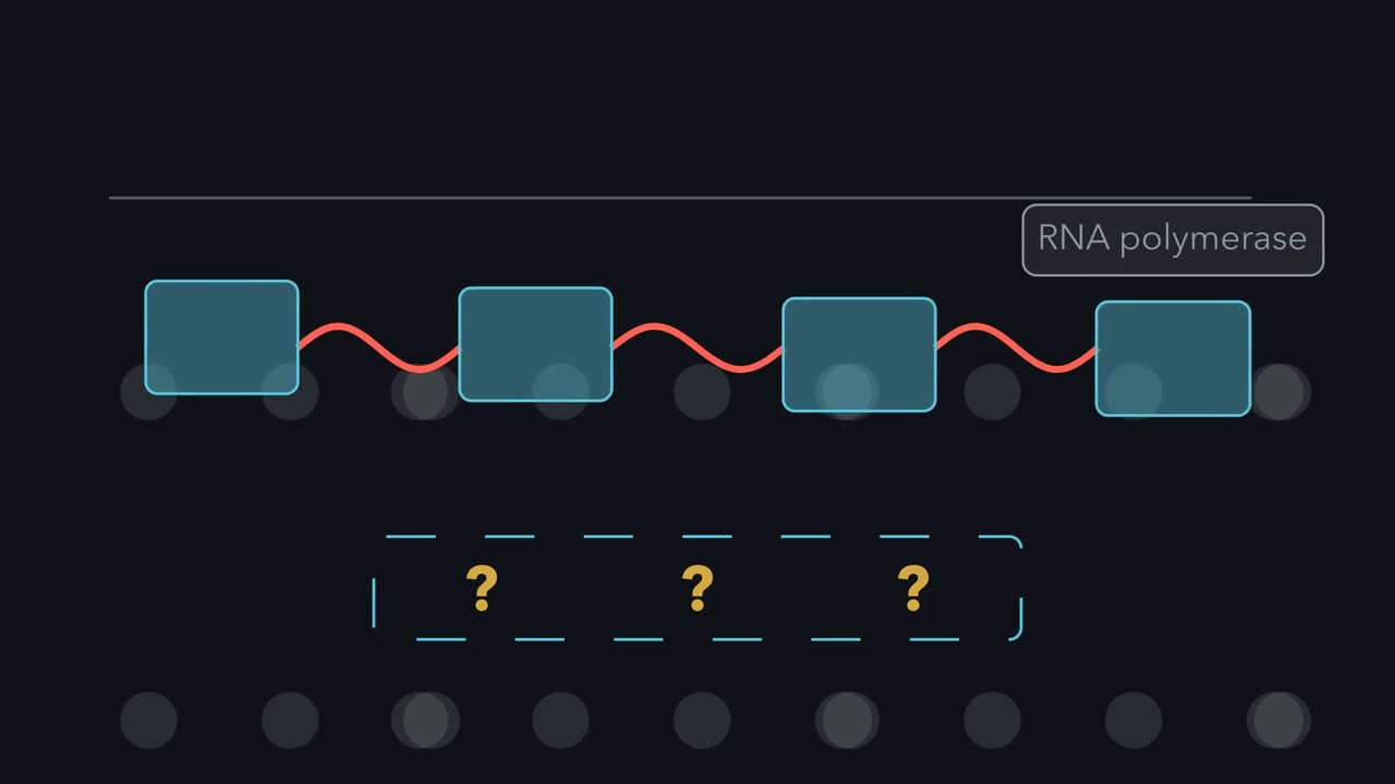 ▶ Watch From Transcript to Message · 3:24
Read the transcript of “From Transcript to Message”
Film transcript: From Transcript to Message
Why can the RNA copied from a gene differ from the message a ribosome reads? Watch one new transcript grow. Some parts will stay, some will leave, and two new end marks will appear. These planned changes are RNA processing.
The first copy is a primary transcript, also called pre-mRNA. Blue exons are retained in the processed RNA. Red introns lie between them and will be removed. Each decision changes this same moving transcript. Processing can begin before transcription ends. A methylated guanosine cap attaches to the 5′ end through an unusual triphosphate bridge. That bridge leaves no ordinary exposed 5′ end for an exonuclease. The cap also recruits proteins for export and translation. Near the other end, an RNA signal guides a cut.
Poly(A) polymerase adds adenosines without copying DNA. The tail is added to RNA, rather than copied from the gene. Its binding proteins support stability and translation. Now zoom into one intron. In the major human class, its ends usually carry GU and AG. A branch point inside contains the adenosine that starts the reaction. A pyrimidine-rich stretch lies before the final site. Small nuclear RNAs help place these sites. U1 recognizes the 5′ splice site. U2 holds the branch region with its adenosine exposed. Other particles join and rearrange the assembly. Together, these RNAs and proteins form a spliceosome anew on each intron. The branch adenosine attacks the 5′ splice site.
The upstream exon comes free, and the intron bends into a lariat. Look at the free edge glowing gold. Which exposed end must move next to join the exons? The freed 3′ end of the upstream exon attacks the 3′ splice site. Both exons join, and the lariat leaves. Two attacks, two bond swaps, and the intron is gone. Pull back to the whole transcript. Introns leave as lariats, and exons meet in order. The transcript is shorter, but the retained exons are connected. The cap and tail remain at opposite ends. A processed message is now ready for later steps. Ends marked; segments chosen. That is how one primary transcript becomes a messenger RNA. The end marks also help protect and use the message.
The rule applies to protein-coding RNA polymerase II transcripts in eukaryotic nuclei. Processing marks transcript ends and chooses which segments join into messenger RNA.
From the film
- Processing marks transcript ends and chooses which segments join into messenger RNA.
- A cap marks the 5' end, while cleavage and a poly(A) tail form the 3' end.
- The branch-point adenosine starts a two-step reaction that joins the exons.
- The spliceosome uses small nuclear RNAs and proteins to recognize sites and rearrange the transcript.
The first film followed one transcript through capping, tailing and splicing; the text adds the chemistry, the evidence and the arithmetic.
An imagined human gene with four exons and three introns runs 5,137 nucleotides from transcription start to the end of its last exon. Its finished mRNA carries 737 of those nucleotides, plus a cap and tail not encoded in the gene.
One transcript, three processing decisions
RNA polymerase II first makes a primary transcript, or pre-mRNA (transcription). Its exons are kept; the introns between them are removed in splicing.
The one idea. Processing marks transcript ends and chooses which segments join into messenger RNA.
Scope: eukaryotic protein-coding RNA polymerase II transcripts. Other eukaryotic RNAs and bacterial RNAs follow different routes.
The three decisions (a 5’ cap, 3’ cleavage with a poly(A) tail, and splicing) need not wait: capping begins when the RNA is only about 25 to 30 nucleotides long, and early introns often splice while later regions are still being copied.
Marking the two ends
The cap, 7-methylguanosine (m7G), is joined to the first transcribed nucleotide 5’ to 5’ through three phosphates, where a normal backbone links 3’ to 5’. That reversed link blocks 5’-to-3’ exonucleases. The film’s helpers are a nuclear cap-binding complex for export and the cytoplasmic translation factor eIF4E, which grips the cap to help recruit the translation machinery.
The film’s 3’ signal is AAUAAA, read in mammals by CPSF. Cross-linking with purified human CPSF by Chan and colleagues in 2014 showed its subunits CPSF30 and Wdr33 contacting it directly. CstF binds a downstream GU-rich stretch, and the nuclease CPSF73 cuts between the two, about 10 to 30 nucleotides beyond AAUAAA. Poly(A) polymerase then adds the tail.
The evidence for split genes
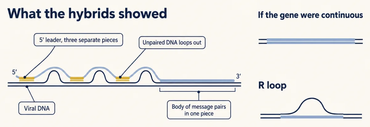
Until 1977 a gene was expected to be a continuous stretch of DNA, as in E. coli. Two groups mapping the messages of adenovirus 2, a DNA virus that grows in human cells, found otherwise. At MIT, Susan Berget, Claire Moore and Phillip Sharp purified hexon mRNA, the message for the virus’s main coat protein, and paired it with mapped viral DNA. At Cold Spring Harbor, Louise Chow, Richard Gelinas, Thomas Broker and Richard Roberts paired viral DNA with RNA from infected-cell polysomes. Both viewed the hybrids by electron microscopy and mapped the messages’ 5’ ends with single-stranded DNA fragments. Chow and colleagues also made R loops, where RNA displaces one DNA strand and pairs with the other.
Predict. If the message came from one continuous DNA stretch, what should its RNA-DNA hybrid look like?
Check your prediction
One unbroken paired stretch. Instead, the message body paired continuously while its 5’ end paired with separated regions, leaving the DNA between them unpaired.
Chow and colleagues mapped a 150-to-200-nucleotide leader shared by many late mRNAs and copied from three separated regions, the tripartite leader. Berget and colleagues proposed that four separate segments may be joined to make hexon mRNA, implying that intervening RNA is removed.
Split genes were soon found in animal genes, including globin genes, and Roberts and Sharp received the 1993 Nobel Prize in Physiology or Medicine for discoveries of split genes. Introns are not simply junk: some contain sequences that control splicing or transcription.
How the spliceosome joins exons
Most major-class (U2-type) human introns follow the film’s GU-AG rule. The branch point usually lies a few dozen nucleotides before the 3’ end, with a pyrimidine-rich run between it and the AG.
Chemically, the film’s two attacks are transesterifications, reactions that exchange one phosphodiester bond for another.
- The branch adenosine’s 2’-OH attacks the phosphate at the 5’ splice site. The intron’s 5’ G is now joined to branch A through a 2’-to-5’ bond, which closes the lariat.
- The freed upstream exon’s 3’-OH attacks the phosphate at the 3’ splice site, joining the exons.
Predict. Does this bond-exchange chemistry itself need ATP?
Check your prediction
No. No bond is lost overall, so the exchanges need no ATP; the machinery spends ATP to assemble and to rearrange between catalytic states.
The film’s guides are small nuclear ribonucleoproteins, or snRNPs (U1, U2, U4, U5 and U6), each a small RNA with proteins; with many other proteins they build the spliceosome anew on each intron. Part of the guiding is base pairing: U1 snRNA pairs with the 5’ splice site, and U2 snRNA with the branch-point sequence, leaving branch A bulged out. After the U4/U6.U5 tri-snRNP joins, U6 folds with U2 into the active site; cryo-EM structures from yeast and humans show U6 positioning catalytic metal ions. The active site is largely RNA, supported and rearranged by proteins.
This fits a 1982 result from Thomas Cech’s laboratory: a Tetrahymena ribosomal RNA group I intron removed itself without protein, using external guanosine rather than branch A for the first attack. Self-splicing group II introns in organelles and bacteria form a lariat as the spliceosome does.
Because splice sites are short, loosely defined signals read by snRNAs and proteins together, a transcript can offer more than one possible site, and the joins can change.
Read the transcript of “When the Joins Change”
Film transcript: When the Joins Change
The first film marked both ends and joined blue exons. If those joins change, must the protein's reading frame change too? Follow different copies of one gene's transcript as each is spliced its own way. First, in a normal cell, regulatory signals can steer the spliceosome to different junctions.
Splicing need not make one pattern. A cell may skip an exon, shift a splice site, choose between exons, or retain an intron. Each choice makes a different messenger RNA. Regulatory signals can make these patterns differ among tissues. A sequence change can weaken a normal splice site. Nearby sites then compete differently. A weakened splice site most often causes exon skipping. Less often, a cryptic site takes over or the intron is retained. One allele can make several products at once. One studied beta-thalassemia allele made two abnormal RNAs in cultured cells. The main product kept part of an intron. A smaller amount joined nonadjacent exons.
One changed splice signal shifted competition in more than one way. Length is only the first consequence. In coding sequence, ribosomes read bases in groups called codons. Changing the length can shift every downstream group, changing many amino acids. A shift after one junction can change every later codon boundary. This outlined exon contains only complete codon groups. Imagine that splicing skips it. Use the ruler, not the exon count. Will the downstream groups stay aligned, or will every boundary move?
They stay aligned. Removing complete codon groups changes the message's length but not its downstream grouping. Whole triplets leave, so the next base begins a codon normally. The new junction can still matter, because sequence determines what it encodes. Now skip a coding segment with one extra base beyond complete groups. Every downstream boundary shifts. The reading marker now meets different base groups. A premature stop becomes likely, although only the sequence can show where one appears. The remainder test applies to net changes inside coding sequence. It does not predict effects of untranslated changes. It also cannot tell whether an RNA is exported, degraded, or translated.
Those fates require evidence beyond a length calculation. Processing still marks transcript ends and chooses which segments join into messenger RNA. For coding consequences, keep a second rule. Coding-length changes keep the downstream frame only in multiples of three. It applies only when the changed nucleotides are coding.
Sequence decides the actual protein outcome. That is the scope of this rule.
From the film
- Coding-length changes keep the downstream frame only in multiples of three.
- Alternative splicing lets one gene make mRNAs with different combinations of exons.
- A weakened splice site most often causes exon skipping; less often a cryptic site takes over or the intron is retained.
- The frame rule applies to net changes in coding sequence; sequence decides the protein outcome.
One gene, several messages
Wang and colleagues (2008) classed the patterns they found in human transcriptomes into skipped exons, alternative 5’ or 3’ splice sites, mutually exclusive exons and retained introns, the four patterns in the second film, and estimated that 92 to 94 percent of human genes undergo alternative splicing. Their regulation belongs to eukaryotic gene regulation.
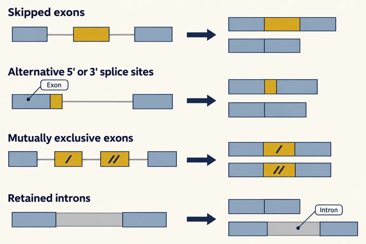
Predict. A splice-junction sequencing read connects exon 1 directly to exon 3. Does that one read show that every message from the gene skips exon 2?
Check your prediction
No. It proves only that this product exists; how often exon 2 is included needs counts from many informative reads and a suitable comparison.
What a splice-site mutation can do
A weakened splice site most often causes exon skipping; less often a cryptic splice site, a sequence not normally used, takes over, or the intron is retained. Competing signals nearby set the outcome, so it cannot be promised for one variant (mutation).
The film’s beta-thalassemia allele changed the GT at the start of intron 2. In HeLa cells its main RNA retained the intron’s first 47 nucleotides, and a smaller amount joined exon 1 directly to exon 3. Because 47 = 3 x 15 + 2, that insertion shifts the frame.
That is the second film’s rule for net changes within coding sequence: coding-length changes keep the downstream frame only in multiples of three.
frame kept if (change in coding length) / 3 leaves remainder 0
Why does losing one site so often drop a whole exon? Vertebrate exons are often recognized as units by the sites at both ends.
Worked problem. An imagined human gene has four exons. Exon 1 has a 60-nucleotide 5’ UTR followed by 90 coding nucleotides beginning with AUG, for 150 total. Exons 2, 3 and 4 have 87, 100 and 400 nucleotides; exon 4 contains the stop codon and 3’ UTR. Introns 1, 2 and 3 have 900, 1,500 and 2,000. Ignore cap and tail.
- Mature mRNA: 150 + 87 + 100 + 400 = 737 nucleotides. Adding 4,400 intron nucleotides gives a 5,137-nucleotide pre-mRNA.
- A 5’ splice-site mutation of intron 2 causes exon 2 to be skipped: 737 - 87 = 650. Because 87 = 3 x 29, the frame stays aligned and the protein loses 29 amino acids.
- Exon 3 skipped: 737 - 100 = 637. Because 100 = 3 x 33 + 1, the frame shifts by one base after the new exon 2-to-exon 4 junction. A premature stop in exon 4 is likely, though sequence is required. In mammalian cells, a premature stop in the last exon usually escapes exon-junction-dependent nonsense-mediated decay, so a truncated protein could be made (genetic code and translation).
- Intron 1 retained: 737 + 900 = 1,637. Such RNAs are often held in the nucleus or degraded.
Rule to keep: the downstream frame is kept only if the change in coding length is a multiple of three. This is a sketch of the logic for an imagined gene, not a prediction for a real one; the sequence decides whether a stop codon appears.
Design problem
A patient variant changes intron 3 position +5 from G to A; the G of GU is +1. The gene is expressed in muscle. Design a minigene test, predict band sizes and state the limits.
Predict. If one band lies close to the normal-splicing size, is its size alone enough to call the junction normal?
Saved in this browser only.
Check your prediction
No. A nearby cryptic site can make a nearly normal-sized band, so sequence every band.
One way to do it
Clone exon 3, intron 3 and exon 4, with enough flanking intron to keep the outer splice sites (assuming exon 4 is not last), between two vector exons of a splicing reporter (hacking the cell). Transfect wild-type and +5 versions into a cell line, ideally muscle-derived, and run RT-PCR with primers in the vector exons.
With invented lengths of 100 vector-exon nucleotides between the primers, 120 for exon 3, 150 for exon 4 and 800 for intron 3, normal splicing gives 370 bp. Exon 3 skipping gives 250 bp; intron 3 retention gives 1,170 bp.
Controls are empty vector at 100 bp, the wild-type minigene, a change known to disrupt the same 5’ splice site, such as +1 G to A, and a no-reverse-transcriptase sample to detect plasmid DNA. The minigene lacks the rest of the gene and the cell line may lack muscle splicing factors, so the result is evidence, not proof; confirm it in patient RNA if available. The assay tests splicing, not the variant’s clinical meaning.
Going deeper
Variations at the ends
Three activities build the cap in order; in mammals, one protein carries the first two. Triphosphatase removes the outermost, gamma phosphate. Guanylyltransferase transfers GMP from GTP through a lysine-bound intermediate. Methyltransferase moves a methyl group from S-adenosylmethionine to guanine N7, making cap 0; in many organisms, 2’-OH methylation of the first nucleotide makes cap 1 (nucleotide metabolism).
Predict. If the triphosphatase fails, can guanylyltransferase still attach GMP normally?
Check your prediction
No. Guanylyltransferase needs the end that triphosphatase prepares by removing the outermost phosphate.
New mammalian tails are commonly described as about 200 to 250 adenosines; yeast tails are shorter, and tails shrink with time, so any tail length needs an organism and whether new RNA or the whole population was measured. In L4-stage C. elegans larvae, sequencing individual tails found a median of 57. Animal replication-dependent histone mRNAs are a textbook exception: they are cut but normally form a 3’ stem-loop instead of receiving a poly(A) tail.
Bacterial mRNAs lack an m7G cap, though some carry other caps such as NAD+, and lack spliceosomal introns, though some genes contain self-splicing introns. Bacterial ribosomes can translate an RNA while it is still being made; in eukaryotes the nuclear envelope separates the two, giving time to process the message first.
The snRNP exchange and the minor spliceosome
After the tri-snRNP joins, U1 leaves and U6 takes over the 5’ splice site. The helicase Brr2 unwinds U4 from U6, and U4 leaves before U6 and U2 form the active site.
A second, minor spliceosome removes well under 1 percent of introns in human genes. It uses U11, U12, U4atac and U6atac, plus the shared U5.
Your notes
Select any text in the lesson to highlight it or add a note to it. Your highlights and notes are saved in this browser and collected in your notebook.
This browser is not letting the site save anything (a private window or strict privacy settings), so highlights and notes will be lost when you leave the page.
How we measure it
R-loop and RNA-DNA hybrid mapping by electron microscopy
Pair a purified RNA with its DNA under conditions that favour RNA-DNA hybrids, then view the molecules in the electron microscope. Paired stretches and the loops of unpaired DNA between them show which parts of the DNA are present in the RNA and which are left out.
Minigene splicing assay
Clone an exon with its flanking intron sequence, wild type or variant, into a reporter plasmid, put it into cultured cells, and read the spliced RNA by RT-PCR with primers in the flanking exons. The band sizes, confirmed by sequencing, tell you whether the exon was included, skipped, or joined at a new site.
RNA sequencing with splice-junction reads
Sequence short fragments of cDNA made from mRNA and map them to the genome. Reads that span two exons record which exons were joined, so the share of messages that include a given exon can be estimated across tissues.
Practice questions on this lesson (4) → MCAT-style, original, each with an explanation
Where this lesson fits
Before thisReview first
- Transcription: From DNA to RNAThis course · Lesson 16
You need to know how RNA polymerase makes the primary transcript that gets capped, tailed and spliced here.
- DNA Is the Genetic MaterialThis course · Lesson 12
This lesson assumes you know DNA carries the gene, so a gene's length can be compared with its message.
After thisStudy next
- The Genetic Code and TranslationThis course · Lesson 18
The reading-frame rule for splice changes builds directly on how codons are read in triplets.
- Eukaryotic Gene Regulation and EpigeneticsThis course · Lesson 20
Alternative splicing and the cap-and-tail steps are regulated there as part of gene control in eukaryotes.
- Sequencing and GenomesThis course · Lesson 23
Reading spliced transcripts, such as junction reads joining exon 1 to exon 3, depends on sequencing methods taught there.
Elsewhere on the siteFilms, simulations and method films in other courses
- From Gene Copy to Protein ChainFilm · Biology
It shows how a gene copy is made into mRNA and read, which the processed message feeds.
- Where the Reader WorksFilm · Cell Biology
It shows how a signal sequence in a processed message's protein directs it to its compartment.
- One Cell, One Shipping LineFilm · Biology
It follows an mRNA copy leaving the nucleus to a ribosome, the step that export of a capped, tailed message enables.
Suggested by an AI model from the lessons themselves, then checked. How we use AI
Finish line
Test yourself
9 questions
Practice and MCAT-style, with explained answers.
Next · Lesson 18
The Genetic Code and Translation
How can one added base regroup every codon that follows, while one substituted base may change no amino acid at all?
Continue →