Cell Biology · Skim version
Nuclear Architecture and Membraneless Compartments
Read the full lesson → Skim the whole course →
The guiding idea
How does a nucleus keep things apart with no walls around its compartments?
The nucleus has no membrane-bound compartments inside it, yet its chromosomes keep to their own regions and its nucleoplasm holds bodies with different jobs.
From the film: Bigger or Brighter?
- A bright spot in the nucleus shows only that a protein is concentrated there, not how.
- Both kinds of spot look round and refill after bleaching, and site clusters that drift together can look fused.
- At equilibrium under fixed conditions, one protein forming a phase on its own adds droplet volume while the outside stays at saturation.
- On a fixed array of sites that does not change size, extra protein mainly brightens the spots until the sites are full.
- Where the extra goes is a clue, not a proof: mixtures, chromatin and tiny spots can blur it.
From the film: Real Bodies, Real Tests
- Chromosomes keep to territories, and Hi-C reveals open A and closed B compartments.
- Mammalian nucleoli have three parts that build ribosomal subunits, read from the inside out.
- Speckles, Cajal bodies, PML bodies and paraspeckles are each known by what they hold.
- The frog oocyte nucleolus provides strong evidence for coexisting liquid phases.
- Chromatin bridging is a third mechanism, and for heterochromatin the verdict splits by system.
-
Why looking harder does not help
-
Add more protein: the numbers
-
What the nucleus holds
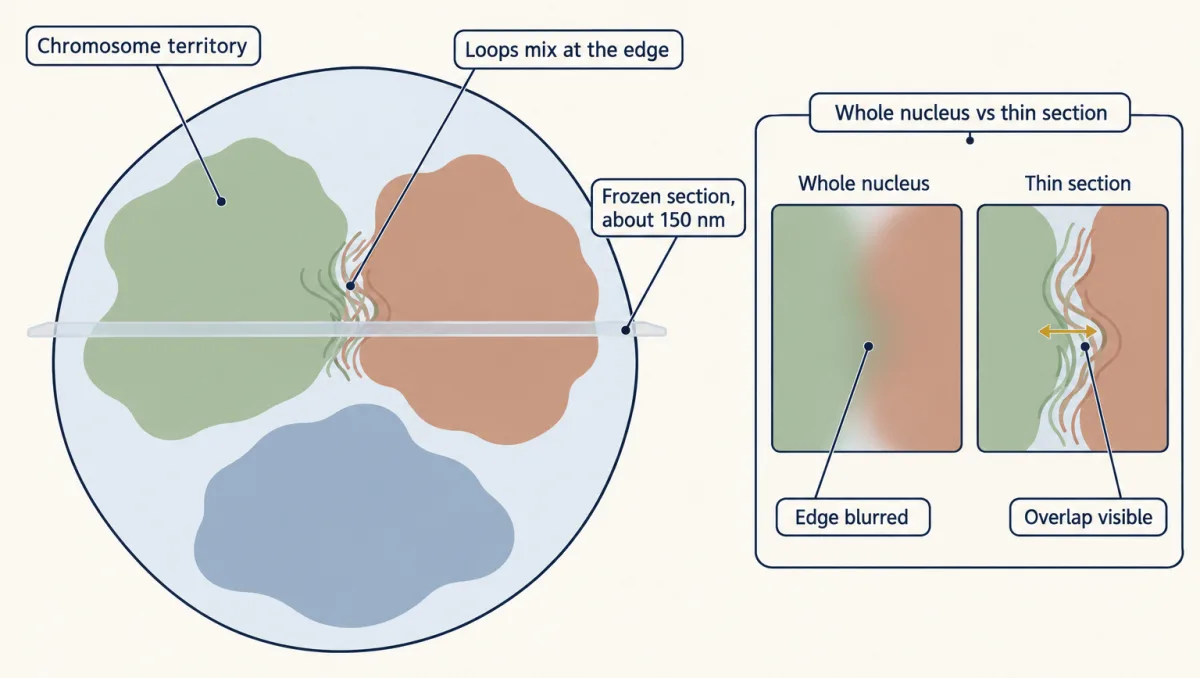
Each chromosome fills a territory, but where two meet, loops of one poke into the other. A frozen section about 150 nm thick removes most of the blur, so the overlap can be seen. AI-generated illustration 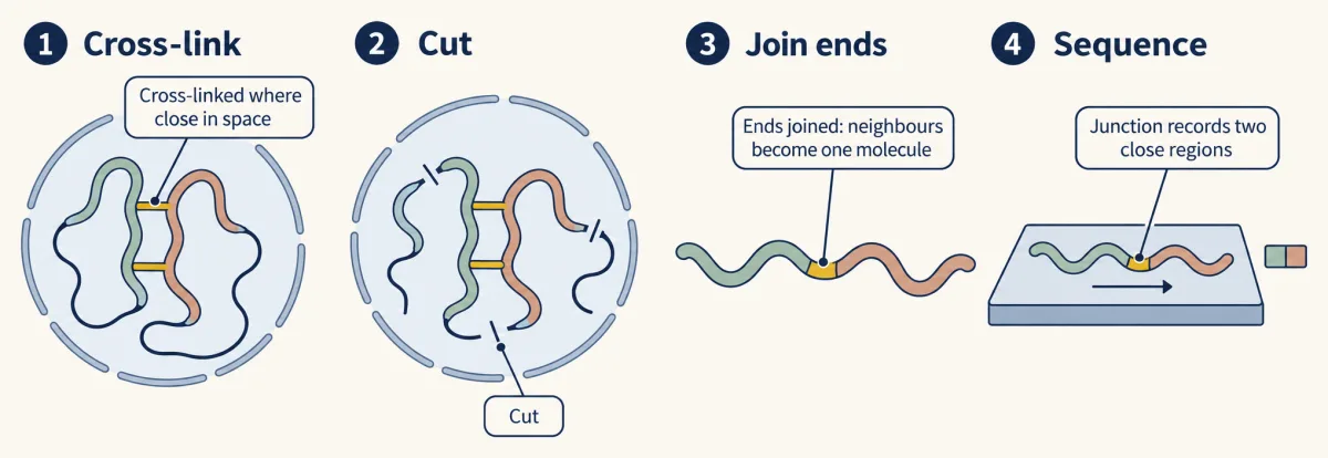
In Hi-C, chromatin is cross-linked, cut and re-joined so that regions close in space end up in one molecule; each sequenced junction records two regions that were near each other. Standard Hi-C data average over millions of cells. AI-generated illustration 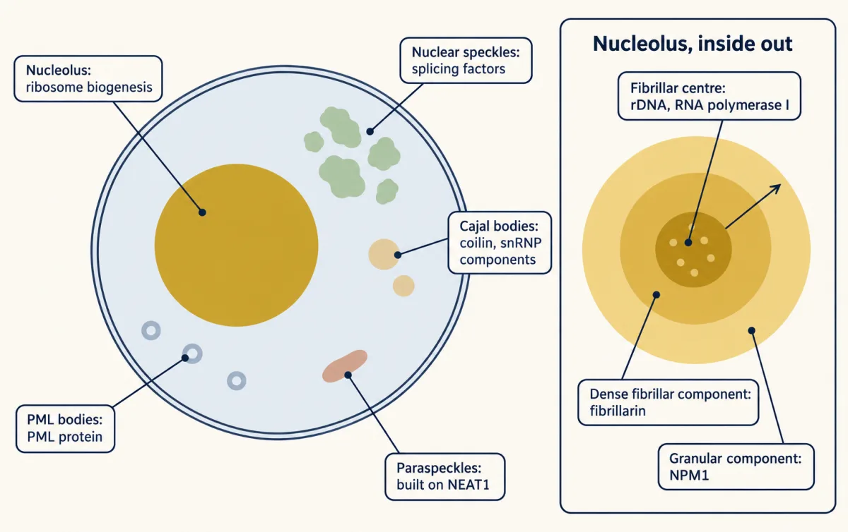
The nucleus holds bodies with no membrane, each known by what it concentrates. The nucleolus has three parts, read from the inside out: fibrillar centre, dense fibrillar component, granular component. AI-generated illustration -
Real bodies: one strong case, one split verdict
-
Where the test is weaker
-
Going deeper
Where this lesson fits
Before thisReview first
- Organelles as Distinct EnvironmentsThis course · Lesson 6
It shows how membrane-bound compartments hold distinct contents, the contrast this lesson draws for compartments with no membrane.
- Experiments That Distinguish MechanismsThis course · Lesson 3
You need its logic of tests that tell mechanisms apart to judge what round shape, FRAP and hexanediol can show.
- The Nuclear Envelope and Nuclear TransportThis course · Lesson 8
It describes the nuclear envelope and the nucleoplasm that this lesson's bodies and territories sit inside.
After thisStudy next
- Mitosis and Chromosome SegregationThis course · Lesson 19
It follows how chromosomes that sit in territories in interphase condense and are segregated in mitosis.
- Cell Identity, Cell State and DiseaseThis course · Lesson 25
It takes up how chromatin and regulation set cell identity, building on compartments and nuclear organization.
- Designing a Cell Biology ExperimentThis course · Lesson 26
It uses the same skill of designing a test that separates mechanisms, as in this lesson's design problem.
Suggested by an AI model from the lessons themselves, then checked. How we use AI