Cell Signaling · Unit 4: Receptors with enzymes attached
PI 3-Kinase, Akt and Insulin
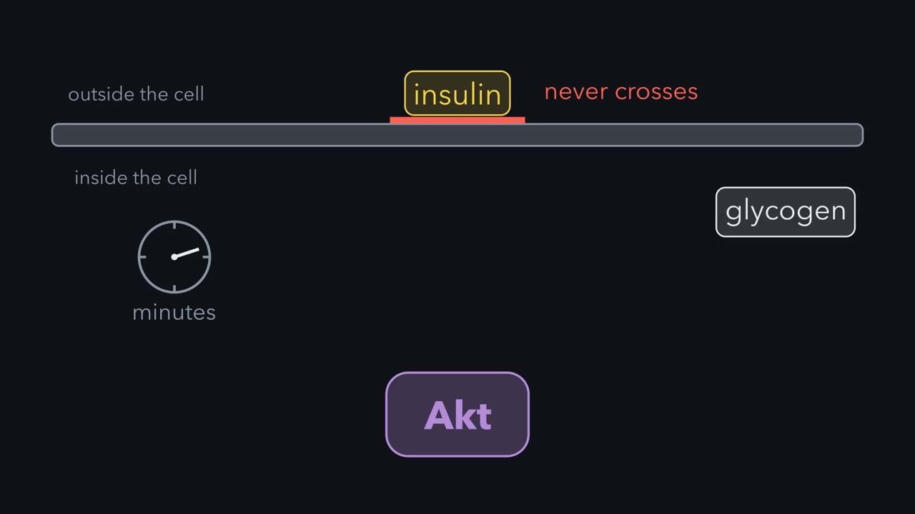
Insulin never crosses the cell membrane. So how does it move glucose transporters to the surface within minutes?
Pause when the reading reaches a film, and offer to play it. Saved on this device.
Watch the film now, or keep listening
Builds on:Adaptors, Docking Proteins and ScaffoldsProtein Modules: SH2, SH3, PTB, PDZ and PHLipids as Anchors and AddressesWhy these?
 ▶ Watch A Membrane Address for Akt · 2:53
Read the transcript of “A Membrane Address for Akt”
Film transcript: A Membrane Address for Akt
Insulin never crosses the cell membrane. Yet within minutes, a muscle cell pulls glucose out of the blood and stores it as glycogen. The signal runs from the receptor to one kinase, called Akt. Before insulin arrives, Akt drifts in the cytosol, inactive. How does a message from outside reach it? Insulin binds its receptor, which is a tyrosine kinase. The receptor adds phosphates to its own tyrosines, and then to those of a docking protein, IRS-1. IRS-1 is held at the membrane beside the receptor, and has a long tail with many tyrosines. Once the receptor has phosphorylated that tail, proteins with SH2 domains dock on it. An SH2 domain binds a phosphorylated tyrosine, and one such protein is PI 3-kinase.
PI 3-kinase now sits at the membrane, where its substrate is. That substrate is a lipid called PIP2. The catalytic subunit of PI 3-kinase adds a phosphate to the 3 position of the lipid. The product is PIP3. PIP3 is a membrane address. Akt has a PH domain at its amino end. This domain binds PIP3. So Akt leaves the cytosol. The lipid holds it against the membrane. Akt is now at the address. A second kinase, PDK1, has its own PH domain, so the lipid holds it there too. PDK1 adds a phosphate to a threonine in the activation loop of Akt. Akt needs a phosphate on that loop to work well. A separate kinase complex, mTORC2, adds a second phosphate near the tail, on a serine. The first phosphate does most of the work, the second adds to it, and together they give full activity.
Why does Akt need a lipid and two phosphates? Each is a check: Akt is on mainly where PI 3-kinase has made its lipids. With no insulin, normal Akt stays in the cytosol. But give Akt a permanent fatty acid anchor, and it sits at the membrane, gains both phosphates, and is active. Keep this rule: PIP3 first puts Akt in the right place, and also opens its folded shape.
Insulin never crosses the cell membrane, yet within minutes of its arrival a muscle cell is pulling glucose out of the blood and storing it as glycogen. The signal gets there by a relay: the insulin receptor, a docking protein called IRS-1, PI 3-kinase, a membrane lipid called phosphatidylinositol 3,4,5-trisphosphate (PIP3), and then the kinase Akt, which the lipid holds at the membrane and switches on. Akt phosphorylates a different protein for each job, so one hormone moves glucose transporters to the surface, speeds glycogen synthesis and changes gene expression in the liver. The same pathway held on too long drives many cancers, and when it is turned down too far it is part of insulin resistance. Insulin has been given to patients since 1922, yet the receptor was not known to be a tyrosine kinase until 1982, and most of the steps after it were found in the 1990s. This lesson follows the signal from the receptor to Akt, then to its outputs, and then asks how the signal is ended and what happens when it is too weak or cannot be shut off.
Recruitment of AKT: the receptor writes an address on the membrane
“Ligand activates receptor…now what?” The insulin receptor phosphorylates its own tyrosines and then those of a docking protein, IRS-1, which you met with the adaptors and scaffolds. IRS-1 has a PH domain and a PTB domain at one end, which hold it at the membrane beside the activated receptor, and a long tail with many tyrosines. Once the receptor has phosphorylated that tail, proteins with SH2 domains dock on it: PI 3-kinase, the adaptor Grb2 that leads to Ras, and the phosphatase Shp2. Adaptors mediate pathway branching, and this lesson takes the PI 3-kinase branch.
PI 3-kinase has two subunits. The regulatory subunit carries two SH2 domains, and the catalytic subunit does the chemistry. When the SH2 domains bind phosphotyrosines on IRS-1, two things follow. The enzyme is now at the membrane, where its substrate is, and the regulatory subunit loosens the hold it keeps on the catalytic one. The catalytic subunit adds a phosphate to the 3 position of the lipid phosphatidylinositol 4,5-bisphosphate (PIP2) and makes phosphatidylinositol 3,4,5-trisphosphate (PIP3), the membrane address from the lesson on lipid anchors.
Akt is a serine/threonine kinase, also called protein kinase B, with a PH domain at its amino end. Before insulin arrives it drifts in the cytosol, inactive. PIP3 binds the PH domain and holds Akt against the membrane. A second kinase with its own PH domain, PDK1, is held there too, and it adds a phosphate to a threonine in Akt’s activation loop (Thr308), the loop that must carry this phosphate before Akt works well. A separate kinase complex, mTORC2, adds a second phosphate to a serine near Akt’s tail (Ser473). The Thr308 phosphate does most of the work, the Ser473 phosphate adds to it, and the two together give full activity. mTORC2 belongs to the next lesson; here you need only its name.
insulin → receptor → IRS-1 (tyrosine-P) → PI 3-kinase → PIP3 → Akt at the membrane → Thr308-P (PDK1) + Ser473-P (mTORC2) → active Akt
Why a lipid and two phosphates? Each is a check, and together they keep Akt on mainly where PI 3-kinase has made its 3-phosphorylated lipids, downstream of an active receptor, and while those lipids last. Two experiments show what the lipid is for. One is a way of visualizing PIP3: fuse Akt’s PH domain to green fluorescent protein, add insulin, and the glow moves from the cytosol to the plasma membrane within minutes. The other starts from a plain fact: lipids can be attached. Give Akt a permanent fatty-acid anchor and it sits at the membrane, gains both phosphates and is active with no insulin present. So PIP3’s first job is to put Akt in the right place. Its binding also opens Akt’s folded, self-inhibited shape so that PDK1 can reach Thr308, and Akt stays most active while it remains on the lipid.
PI3K–AKT regulation of metabolism: one kinase, three substrates
Different tissues need different things from the same hormone, and Akt provides them by phosphorylating a different protein for each job.
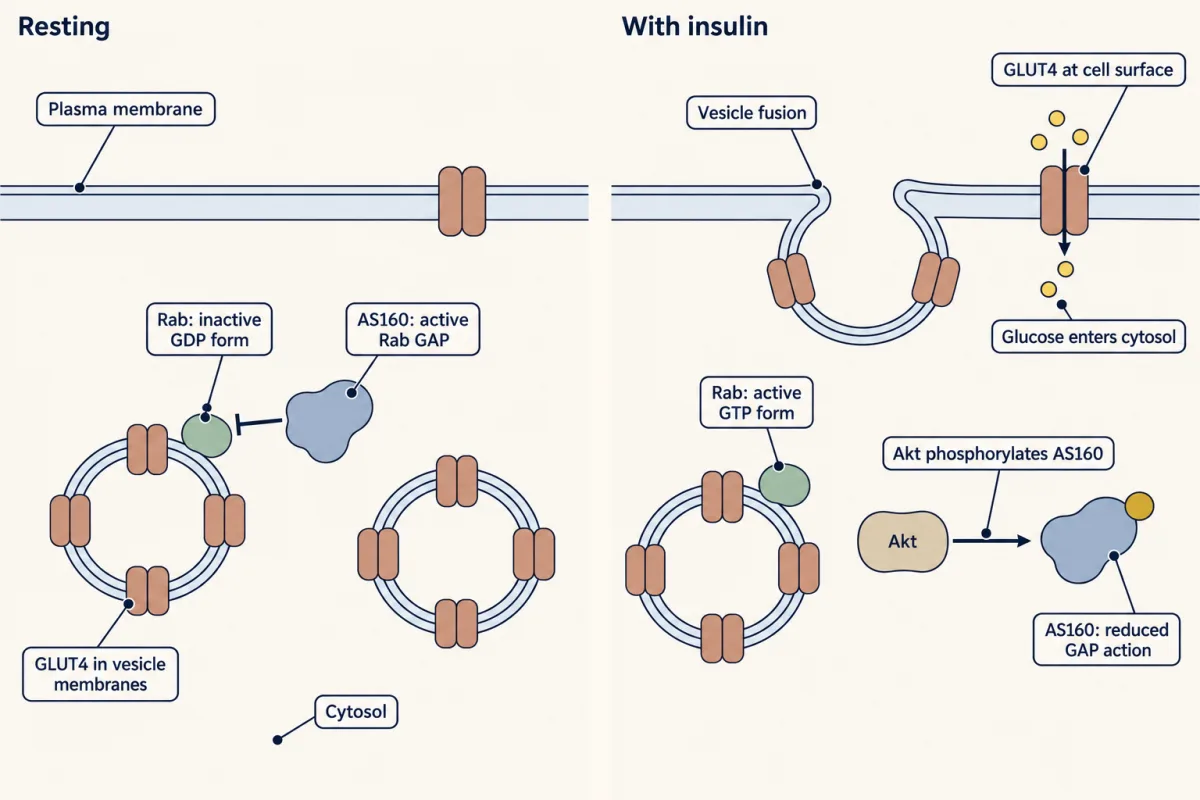
Glucose uptake in muscle and fat. GLUT4 is the main insulin-responsive glucose transporter of these two tissues. In the resting cell most of it sits in small vesicles inside the cell. Insulin moves transporters from an internal membrane pool to the cell surface, as was shown in rat fat cells. Akt’s part, found later, is to phosphorylate AS160, a GAP for the Rab GTPases that direct those vesicles. A GAP switches its GTPase off, as you saw with Ras, and Akt’s phosphate stops the GAP acting on those Rabs. With the Rabs left in their GTP form, the vesicles move to the plasma membrane and, helped by other insulin-regulated proteins, fuse with it, and glucose enters. This step was worked out mostly in fat cells.
Glycogen synthesis. An enzyme called glycogen synthase kinase 3 (GSK3) phosphorylates glycogen synthase and keeps it slow. Akt phosphorylates GSK3 and inhibits it, a link reported in 1995 by the laboratories of Philip Cohen and Brian Hemmings. Phosphatases then strip the inhibitory phosphates from glycogen synthase, and it speeds up. This is one of several ways insulin raises glycogen synthesis.
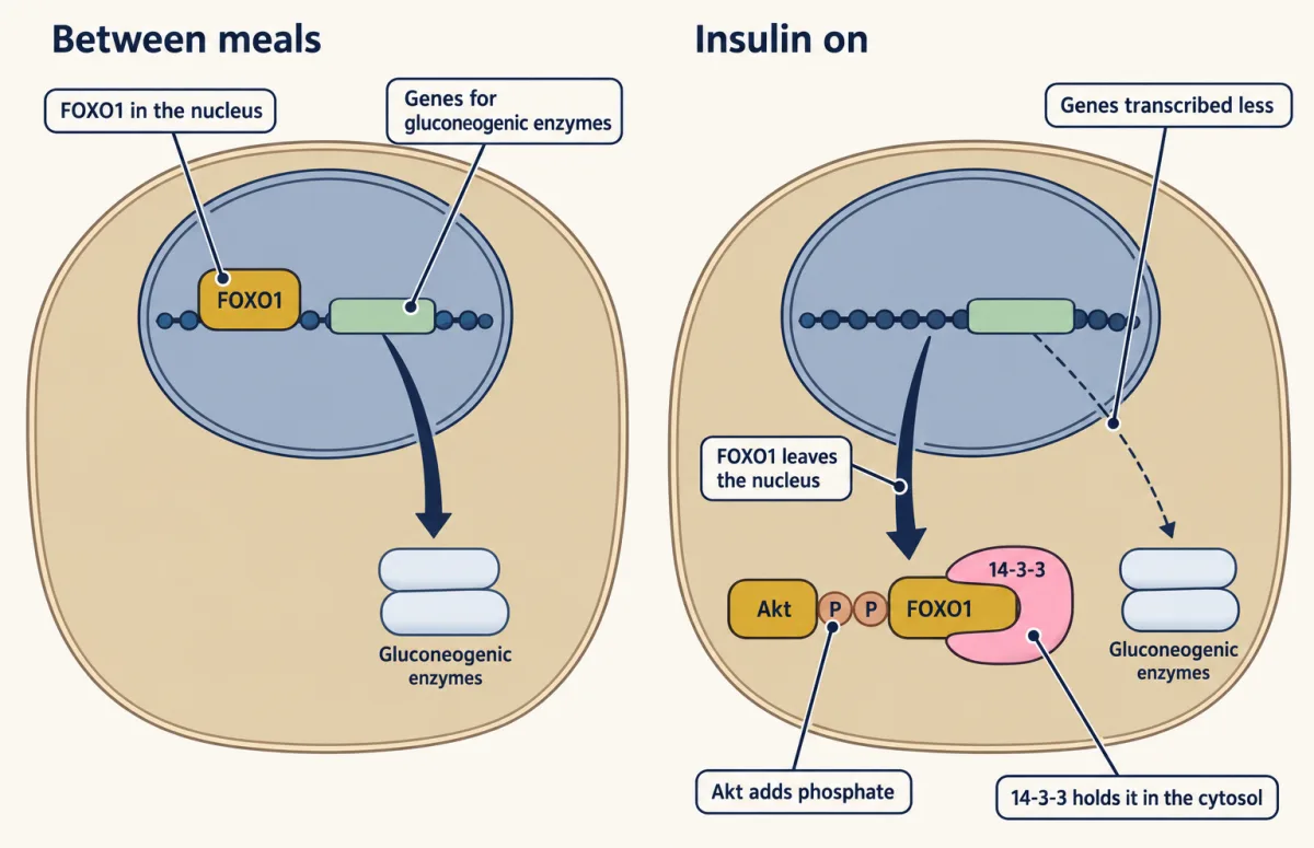
Gene expression in the liver. FOXO1 is a transcription factor, a protein that binds DNA and switches genes on. Between meals it sits in the nucleus of a liver cell and helps drive the genes for enzymes of gluconeogenesis, including glucose 6-phosphatase and phosphoenolpyruvate (PEP) carboxykinase. Akt phosphorylates FOXO1, the phosphates become binding sites for 14-3-3 proteins, and FOXO1 is moved out of the nucleus and held in the cytosol. Those genes are then transcribed less. FOXO1 leaves the nucleus quickly; the enzymes already made take longer to disappear. So this is not how insulin cuts the liver’s glucose output within minutes of a meal. Experiments in mice show that this fast effect can work even without insulin signaling in the liver cell. It depends largely on insulin’s brake on fat breakdown in adipose tissue, which lowers the fatty acids reaching the liver.
Akt also switches on mTORC1, a controller of cell growth, which is the subject of the next lesson.
More AKT: the same pathway says survive and grow
Insulin is one of many signals that use this route. Growth factors such as platelet-derived growth factor (PDGF) switch on PI 3-kinase through their own receptor tyrosine kinases, and the PDGF receptor binds the regulatory subunit directly on its own phosphotyrosines, with no IRS-1 in between. In those cells the output of Akt is read as an instruction to stay alive and grow. Its substrates include proteins that would otherwise start the cell-death programme, and FOXO again, which in many cell types switches on genes that stop division.
A tumour gains from holding such a pathway on, and two breaks are common. PIK3CA, the gene for a catalytic subunit of PI 3-kinase, is among the most often mutated oncogenes in human cancer, including cancers of the breast, colon and endometrium. The mutations cluster at a few positions and raise the enzyme’s activity, so that it needs less of the proper signal. Phosphatase and tensin homolog (PTEN), the phosphatase that removes the 3-phosphate, is a tumour suppressor that many tumours have lost. Either way the membrane carries more phosphatidylinositol 3,4,5-trisphosphate (PIP3) and holds more Akt.
Ending the signal, and what insulin resistance means
Every step above has an eraser. Tyrosine phosphatases reset the receptor and IRS-1. Phosphatase and tensin homolog (PTEN) removes the 3-phosphate from phosphatidylinositol 3,4,5-trisphosphate (PIP3) and returns it to phosphatidylinositol 4,5-bisphosphate (PIP2). With the address gone, Akt leaves the membrane and protein phosphatases take off its two phosphates.
The pathway also turns itself down. Kinases that it switches on, among them mTORC1 and its target S6 kinase from the next lesson, phosphorylate IRS-1 on serines. Phosphate on these serines makes IRS-1 a poorer substrate for the receptor and can mark it for destruction by the proteasome. Not every serine site works this way, and a few support signaling, so the effect depends on the site. This is negative feedback of the kind you met in the signaling circuits lesson: the output reduces its own input, so a long signal fades.
Insulin resistance means that a given concentration of insulin produces less response than it should. In muscle and fat from insulin-resistant people and animals, the receptor is usually present and binds insulin, but the steps after it are weak, and the receptor itself can also be fewer or signal less. Less phosphotyrosine appears on IRS-1, less PI 3-kinase is recruited, and fewer GLUT4 vesicles reach the surface. One widely studied explanation is the feedback loop driven too hard. With a chronic excess of nutrients, lipids and inflammatory signals keep serine kinases active, IRS-1 collects phosphoserine, and the docking protein docks less. Some lipid signals act on the receptor itself instead. The dose-response curve for insulin shifts to the right, so the same effect needs more hormone, and when the defect lies after the receptor the maximal response also falls. The pancreas supplies it, often for years, and type 2 diabetes begins when it no longer can.
How much of human insulin resistance this one mechanism explains is still argued, and the break is not always at IRS-1. The next lesson takes up the kinase mTORC1, which this pathway switches on.
Design problem
Twenty minutes after a meal a muscle cell must take up glucose and store it as glycogen. Over the following hours a liver cell must also make fewer of the enzymes that build new glucose. Using one kinase, Akt, choose a substrate for each job and say whether Akt switches that substrate on or off. Then predict what happens to each job in a person with one faulty copy of the Akt gene that muscle and liver use most.
Saved in this browser only.
One way to do it
Uptake: AS160, switched off. It is the GAP that keeps GLUT4 vesicles inside the cell.
Storage: GSK3, switched off. It was holding glycogen synthase in its slow, phosphorylated form.
Liver: FOXO1, switched off by removal from the nucleus. The gluconeogenic genes lose an activator. This is the slow arm; the fast fall in output depends on routes outside the liver cell.
In every case Akt turns its substrate off, and the substrate was a brake on that job.
The gene is AKT2. With one faulty copy you might expect half the Akt and a mild defect. Predict a larger one if the faulty protein also interferes with the normal one. Then each job weakens: fewer transporters reach the surface after a meal, glycogen is stored more slowly, and the liver keeps making glucose while insulin is present. Blood glucose rises and the pancreas answers with more insulin. A family with this defect was described in 2004. One copy of AKT2 carried a single amino acid change in the kinase domain, the change was inherited together with severe insulin resistance and diabetes, and in cultured cells the mutant kinase blocked the normal one.
Going deeper
The hormone before the pathway: Toronto, 1921 to 1922
Read the transcript of “Four Names Behind the First Insulin”
Film transcript: Four Names Behind the First Insulin
The pancreas makes digestive enzymes and, in islets, a hormone. By 1920, extracts lowered blood sugar, but none was safe for patients. In November 1920, Frederick Banting took an idea to John Macleod, a Toronto professor. Tie off the pancreatic duct in dogs. The enzyme-making tissue wastes away. Extract what is left. Macleod gave space, dogs, and two student assistants. One was Charles Best. In summer 1921, Banting and Best made extracts that lowered blood sugar in diabetic dogs. On 11 January 1922, their extract went into Leonard Thompson, then in his early teens. His blood sugar fell a little, and an abscess formed. The extract was too impure.
James Collip raised the alcohol concentration in steps. Impurities dropped out of solution, while the active substance stayed in. He tested each batch on rabbits. It went into the same boy on 23 January. His blood sugar fell by about three quarters. The 1923 Nobel Prize named Banting and Macleod. Banting shared his prize money with Best, Macleod with Collip. The two-name story misses half the people. The receptor was not known to be a tyrosine kinase until 1982. The drug came first, and the mechanism came later.
In January 1922 a teenage boy in Toronto was injected with a purified extract of beef pancreas, and his blood sugar fell by about three quarters. Nobody could have said how.
The pancreas makes digestive enzymes and, in small clusters of cells called islets, a hormone released into the blood. By 1920 several researchers had made pancreatic extracts that lowered blood sugar, and none of those extracts could be given safely to a patient.
Frederick Banting, a surgeon with no research training, took an idea to John Macleod, professor of physiology at Toronto, in November 1920: tie off the pancreatic duct in dogs so that the enzyme-making tissue wastes away, then extract what is left. Macleod gave him laboratory space, dogs and two student assistants, one of them Charles Best. Through the summer of 1921 Banting and Best prepared extracts that lowered blood sugar in diabetic dogs. On 11 January 1922 their extract was injected into Leonard Thompson, then in his early teens. His blood sugar fell a little, and an abscess formed at the injection site.
The extract was active and too impure to use. James Collip, a biochemist visiting from Alberta who had joined the group in December, raised the alcohol concentration of the extract in steps so that impurities dropped out of solution while the active substance stayed in, until at the highest concentration the active substance itself came out and could be collected, and he tested each batch on rabbits. His preparation went into the same boy on 23 January, the injection described at the top of this section. The 1923 Nobel Prize named Banting and Macleod. Banting shared his prize money with Best and Macleod shared his with Collip, so the two-name story you may know leaves out half the people.
They had a drug and no mechanism. The mechanism came later, and the rest of this lesson is that story.
Who showed that insulin moves GLUT4
The 1980 result came from two laboratories working with rat fat cells (Cushman and Wardzala; Suzuki and Kono).
Insulin-resistant liver: a puzzle
There is also a puzzle: the insulin-resistant liver fails to shut off glucose production, yet goes on making fat. Whether insulin signaling in the liver is still driving that fat synthesis, or the supply of nutrients and fatty acids is, is debated. One proposed explanation for that split involves the kinase in the next lesson, mTORC1.
Your notes
Select any text in the lesson to highlight it or add a note to it. Your highlights and notes are saved in this browser and collected in your notebook.
This browser is not letting the site save anything (a private window or strict privacy settings), so highlights and notes will be lost when you leave the page.
How we measure it
Bioassay-guided purification
You split a crude extract into fractions, here by raising the alcohol concentration in steps, and test every fraction on an animal for the activity you want. Collip's rabbit test for a fall in blood sugar told him which fraction held the hormone and turned a crude pancreatic extract into a preparation fit to inject.
Subcellular fractionation with a transporter count
You break cells open gently, separate plasma membranes from internal membranes in a centrifuge, and count glucose transporters in each fraction, for example by the binding of cytochalasin B. Comparing cells with and without insulin shows whether the hormone made new transporters or moved existing ones.
Kinase assay on lipid vesicles
You mix a purified kinase, its substrate protein, radioactive ATP and small artificial membranes of known lipid composition, then measure the phosphate transferred. Leaving PIP3 out of the vesicles showed that PDK1 phosphorylates Akt only when the lipid is present.
PH domain reporter for PIP3
You fuse the PH domain of Akt to green fluorescent protein and watch living cells under a microscope. The fluorescence moves from the cytosol to the plasma membrane where 3-phosphorylated lipids are made. The same domain also binds PI(3,4)P2, so the reporter reads both lipids.
Method films · the methods library
- Binding assays: immunoprecipitation, pull-down and gel shift 3:43
- Centrifugation and subcellular fractionation 2:50
- Cryo-electron microscopy 4:06
- Engineered mutants: site-directed, dominant-negative and constitutively active 4:25
- Family linkage and positional cloning 3:52
- Gene cloning: vectors, libraries and screening 4:04
- Protein purification and column chromatography 4:14
- Radioactive and stable isotope tracers 4:03
- X-ray crystallography 4:06
Practice questions on this lesson (3) → MCAT-style, original, each with an explanation
Where this lesson fits
Before thisReview first
- Adaptors, Docking Proteins and ScaffoldsThis course · Lesson 10
IRS-1 is the docking protein here, and you need how its PH and PTB domains hold it at the membrane.
- Protein Modules: SH2, SH3, PTB, PDZ and PHThis course · Lesson 9
PI 3-kinase's SH2 domains and Akt's PH domain read phosphotyrosines and lipids, as taught there.
- Lipids as Anchors and AddressesThis course · Lesson 11
PIP3 works as a membrane address that holds Akt, which this lesson uses without re-teaching.
After thisStudy next
- mTOR and AMPK: Reading Nutrients and EnergyThis course · Lesson 21
Akt switches on mTORC1, and mTORC2 phosphorylates Akt Ser473, so this lesson takes the relay further.
- Oncogenes: When the Switch SticksThis course · Lesson 41
Mutant PIK3CA and lost PTEN are examples of a stuck switch driving cancer.
- Endocytosis and Signaling EndosomesThis course · Lesson 36
Insulin-driven GLUT4 vesicle traffic and receptor internalization show how membrane trafficking shapes a signal.
Elsewhere on the siteFilms, simulations and method films in other courses
- What the pump rate measuresSimulation · Physiology
It shows how insulin and glucagon set blood glucose, the whole-body result of this cellular pathway.
- A Beta Cell Burns Glucose to Read ItFilm · Physiology
It shows how a beta cell reads glucose to release the insulin that starts this pathway.
- How the body switches fuelsFilm · Biochemistry
It shows how insulin switches the body's pathways between fed and fasting states.
Suggested by an AI model from the lessons themselves, then checked. How we use AI
Finish line
Test yourself
8 questions
Practice and MCAT-style, with explained answers.
Next · Lesson 21
mTOR and AMPK: Reading Nutrients and Energy
How does a cell decide it can afford to grow?
Continue →