Sensory Systems · Unit 2: Touch
Merkel Cells: Touch Receptors That Are Not Neurons
Rewritten on : new text, flashcards and film. Your notes and highlights stay, but the text around them may have moved.
A skin cell that is not a neuron helps a nerve keep firing for as long as you press. How does it hold the press, and what does it tell the nerve?
Pause when the reading reaches a film, and offer to play it. Saved on this device.
Watch the film now, or keep listening
Builds on:Piezo Channels and the Physics of ForceFive Ways to Turn a Stimulus into a CurrentSensory Neurons by Their Genes: the Atlas EraWhy these?
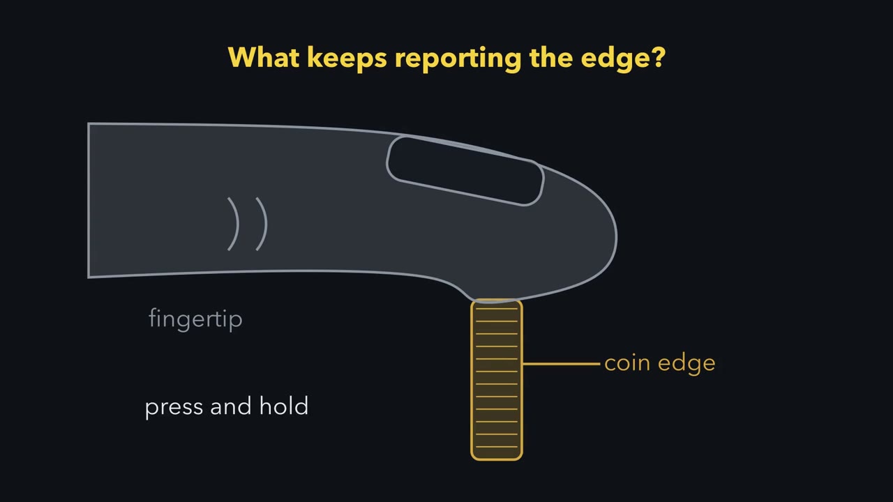 ▶ Watch Two Receptors, One Nerve · 2:24
Read the transcript of “Two Receptors, One Nerve”
Film transcript: Two Receptors, One Nerve
Press a fingertip on the edge of a coin, and hold it still. What keeps telling your brain where that edge is? Some nerve fibres from that skin fire only while the skin is moving. Then they go quiet. Others keep firing for as long as you hold. One such class is the slowly adapting type I afferent, or SA1. In macaque fingertips, its firing carried the spatial layout of millimetre-scale features, such as raised patterns. Its firing has two parts. First, a fast burst as the press begins. That is the dynamic phase. Then firing through the hold, with irregular gaps between spikes. That is the static phase. Now the surprise. The cell that helps make the hold is not a neuron.
It is the Merkel cell. In mice, it comes from epidermal progenitors, not from neural crest. In hairy mouse skin, Merkel cells cluster in touch domes beside guard hairs. An afferent's branched ending makes disc-like contacts on them. So force can reach this fibre by two routes. The ending makes its own Piezo2. Even without Merkel cells, it still responds to touch. Or the Merkel cell feels force through its own Piezo2, and passes the signal across a synapse. Predict. One route reports change. One route holds the press. Which would you guess holds the press?
In mouse touch domes, the ending mainly carries the onset. The Merkel cell mainly holds the press. That is the idea for this lesson. The ending mainly feels the change, and the Merkel cell mainly holds the press. This is mouse hairy skin. And the word mainly matters. The Merkel cell may help with the onset too. Rat whiskers give a different reading. There, another laboratory argues that Merkel cells drive both phases. Keep this. Two receptors feed one nerve, and each mainly makes one phase.
From the film
- In macaque fingertips, SA1 firing carried the spatial layout of millimetre-scale features, with a dynamic onset burst and a static hold phase.
- In mouse touch domes, the ending mainly feels the change and the Merkel cell mainly holds the press.
- The word mainly matters, because rat whiskers give a different reading in which Merkel cells drive both phases.
If a cell in the skin is not a neuron, how can it be the receptor for a nerve that keeps firing as long as you press?
Press a fingertip on the edge of a coin and hold it still. Some of the nerve fibres from that skin fire only while the skin is moving. Others keep firing for as long as you hold, and one such class is the slowly adapting type I (SA1) afferent, named in the touch and pain lesson. In macaque fingertips, SA1 firing carried the spatial layout of millimetre-scale features, such as raised patterns. That is why the SA1 afferent and the cell it contacts are the receptor classically linked to sustained, spatially detailed touch.
The touch and pain lesson also noted that Piezo2, a force-gated channel, is needed in the skin cell that an SA1 afferent contacts: the Merkel cell. But a Merkel cell is not a neuron. That raises two questions. Which part of the SA1 response does a non-neuronal cell make? And how does it pass the signal to the nerve? The answers come mostly from mice, and the second one is still disputed.
An epithelial cell that feels force
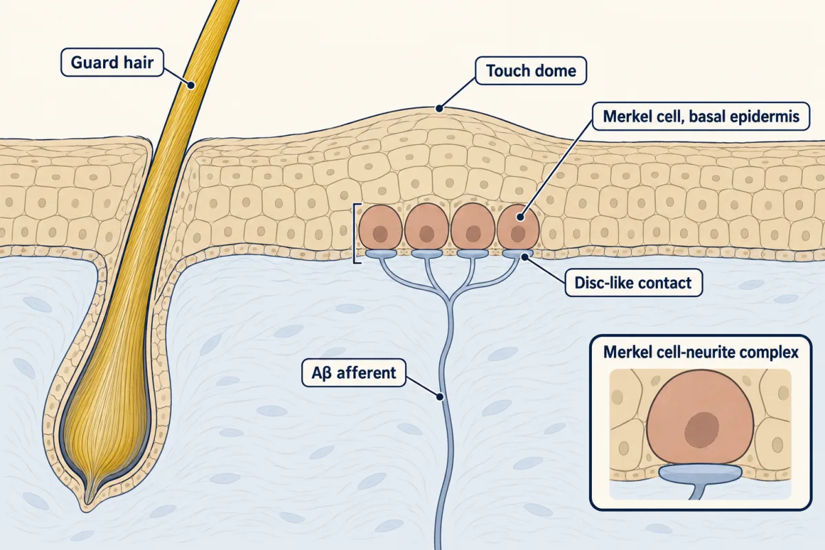
A Merkel cell-neurite complex is a Merkel cell in the basal epidermis together with the terminal of an Aβ low-threshold afferent that contacts it. Aβ fibres are large, myelinated, fast-conducting axons, and low-threshold means that light touch is enough to excite them. In hairy mouse skin, Merkel cells cluster in touch domes beside guard hairs. One afferent’s branched terminal usually contacts a whole dome’s cluster, ending in disc-like contacts on the cells, though some touch domes receive more than one afferent.
The cell looks neuronal. It carries neuronal markers, and in rat whisker follicles it fires spikes. But genetic lineage tracing in mouse embryos (Van Keymeulen and colleagues, 2009) showed that Merkel cells arise from epidermal progenitors. In mouse skin, then, a Merkel cell is an epidermal cell. It belongs with the cells described in epithelia. Its afferent is a sensory neuron, one of the derivatives of neural crest.
The signature response: a burst, then a hold
The SA1 response has two phases, defined in mouse touch-dome recordings (Maksimovic and colleagues, 2014). Picture an afferent’s spikes as ticks on a time line while a probe presses the skin, holds it, and lifts.
| SA1 afferent | Rapidly adapting (RA) afferent | |
|---|---|---|
| As the press begins | a high-frequency burst: the dynamic phase | fires while the skin moves |
| Through the hold | keeps firing, with variable intervals between spikes: the static phase | little or no firing |
| As the probe lifts | fires again while the skin moves |
The SA1 column is the signature to remember: a burst that reports the onset, then irregular firing that lasts as long as the press. A response that fires through the onset and into the hold, but stops before the press ends, is called intermediately adapting. Adaptation and rate coding are covered in sensory coding. Here the two phases are two measurements, and the question is which cell makes each.
Two receptors, one axon
Read the transcript of “Take One Route Away”
Film transcript: Take One Route Away
Two routes feed one axon. How can you tell which route makes which part of the firing? Take one route away, and watch which phase is left. Here, the route taken away is always the Merkel cell's. The recordings come from mouse skin, kept alive in a bath with its nerve attached. An electrode records one afferent while a probe presses the skin. The recorded afferent lacks the light-activated proteins. Light aimed at the touch dome, plus controls, ties the effects to Merkel cells. First test. Light aimed at the Merkel cells depolarizes them, with no touch at all. The afferent fires. The Merkel cell is enough to drive it. Second test. Light silences the Merkel cells during a held press.
What do you expect to lose? The sustained firing is suppressed. The hold needs the Merkel cell in the moment. Third test. Delete Piezo2 from the skin's epithelium, but not from the nerve. In culture, Merkel cells without epithelial Piezo2 lose their touch-evoked current. The ending keeps its channel. Predict what is left.
The onset stays similar to controls. The hold loses its normal sustained firing. That is the cleanest split. One limit. This deletion covers all of the skin's epithelium, not only the Merkel cells. Fourth test. Remove the Merkel cells altogether, by deleting the gene Atoh1 during development. The afferents still respond to touch. But the hold is cut short, before the press ends. That is called intermediately adapting. And the onset falls too. Peak firing drops to about a third of normal. That row only partly fits. A developmental loss cannot say whether the cell's own signal, or a changed skin, caused it. Blocking vesicle release in the epidermis, with tetanus toxin, also reduced both phases.
So release, probably from Merkel cells, may help with the onset too. That is why the idea says mainly. In mouse touch domes, the ending mainly feels the change, and the Merkel cell mainly holds the press. Neither acts alone.
From the film
- Taking one route away, the Merkel cell's, and watching which phase is left shows what each receptor contributes.
- In mouse skin, light on Merkel cells was enough to make the afferent fire, and silencing them suppressed sustained firing during a held press.
- Deleting epithelial Piezo2 left the onset similar and the hold weakened, but developmental loss of Merkel cells also lowered onset, so each route acts mainly, not only, on one phase.
Start with the wiring. Force can reach an SA1 afferent by two routes. The ending makes its own Piezo2, and afferents in mice without Merkel cells still respond to touch, so the ending can transduce force itself. Or a Merkel cell can transduce through its Piezo2 and pass the signal across a synapse. Both routes converge on one axon, so a recording from the nerve mixes them. To pull them apart, take one route away and see which phase of the response is left. In practice, every experiment below takes away or perturbs the Merkel-cell route; none removes the ending’s own transduction. That is one reason the source of the onset is still argued.
The lineage difference makes this possible. A Cre driver is a mouse line that makes the enzyme Cre recombinase under a particular promoter. Crossed to a line in which the target gene is flanked by loxP sites (floxed), it deletes that gene in cells with Cre activity, and their descendants keep the deletion even after the promoter switches off. Each driver’s actual recombination pattern has to be checked. A driver active in skin epithelium, such as Krt14-Cre, can delete a gene from the Merkel cell, along with the rest of the epithelium, and spare the nerve. A driver active in neural-crest derivatives, such as Wnt1-Cre, can delete a gene from the afferent instead, along with other neural-crest derivatives.
First, does the Merkel cell have a force-gated current of its own? Woo and colleagues, from the Patapoutian laboratory, purified Merkel cells from the dorsal skin of newborn mice (P0 to P5) and cultured them overnight. The purified cells contained Piezo2 transcripts. When Piezo2 was deleted from skin epithelium, none of 13 cultured Merkel cells had a detectable mechanically activated current. The afferents that contact Merkel cells still expressed Piezo2, and the complexes appeared anatomically normal. The epithelial driver is not specific to Merkel cells, so the deletion removes the channel from the whole epidermal lineage, and only the Merkel cells were recorded. Still, both routes now have a channel: Piezo2 in the cell, and Piezo2 in the ending.
Three papers took the response apart in 2014, each in a different preparation. This section uses the two mouse papers.
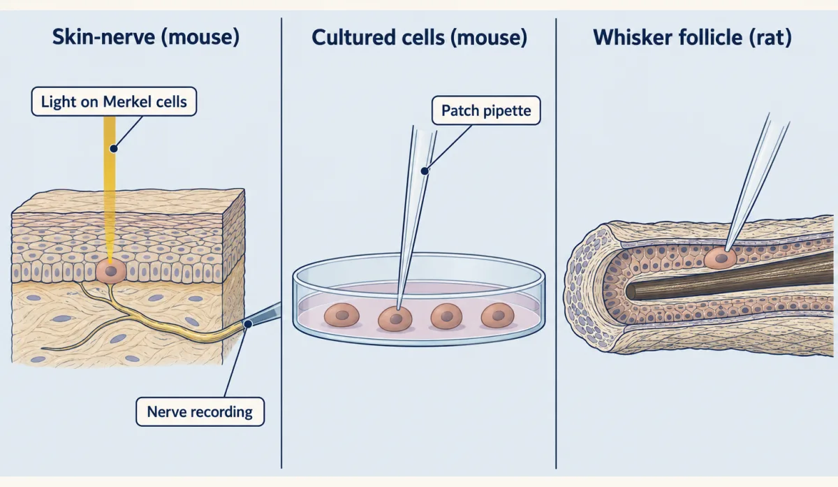
Maksimovic and colleagues recorded single touch-dome afferents in an ex vivo mouse skin-saphenous nerve preparation: a patch of skin kept alive in a bath with its nerve attached, so that one afferent can be recorded while the skin is pressed. They used optogenetics: light-activated proteins that depolarize or silence a cell. The recorded afferents lacked these proteins. The drivers did not confine them to Merkel cells, so the light was aimed at the touch dome and control experiments were used to attribute the effects to Merkel cells. Driving the cells tests whether they are sufficient to make the afferent fire. Silencing them during a touch tests whether they are needed in the moment (acute necessity).
Predict. Suppose the ending mainly reports the onset of a touch and the Merkel cell mainly reports the hold. What should each manipulation leave behind?
- (a) Light aimed at the Merkel cells that silences them, during a held touch.
- (b) Deleting Piezo2 from skin epithelium, so that the Merkel cell cannot transduce but the ending still can.
- (c) Removing the Merkel cells altogether during development, by deleting Atoh1, a gene Merkel cells need in order to form.
Check your prediction
| Manipulation (mouse touch domes, ex vivo skin-nerve) | What it removes | Onset (dynamic) | Hold (static) |
|---|---|---|---|
| Light depolarizes Merkel cells, with no touch (Maksimovic) | nothing; it drives the cell route alone | no touch given | the afferent fires: the cell is sufficient to evoke SA1 action potentials |
| (a) Light silences Merkel cells during touch (Maksimovic) | the cell route, acutely | sustained firing suppressed; median firing fell tenfold (3 units, meaning recorded afferents) | |
| (b) Piezo2 deleted from skin epithelium (Maksimovic) | the cell’s transduction; the ending keeps Piezo2 | similar to controls | normal sustained firing lost; firing in the hold reduced |
| (c) Atoh1 deleted in development, leaving no Merkel cells (Maksimovic) | the Merkel cells themselves | lower: peak fell from 238 to 79 Hz (group means) | cut short: the response becomes intermediately adapting |
The first three rows fit the idea: the Merkel cell is enough to drive the afferent, it is needed in the moment for the hold, and without its channel the onset stays similar to controls while normal sustained firing is lost. Woo and colleagues’ skin-nerve recordings from these knockouts also found lower static firing, though their cultured-cell experiments did not test the split. The Piezo2 row has the limit of its epithelial driver, which acts beyond Merkel cells.
The fourth row fits only partly. Without Merkel cells the afferents still responded to touch, and the hold was cut short, as predicted. But firing rates, including peak firing at onset, were also lower. Atoh1 loss is developmental, so it cannot say what the cells do acutely in an adult. Blocking vesicle release in the epidermis with tetanus toxin, which cuts a SNARE protein that vesicles need to fuse, also reduced both phases (Hoffman and colleagues, 2018). So release from the epidermis, probably from Merkel cells, matters for the onset too, though neither manipulation isolates the Merkel cell’s acute contribution. The split is a matter of which input dominates each phase, not an exclusive division.
The one idea to carry through this lesson. The ending feels the change; the Merkel cell holds the press. In mouse touch domes, the afferent ending’s own transduction mainly carries the onset of a touch. The Merkel cell transduces through its own Piezo2 and, by releasing transmitter onto the ending, mainly sustains firing through the hold.
The idea is the two-receptor model that Maksimovic, Woo and their colleagues proposed from the mouse data. It covers mouse touch domes. “Mainly” rests on the epithelial Piezo2 deletion, which left the onset similar to controls; the Atoh1 deletion cut peak firing by about two thirds, so how much of the onset depends on Merkel cells is still open. The idea says nothing about which transmitter crosses the synapse, and nothing about the human fingertip, where no one can delete a gene. A rat whisker laboratory reads its data differently.
Worked problem: how much does losing Merkel cells cost the dynamic phase?
In an ex vivo skin-nerve preparation, Maksimovic and colleagues recorded touch-dome afferents from female mice aged 8 to 15 weeks, with or without a developmental Atoh1 deletion that removes Merkel cells. Peak SA1 firing was 79 ± 40 Hz in the knockouts and 238 ± 69 Hz in controls (mean ± SD).
- Remaining fraction: 79 Hz / 238 Hz = 0.332, so the knockout afferents reached 33.2% of the control peak.
- Reduction: (238 - 79) Hz / 238 Hz = 159/238 = 0.668, a 66.8% fall. As a check, 33.2% + 66.8% = 100%.
- Scope of the number: this is a ratio of two group means, not the change within any one afferent. The standard deviations (40 and 69 Hz) tell you the groups are spread widely.
Peak firing belongs to the dynamic phase, so the popular summary that Merkel cells do only the static phase does not survive this number. How does it square with the Piezo2 deletion, which left dynamic responses similar to controls? They are different manipulations. Either Merkel cells support onset firing by some route other than their own Piezo2 current, or the skin and ending developed differently without them. Merkel cells affect dynamic firing, and a developmental deletion cannot say through which mechanism.
| Phase | Remaining fraction = manipulated ÷ control | Reduction = (control − manipulated) ÷ control |
|---|
- Peak current
- Steady current
- Steady ÷ peak (ratio of group means)
In this sketch: ratios of group means, not changes within one afferent; bars use the lesson’s values, or relative units where the lesson gives none; the transmitter is not modelled; the current is drawn as one exponential with the lesson’s 8 ms time constant.
Rule to keep: remaining fraction = manipulated ÷ control, reduction = (control − manipulated) ÷ control, and the two add to 100%. Compare the phases by fraction rather than by Hz, and read the preparation before the result.
A brief current that holds a press
Read the transcript of “A Brief Current That Holds a Press”
Film transcript: A Brief Current That Holds a Press
If the Merkel cell holds the press, you might expect its own current to last. Does it? In cultured Merkel cells from mouse pups, pressing evokes a tall, brief current. It inactivates within milliseconds, then settles to a small steady current. The steady part is only about one twentieth of the peak. So how can a mostly brief current hold a press? Compare charge, not peaks. Charge is the area under the current trace. Predict. Suppose the steady part lasts through a one-second hold. Which part brings in more charge: the tall peak, or the steady part?
The small steady part, by far. In this thought experiment, it carries nearly nine tenths of the charge. The peak's charge is all in within tens of milliseconds. A steady current keeps adding charge for as long as it flows. Peaks mislead about holds. But the membrane leaks, so the brief charge drains away. A steady current holds the cell depolarized while it flows. The authors proposed that voltage-gated calcium channels amplify it. That is a proposal, not proven. There is a second clue, in the timing of the afferent's spikes. Irregularity can be measured. Divide the standard deviation of the gaps between spikes by their mean. Predict. In a noise-free integrate-and-fire model with a fixed threshold and reset, a steady current above threshold makes it fire like a metronome.
What could randomly timed packets of transmitter do instead? One possible result is irregular firing, depending on the timing and size of the packets. And light on the Merkel cells gave an irregular hold. That is a clue, not proof. A branched ending with several spike sites could add irregularity of its own. So what crosses that synapse? That is still argued. In mouse touch domes, the evidence points toward noradrenaline on beta β2 receptors. The channel it opens is unknown. In rodent whiskers, fast synaptic currents were attributed to proton-gated ASIC channels instead. Neither account is settled, and neither is refuted. Keep the idea. The ending feels the change.
The Merkel cell holds the press. How it signals is still open.
From the film
- Compare charge, not peaks: in a thought experiment, a small steady current through a one-second hold carried most of the charge.
- Irregular spike timing in the hold is a clue, not proof, that packets of transmitter help sustain firing.
- How the Merkel cell signals to the nerve is still argued, with noradrenaline on beta-2 receptors in mouse touch domes and protons acting on ASIC channels in rodent whiskers.
There is a puzzle in the cell’s own current. In cultured Merkel cells from mouse pups aged P2 to P5 (Maksimovic and colleagues), pressing evoked a current that averaged 370 pA at its peak (±80 pA), inactivated with a time constant of 8 ms (the excess above the steady level falls to about 37% every 8 ms) and settled to a steady 20 pA (±6 pA). As a ratio of the two group means, the steady current is 20/370, about 5.4% of the peak. The cell’s own current is mostly transient, yet the afferent it drives fires for as long as the touch lasts. How?
Predict. Suppose the steady 20 pA lasted through a one-second hold. The cultured-cell recordings did not measure a press that long, so this is a thought experiment. Which part of the current would deliver more charge into the cell: the large brief peak, or the small steady part?
Check your prediction
Charge is the area under the current trace. For a constant current, that area is current × time. For a transient that decays exponentially, it is about amplitude × time constant, once the hold is much longer than the time constant.
- The transient part delivers about (370 - 20) pA × 8 ms ≈ 2.8 pC, nearly all of it in the first few tens of milliseconds.
- The steady part delivers 20 pA × 1 s = 20 pC.
In this thought experiment, the steady current carries about 88% of the charge over a one-second hold, 20 of about 22.8 pC. It overtakes the transient part after about 140 ms (2.8 pC / 20 pA), and its share keeps growing with longer holds: about 78% for half a second and 93% for two seconds. “Mostly transient” describes the peak, not the hold.
Charge is bookkeeping here, not what drives release. The membrane leaks, so the charge from the brief peak drains away. What a steady current does is hold a steady depolarization for as long as it flows, and voltage-gated calcium channels respond to voltage. So a sustained afferent response is not a copy of a large sustained receptor current. In a cell this small, 20 pA is not negligible. For illustration only, imposing a constant 20 pA on a passive membrane with an assumed input resistance of 1 GΩ (not a Merkel-cell measurement) gives a steady change of IR = 20 mV above rest. A Piezo2 current is not imposed: it depends on voltage. If it were 20 pA at rest through a fixed open conductance, the depolarization would be smaller, because the driving force shrinks as the cell depolarizes. The measured 20 pA came from voltage clamp, so predicting the real cell’s voltage needs its resting potential, the channel’s reversal potential and its conductances. Maksimovic and colleagues proposed that voltage-gated calcium channels amplify the small steady current. When Woo and colleagues followed a short current pulse with a very small continuing current, about the size of the residual Piezo2 current, cultured Merkel cells stayed depolarized; without it, the voltage decayed. These data do not establish each step from that current to sustained firing, and cultured neonatal cells may not behave exactly like cells in adult skin.
The afferent’s firing during the hold carries a second clue. Its intervals are irregular, and irregularity can be measured. The coefficient of variation (CV) of the interspike intervals is their standard deviation divided by their mean: 0 for a metronome, larger for more irregular firing.
Predict. Picture a noise-free integrate-and-fire model neuron with a fixed threshold that resets after each spike. Driven above threshold by a steady current, it fires at perfectly regular intervals, a CV of 0. Now drive it with randomly timed packets of transmitter instead, as a synapse would. What happens to the intervals, and which pattern would you expect during an SA1 hold driven by Merkel cells?
Check your prediction
Randomly timed packets can make firing irregular, but the CV depends on the packet statistics and on the spike generator. Two limiting cases, not recordings, show the range. If equal packets arrive independently at a constant rate (a Poisson process) and each one triggers a spike, with no refractory period, the intervals have a CV of 1. If instead a perfect integrator with no leak resets after exactly k equal packets, the CV is 1/√k, or 0.5 for four packets per spike. Neither is a general prediction for a leaky neuron or a real afferent. Some patterns of clustered release can push the CV above 1. Under prolonged photostimulation of the Merkel cells, the afferent’s CV averaged 1.17 (12 units). So the irregular SA1 hold is the kind of pattern a synapse could produce, though it does not identify the release statistics. That makes it consistent with the idea, not proof of it, because a branched ending with several spike-initiation sites, contacting many Merkel cells, can add irregularity of its own.
A different reading from rat whiskers
Ikeda and colleagues, from the Gu laboratory, chose a different preparation: they patch-clamped Merkel cells in place inside excised rat whisker follicles (age not stated in the accessible text). Mechanically activated currents were rapidly adapting, with a small steady component. Tetrodotoxin did not block the action potentials evoked by depolarizing the cell, and cadmium, which blocks voltage-gated calcium channels, abolished them. So these Merkel cells fire calcium spikes, and pressing the cells could evoke spikes too. Excitability does not make them neurons: many non-neuronal cells fire calcium spikes. Knocking down Piezo2 in the follicle reduced slowly adapting responses recorded from the afferent bundle, the nerve fibres leaving the follicle.
Predict. The same study recorded the afferent while cadmium stopped the Merkel cells’ calcium spikes. The static phase disappeared, and most of the dynamic phase went too. Cadmium applied to the follicle can reach any cell in it. What does the result show, and what can it not show?
Check your prediction
It shows that the static phase, and most of the dynamic phase, depend on calcium-channel steps. Ikeda and colleagues therefore proposed that Merkel cells transduce and encode both phases. But cadmium can act on calcium channels in any cell it reaches, not only on those the Merkel cell needs for release, so it cannot say in which cell each phase depends on calcium. A rat whisker follicle is also not a mouse touch dome, so a difference between this paper and the mouse papers could be a difference of preparation. Where onset firing mainly comes from is still argued.
What SA1 input is for
Knowing that the cell transduces does not tell you what the brain does with SA1 spikes. In anaesthetized rhesus macaques, Weber and colleagues (2013) recorded 15 SA1, 13 rapidly adapting (RA) and 7 Pacinian (PC) fingertip afferents while textures moved across the skin at 40, 80 and 120 mm/s, and compared the responses with roughness judgments by human volunteers aged 18 to 31. In this texture set, SA1 firing represented millimetre-scale features spatially, and fine texture was carried by the timing of RA and PC responses. Those recordings measured afferents and did not manipulate Merkel cells, so they show what SA1 fibres carry, not what Merkel cells are needed for. Merkel-associated signalling should not be taught as the sole code for fine natural texture. The vibration-sensitive afferents are the subject of vibration corpuscles.
The synapse nobody agrees on: noradrenaline, serotonin or protons
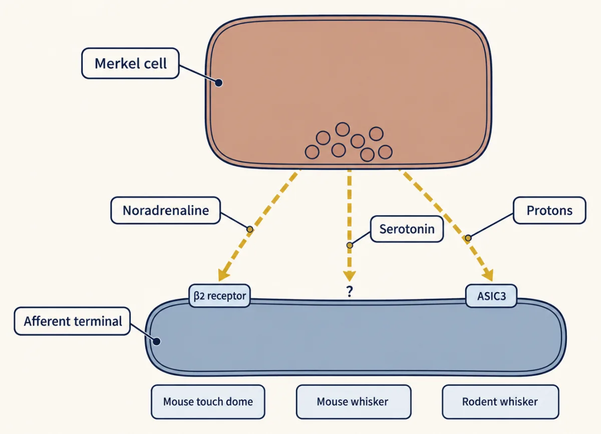
The idea says the Merkel cell holds the press by releasing transmitter onto the ending, and the tetanus toxin result shows that release matters. What is released is the open problem. Three accounts say what, and each rests on its own preparation.
Noradrenaline, mouse touch dome. Hoffman and colleagues, from the Lumpkin laboratory, found transcripts for presynaptic machinery and adrenergic biosynthesis in purified adult Merkel cells. The cells loaded a fluorescent vesicle tracer, FFN206, and calcium-channel blockers prevented its release on depolarization. Local noradrenaline evoked spikes in touch-dome afferents without touch (half-maximal concentration 2.5 mM in this bath assay; drug applied to tissue has to diffuse in, so this is not an estimate of the concentration at the synapse). Deleting Adrb2, the gene for the β2-adrenergic receptor, in neural-crest derivatives with Wnt1-Cre attenuated dynamic and static firing. That Cre driver is broad and the deletion is developmental. A β2 receptor is a G protein-coupled receptor (see GPCRs and G proteins and cAMP and PKA), but nothing here shows which conductance follows it; the authors left the channel that β2 signalling opens for future work.
Serotonin, mouse whisker. Chang and colleagues (2016) reported mechanically triggered serotonin release from mouse whisker Merkel cells. They also found serotonin receptors of two kinds (5-HT3 and 5-HT2) on whisker Aβ endings, and blocking them reduced afferent firing and whisker touch behaviour. Against this, Hoffman and colleagues found that adult mouse Merkel cells lack transcripts for the enzymes that make serotonin, that serotonin did not excite touch-dome afferents, and that the release method used cannot tell serotonin from noradrenaline. Yamada and colleagues found that a 5-HT3 blocker left fast excitatory postsynaptic currents (EPSCs) unchanged.
Protons, rodent whisker. Yamada and colleagues (2024), also from the Gu laboratory, recorded with pressure-clamped patch electrodes from the afferent axon right next to the complex, in rat and mouse whisker complexes: mostly at its first node of Ranvier, sometimes at the heminode just before it, where the myelin ends. That measures the excitatory postsynaptic current (EPSC), the current that transmitter evokes in the afferent, without passing through spike generation.
Predict. If noradrenaline acting on β2 receptors carried the fast signal from Merkel cell to afferent, what should a β2 blocker do to the fast EPSCs evoked by touch?
Check your prediction
It should shrink them. It did not: a β2 blocker left the mechanically evoked fast EPSCs unchanged, and ASIC3 deletion diminished them. In ASIC3-deficient mice, whisker SA1 afferents also fired fewer impulses in both phases, and puffing noradrenaline or serotonin onto the complexes evoked no current. Yamada and colleagues attributed fast transmission to ASICs on the afferent, channels gated by protons. That argues against β2 carrying the fast EPSC at whisker complexes, though not against a slower adrenergic effect.
So both deletions reduce SA1 firing, Adrb2 in touch domes and ASIC3 in whiskers. The whisker blocker and puff results are the sharpest tension, but even they are not a matched test: the sites and endpoints differ, and a β2 receptor, being a G protein-coupled receptor, could act more slowly than a fast EPSC. A companion 2024 study from the same laboratory found that ASIC blockers and ASIC3 deletion also reduced SA1 firing in hindpaw glabrous skin, so the difference is not only whisker versus touch dome. The accounts compete as much as they complement each other. Calling the adrenergic synapse settled is wrong, and so is calling it refuted.
Design problem
Design experiments that could decide whether Hoffman’s touch-dome evidence for β2 receptors and Yamada’s whisker evidence for ASICs conflict.
Saved in this browser only.
One way to do it
First, remove the confounds of site and age. Record both touch domes and whisker follicles in the same hands, from mice of one age, so that a difference in outcome is not a difference of lab or developmental stage.
Second, use the same endpoint at both sites. Pressure patch the afferent at its first node or heminode in each preparation and measure the postsynaptic current directly, so that transmission is separated from spike generation. Then ask separately about fast EPSCs and slower modulation, since a β2 receptor and a proton-gated channel could each act on a different time scale.
Third, replace Wnt1-Cre with an acute, adult, afferent-specific deletion of Adrb2 using an inducible Cre, and do the same for Asic3, so that the loss of each receptor is tested on the same afferents.
Fourth, use blocking vesicle release in Merkel cells as the common test: if both currents vanish when release is blocked, both depend on the cell. Prediction: if the adrenergic and proton accounts describe different time scales, β2 deletion would alter slow modulation and ASIC3 deletion the fast EPSC at both sites; if they describe different sites, each deletion would act at one site only. The hindpaw ASIC result already strains the site explanation. Recording spikes as well as currents, and measuring what is released at each site, would complete the test.
A human fingertip needs other methods: microneurography can record afferents but cannot delete a gene.
Going deeper
Preparation ages
Hoffman and colleagues blocked release with tetanus toxin in an ex vivo hindlimb skin-saphenous nerve preparation from mice aged 7 to 10 weeks. Yamada and colleagues recorded from rats aged 24 to 60 days and mice aged 6 to 13 weeks.
Edge orientation in the human fingertip
In human microneurography at the fingertip, Pruszynski and Johansson (2014) found that first-order neurons signalled edge orientation through both response intensity and temporal structure.
A cancer that borrowed the name
A skin cancer, Merkel cell carcinoma, is named after the cell, but the name is not evidence that the cell of origin is a touch-receptor Merkel cell.
Your notes
Select any text in the lesson to highlight it or add a note to it. Your highlights and notes are saved in this browser and collected in your notebook.
This browser is not letting the site save anything (a private window or strict privacy settings), so highlights and notes will be lost when you leave the page.
How we measure it
Ex vivo skin-nerve recording with optogenetics
A patch of skin is dissected with its nerve, kept alive in a bath, and single afferents are recorded from the nerve while the skin is pressed. Expressing a light-activated depolarizing or hyperpolarizing protein in Merkel cells but not in the recorded afferent, and aiming the light at the touch dome, lets you drive or silence them on demand, which tests sufficiency and acute necessity for afferent firing.
Conditional (Cre-lox) gene deletion
A Cre driver is a mouse line that makes Cre recombinase in cells that express a chosen promoter; crossed to a floxed gene, it deletes that gene only in those cells and their descendants. Krt14-Cre removed Piezo2 from skin epithelium but not from sensory neurons, and Wnt1-Cre removed Adrb2 from neural-crest derivatives. A driver's scope, and whether the loss is developmental or acute, limit what the result can show.
Patch clamp of Merkel cells and afferent terminals
Whole-cell recording measures the mechanically activated current and the spikes of a Merkel cell, either after dissociation and culture or in place in an excised whisker follicle. Pressure-clamped patch recording at the afferent's first node or heminode, next to the complex, records the postsynaptic current itself, so transmission can be measured without passing through spike generation.
Genetic lineage tracing
A Cre driver active in a progenitor population permanently switches on a reporter in those cells and all their descendants. Finding the reporter in Merkel cells after marking epidermal progenitors in mouse embryos showed that Merkel cells are epidermal, not neural crest, in mice.
Practice questions on this lesson (3) → MCAT-style, original, each with an explanation
Where this lesson fits
Before thisReview first
- Piezo Channels and the Physics of ForceThis course · Lesson 2
You need how Piezo2 turns force into current to follow the Merkel cell's own mechanically activated current.
- Five Ways to Turn a Stimulus into a CurrentThis course · Lesson 1
Its ways of turning a stimulus into a current frame how a non-neuron can act as the receptor.
- Sensory Neurons by Their Genes: the Atlas EraThis course · Lesson 3
It introduces the touch afferent classes and the Cre driver lines this lesson uses to separate cell from ending.
After thisStudy next
- Where Touch Is First Processed: Dorsal Horn and Dorsal Column NucleiThis course · Lesson 8
It shows how the sustained SA1 signal is first processed once it reaches the spinal cord.
- Touch Over-reactivity in Autism ModelsThis course · Lesson 9
It uses the Merkel cell-afferent touch route and its Piezo2 dependence in a disease model.
Elsewhere on the siteFilms, simulations and method films in other courses
- Quantal analysisMethod film
The lesson reads irregular spike timing as a clue for packets of transmitter, and quantal analysis is the method for testing packets.
- Pharmacological blockade and inhibitorsMethod film
The debate over noradrenaline, serotonin and ASIC protons rests on blocking each route with drugs.
Suggested by an AI model from the lessons themselves, then checked. How we use AI
Finish line
Test yourself
8 questions
Practice and MCAT-style, with explained answers.
Next · Lesson 6
Meissner and Pacinian Corpuscles: Flutter and Vibration
Slide a fingertip across cloth and Meissner corpuscles report the movement; hold a buzzing phone and Pacinian corpuscles report the hum. Each is a nerve ending wrapped in cells that help it feel. How do the two split the work of sensing change?
Continue →← Lesson 4 · The Low-Threshold Mechanoreceptors: One Touch, Several Signals