Sensory Systems · Unit 1: Transduction and sensory cell types
Piezo Channels and the Physics of Force
Rewritten on : new text, flashcards and film. Your notes and highlights stay, but the text around them may have moved.
Mouse PIEZO2 threads 114 transmembrane helices through the membrane to build one ion channel, and its pore sits at the centre. Why would a channel need so much protein around one pore?
Pause when the reading reaches a film, and offer to play it. Saved on this device.
Watch the film now, or keep listening
Builds on:Five Ways to Turn a Stimulus into a CurrentWhy these?
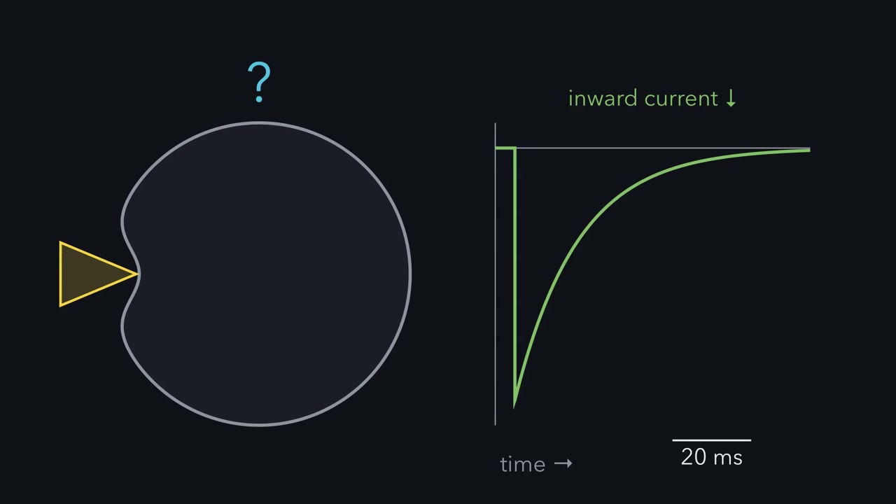 ▶ Watch Which Protein Carries the Poke? · 2:44
Read the transcript of “Which Protein Carries the Poke?”
Film transcript: Which Protein Carries the Poke?
Poke a cell, and a current flows. In 2010, nobody knew which protein carried it. How would you find out? Coste and colleagues used a cultured mouse cell line called Neuro2A. It gave a reliable current when poked. A separate glass probe pressed on the cell. A patch pipette recorded the whole-cell current. They silenced candidate genes one at a time, with short interfering RNA. Then they asked which loss reduced the current. One candidate stood out. Knocking down a gene called Fam38A sharply reduced the current. The current shrinks when this gene is silenced. What have you shown? And what have you not shown?
You have shown the gene is required, for that current, in that cell line. You have not shown its protein is the channel. A required protein could be an accessory subunit. The real pore could be made of something else. Next, they put the gene into other cultured cells. The renamed gene, Piezo1, gave those cells mechanically activated currents. So did a related gene, Piezo2. The two currents had different kinetics. Ion substitution pointed to a non-selective cation current for Piezo2. And its current inactivated quickly. Inactivation means the current decays while the stimulus is held. Keep that word. It returns at the end. Notice the logic. Silencing showed the gene is necessary.
Expression showed it is sufficient. Neither showed that it forms the pore. The authors said so. They kept open the possibility that Piezo proteins were subunits of some other channel. Necessity, sufficiency and pore identity are three separate claims. Each needs its own experiment. The 2010 paper earned two. Later work earned the third. In 2012, purified Piezo1 formed ion channels in artificial bilayers and liposomes. Structures then placed the pore. The channel's carboxy-terminal domains line a central ion pore. So the protein is the channel. Now a stranger question. Why build so much protein around one central pore? Required, sufficient, and the pore itself.
Three claims, three experiments.
From the film
- Silencing a gene in a cell line showed it was required for the poke-evoked current there, not that its protein is the channel.
- Expressing the Piezo genes in other cultured cells gave mechanically activated currents, which showed sufficiency in those cells.
- Necessity, sufficiency and pore identity are three separate claims, and later work on purified protein and structures earned the third.
Mouse PIEZO2 threads 114 transmembrane helices through the membrane to build a single ion channel. The pore it opens sits at the centre.
Why would a channel need so much membrane-spanning protein around one pore? Piezo channels turn mechanical force directly into ionic current, and Piezo2 is a principal channel for touch and limb position: mice lacking it in sensory neurons lose almost all light-touch behaviour, and two people lacking working PIEZO2 had selective deficits in touch and especially limb position. Touch and pain names Piezo2 as the channel mice need for gentle touch, and transduction strategies places direct mechanical gating among the ways a stimulus becomes a current. This lesson asks the mechanical question those lessons leave alone.
Three questions and one idea
Three questions run through the lesson. Which protein carries the current when a cell is poked? What shape is it? And how does force open it: through the lipid around it, through a protein tether, or both?
The leading structural model, still debated, answers with a shape. The channel is a shallow dome, force favours flattening it, and flattening is thought to help open the pore. One idea turns that picture into numbers you can compute yourself.
The one idea to carry through this lesson. Tension pays for area. If a channel takes up more membrane area when it is open than when it is closed, membrane tension pushes it open. The push is an energy: tension times the area gained, T·ΔA. In a two-state model, that energy gives both numbers in a current-tension fit a meaning, and more area means a steeper switch. Piezo’s curved dome is a way to gain a lot of area when it flattens; in the leading model, that is what the 114 helices are for.
Which protein? A screen, and the claim it refused to make
The 2010 experiment was built around a cultured mouse neuroblastoma line, Neuro2A, that gave a reliable current when a separate glass probe indented the cell while a patch pipette recorded whole-cell current, the current across the entire cell membrane. Coste and colleagues silenced candidate genes one at a time with short interfering RNA and asked which loss reduced the current.
Predict. Knocking down one candidate, Fam38A, sharply reduces the indentation current. What have you shown, and what have you not?
Check your prediction
You have shown that Fam38A, renamed Piezo1, is required for that current in that cell line. You have not shown that its protein is the channel. A protein required for a current could still be an accessory subunit of a channel made of something else.
Coste and colleagues then expressed mouse Piezo1, or the related Piezo2, in heterologous cultured cells, meaning other cultured cells given an introduced copy of the gene. Each produced mechanically activated currents, and their kinetics differed between the two. Ion-substitution experiments pointed to a non-selective cation conductance for Piezo2, and its current inactivated quickly. Inactivation is the decay of a mechanically activated current while the stimulus is held; human variants change it.
This logic recurs across the unit. The screen showed necessity in one cell line; expression showed sufficiency to confer a current in another. Neither shows that the protein lines the pore, and the authors said so. Necessity, sufficiency and pore identity are three separate claims, and the 2010 paper earned two.
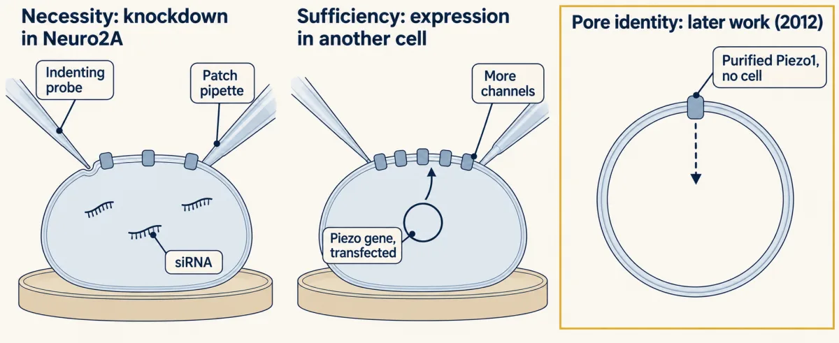
Later work earned the third. Purified mouse Piezo1 reconstituted into lipid bilayers and liposomes formed ion channels sensitive to ruthenium red, a broad blocker of cation channels (Coste and colleagues, 2012). Structural work then placed the pore: in the purified mouse Piezo1 reconstruction of Saotome and colleagues, the carboxy-terminal domains line the central ion pore.
Blades, beam and dome
Read the transcript of “A Channel Shaped Like a Dome”
Film transcript: A Channel Shaped Like a Dome
Why would one ion channel need 114 transmembrane helices? The first map came in 2015, from Ge and colleagues. It showed a trimer with three propeller-like arms. Zhao and colleagues resolved each arm into repeating units of four helices. Those units form curved blades around a central pore. They also mutated single residues of a long beam inside the cell. Mechanical activation was impaired. So that part of the structure matters for gating, not just for building the channel. By 2019, a structure of mouse Piezo2 was sharp enough to count the helices. Each subunit holds 38 transmembrane helices. The three-subunit channel holds 114.
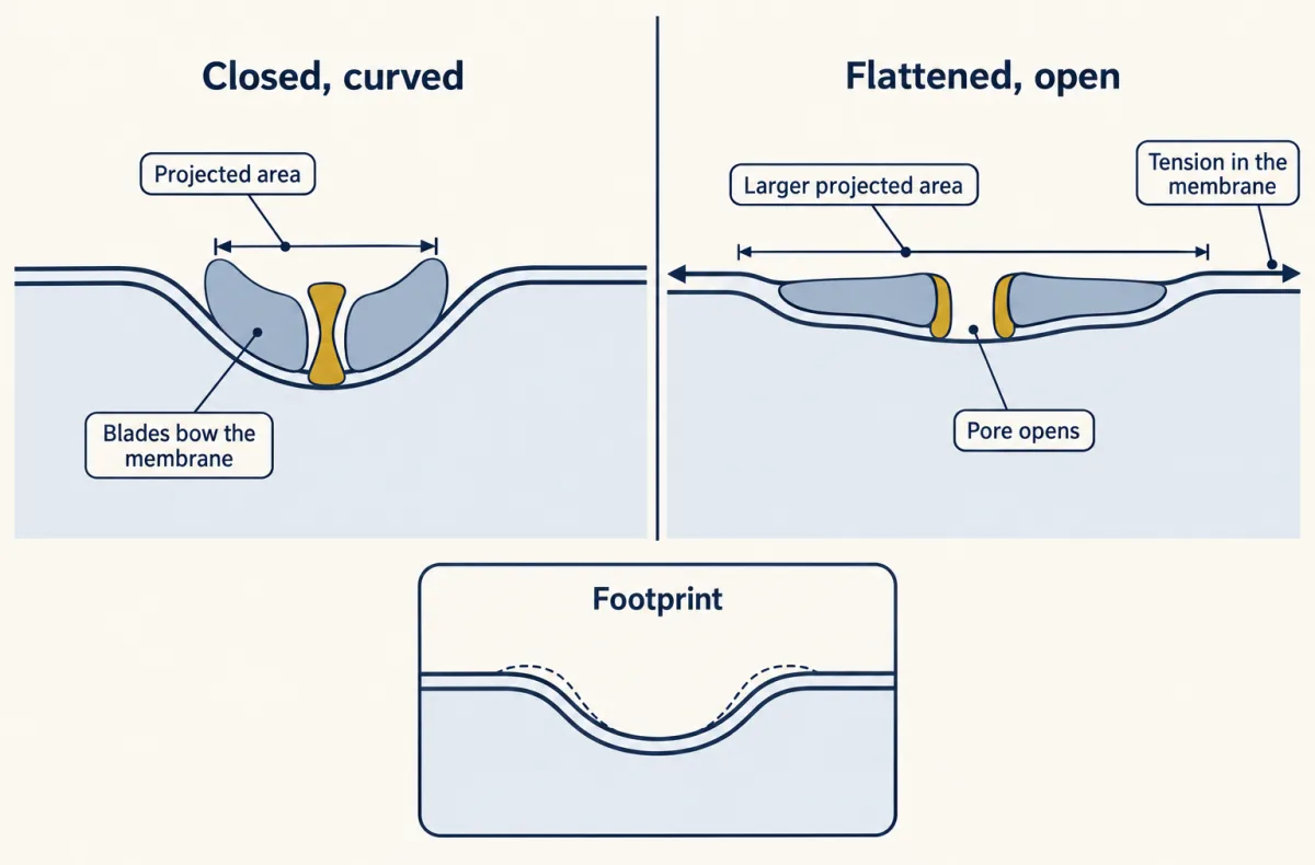
Keep the two numbers apart. Thirty-eight describes one subunit. One hundred and fourteen describes one channel. Those helices do something unusual. The curved blades bow the membrane into a dome. In purified mouse Piezo2, the dome is reported as about twenty-eight nanometres across and ten deep.
A dome has two areas. Its surface area is the membrane actually in it. Its projected area is its shadow on the flat plane around it. That is the area it occupies, seen from above. Treat the dome as a smooth cap. Flatten it without stretching it. Does its shadow grow or shrink? By roughly how much?
It grows. The curved cap casts a shadow of about six hundred and sixteen square nanometres. Flattened, its whole surface lies in the plane, about nine hundred and thirty. The gain is about three hundred and ten square nanometres.
The gain depends on how deep the dome is, not on how wide. This is idealized geometry, not a measurement. The real dome is protein blades and bent lipid, not a smooth cap. So flattening increases projected area. Anything that favours more projected area favours the flat state. In the leading model, still debated, flattening is thought to help open the pore. In the leading model, a dome is a device for gaining area.
From the film
- Mouse PIEZO2 has 38 transmembrane helices per subunit and 114 per channel, and the two numbers are kept apart.
- In the leading model, still debated, the curved blades bow the membrane into a dome.
- For an idealized smooth cap, flattening gains about 310 square nanometres of projected area, which depends on how deep the dome is, not how wide.
The first map, from Ge and colleagues in 2015, was a trimer (three identical subunits, or protomers) with three propeller-like arms at 4.8 angstroms. Zhao and colleagues then resolved the arms into repeating four-helix units assembled into curved blades around the central pore. They also found that mutating single residues of the long intracellular beam impaired mechanical activation, which ties a specific part of the architecture to gating rather than to assembly.
By 2019 the picture was sharp enough to count. Mouse PIEZO2, reconstructed by Wang L and colleagues (Nature, 2019), holds 38 transmembrane helices per protomer, which means 114 in the trimer. Keep them apart: 38 describes one subunit, 114 the whole channel.
The curved blades bow the bilayer into a dome, reported as about 28 nm in diameter and 10 nm deep for purified mouse PIEZO2.
A dome has two areas, and the difference between them is the whole argument. Its surface area is the membrane actually in it. Its projected area is its shadow on the flat plane of the surrounding membrane: the area it occupies seen from above. A curved cap projects less area than its surface area. Flatten the same cap and the two become equal.
Predict. Treat the PIEZO2 dome as an idealized spherical cap, 28 nm across and 10 nm deep. Flatten it without stretching it. Does its projected area grow or shrink, and by roughly how much?
Check your prediction
It grows, by about 310 nm². A spherical cap whose rim has radius a and whose depth is h has a surface area of π(a² + h²). Here a = 14 nm and h = 10 nm, so the surface is π(196 + 100) nm² ≈ 930 nm². Its shadow is the disc under the rim, πa² = π × 196 nm² ≈ 616 nm². Flattened, all 930 nm² lies in the plane, so the projected area rises by about 930 − 616 ≈ 310 nm².
The gain is simply πh² (π × 10² ≈ 314 nm²). It depends on how deep the dome is, not on how wide. This is idealized geometry, not a measurement: the real dome is protein blades and bent lipid, not a smooth cap of membrane.
So anything that favours more projected area favours the flat state.
Tension pays for area: from an energy to a curve
Read the transcript of “Tension Pays for Area”
Film transcript: Tension Pays for Area
Why should stretching a membrane open a channel at all? Start with the simplest picture that could work. The channel is either closed or open. The open state takes up more room in the plane of the membrane. Call the extra amount the area gained. Membrane tension is an in-plane stretch. It is force per unit length of membrane. Stretching a membrane does work on anything in it that gets wider. That work is tension times the area gained. So tension lowers the energy of the open state, relative to the closed one. Write it out. The energy difference equals the resting preference for closed, minus tension times the area gained. In a slack membrane, the channel prefers to be closed.
As tension rises, that preference shrinks. At one particular tension, the preference disappears. Above it, the open state wins. Two channels are each open half the time at the same tension. One gains five square nanometres on opening. The other gains three hundred. Which one goes from mostly closed to mostly open over the narrower range of tension?
The three hundred channel. Each small rise in tension gives it sixty times the energy advantage. So a small change in tension swings it all the way from closed to open. To turn the energy into a curve, use the Boltzmann relation from statistical physics. For a two-state channel, the ratio of open to closed depends on the energy difference, compared with thermal energy. For a two-state channel at equilibrium, this gives the fraction of channels open. Patch papers fit the same curve to current. That reading assumes current tracks the fraction open. The curve has two fitted numbers: a half-activation tension and a slope factor. Match the two forms, and each number gets a meaning.
The half-activation tension is the resting preference for closed, divided by the area gained. The slope factor is thermal energy, divided by the area gained. More area means a smaller slope factor, and a steeper switch. One unit note. Piconewtons per nanometre and millinewtons per metre are the same quantity. That is the one idea. Tension pays for area. In a two-state model, the area sets the steepness.
From the film
- Tension pays for area: if a channel takes up more membrane when open, tension lowers the energy of the open state by tension times the area gained.
- In a two-state model, the half-activation tension is the resting preference for closed divided by the area gained, and the slope factor is thermal energy divided by the area gained.
- More area gained means a steeper switch.
Take the simplest picture: the channel is either closed or open, and the open state has a larger projected area, by an amount ΔA. Membrane tension, T, is an in-plane stretch: force per unit length of membrane. Stretching a membrane does work on anything in it that gets wider, and that work is the tension times the area gained. So tension lowers the free energy of the open state relative to the closed one:
In words The energy cost of opening is the cost in a slack membrane, minus the work tension does as the channel widens: tension times the area gained.
- ΔG“delta G”
- the free energy of the open state minus that of the closed state. Positive means closed is favoured.J
- ΔG₀“delta G nought”
- how strongly the channel prefers to be closed in a slack membrane, with no tensionJ
- T“T”
- membrane tension: in-plane stretch, force per unit length of membrane. Here T is tension, not temperature.N/m (1 mN/m = 1 pN/nm)
- ΔA“delta A”
- the extra projected area of the open state compared with the closed statem² (1 nm² = 10−18 m²)
Sanity check With no tension (T = 0), ΔG = ΔG₀ and the channel keeps its resting preference for closed. When T × ΔA grows equal to ΔG₀, ΔG = 0 and open and closed are equally likely. Above that tension, ΔG is negative and the open state wins.
Try one number
Take a round channel that gains 10 nm² (10−17 m²) on opening, in a membrane at 1 mN/m (10−3 N/m). Both are round values to keep the arithmetic easy.
Work done by tension: T × ΔA = 10−3 N/m × 10−17 m² = 10−20 J.
Units: newtons per metre times square metres is newton-metres, which are joules. So each 1 mN/m of tension lowers this channel’s ΔG by 10−20 J.
Need a refresher? subscripts and Greek letters · units and prefixes · powers and roots
Predict. Two channels are each open half the time at the same tension. One gains 5 nm² on opening and the other 300 nm². Which one goes from mostly closed to mostly open over the narrower range of tension?
Check your prediction
The 300 nm² channel. Each small increase in tension gives its open state 60 times as much of an energy advantage as it gives the 5 nm² channel, so a small change in tension swings it all the way from closed to open.
To turn the energy into a curve, use the Boltzmann relation from statistical physics. For a channel with two states at equilibrium, the ratio of open to closed is exp(−ΔG/kBTabs), where kBTabs is thermal energy, about 4.1 × 10⁻²¹ J at room temperature (kB is Boltzmann’s constant and Tabs absolute temperature; T stays tension, and neither is the fitted slope factor k). The fraction of channels open then follows a curve that rises from 0 to 1 as tension rises, written here in the form fitted to patch data:
In words The open fraction is one over one plus e raised to the power of the half-activation tension minus the tension, divided by the slope factor.
- P(open)“P open”
- the fraction of channels open, from 0 to 1
- exp“exp, or e to the power”
- e raised to the power in the brackets; e ≈ 2.718
- T50“T fifty”
- the half-activation tension: the tension at which half the channels are openmN/m
- T“T”
- membrane tensionmN/m
- k“k”
- the slope factor: each rise of k in tension multiplies the ratio of open to closed channels by e. A smaller k gives a steeper switch. It is not Boltzmann’s constant kB.mN/m
Sanity check At T = T50 the exponent is zero, exp(0) = 1, and P(open) = 1/2. Far above T50 the exponent is very negative, exp is near 0, and P(open) approaches 1. Far below T50, exp is huge and P(open) approaches 0.
Try one number
Use the patch fit given below for Piezo1, T50 = 2.7 mN/m and k = 0.8 mN/m, and set the tension one slope factor above half-activation: T = 2.7 + 0.8 = 3.5 mN/m (a tension, not a range of tensions).
Exponent: (2.7 − 3.5)/0.8 = −1. And e−1 ≈ 0.37.
P(open) = 1/(1 + 0.37) = 1/1.37 ≈ 0.73. One slope factor above T50, about 73% of channels are open; one slope factor below (T = 1.9 mN/m), the exponent is +1, e1 ≈ 2.72, and about 1/3.72 ≈ 27% are open.
Where it comes from optional
Step 1, the Boltzmann relation: open ÷ closed = exp(−ΔG/kB Tabs), with ΔG = ΔG₀ − T × ΔA.
Step 2, from a ratio to a fraction: P(open) = open ÷ (open + closed) = 1 / [1 + closed ÷ open] = 1 / [1 + exp((ΔG₀ − T × ΔA)/kB Tabs)].
Step 3, divide the top and bottom of the exponent by ΔA: (ΔG₀/ΔA − T) / (kB Tabs/ΔA). Name ΔG₀/ΔA as T50 and kB Tabs/ΔA as k, and the exponent becomes (T50 − T)/k.
Need a refresher? e and ln · subscripts and Greek letters
Written with energies, the same curve is P(open) = 1 / [1 + exp((ΔG₀ − T·ΔA)/kBTabs)] (the card’s optional fold shows the steps). At T = T50 the energy difference ΔG is zero, so half the channels are open.
The tension form is the one fitted to patch data, usually as a normalized current. Reading a current fit this way assumes that normalized current tracks the fraction of channels open: constant conductance and availability, and a plateau where the available channels are open. With that assumption, matching the tension form with the energy form gives each fitted number a physical meaning:
- T50 = ΔG₀ / ΔA. The half-activation tension is the channel’s resting preference for closed, divided by the area it gains on opening.
- k = kBTabs / ΔA. The slope factor is thermal energy divided by that area. A larger area gives a smaller k and a steeper switch.
One unit note before the numbers. Tension is force per length, so 1 pN/nm and 1 mN/m are the same quantity. Structural papers tend to use the first and patch papers the second.
Reading a patch: from pressure to current, and back to an area
Read the transcript of “Reading a Patch”
Film transcript: Reading a Patch
A patch recording gives a curve of current against tension. Can you read an area out of it? Lewis and Grandl expressed mouse Piezo1 in a human cell line. They recorded cell-attached patches. In a cell-attached patch, the pipette seals onto an intact cell. It records the channels in the membrane under its tip. They pressurized the patch and imaged it at the same time. So the patch radius was measured, not assumed. For a patch shaped like part of a sphere, pressure becomes tension. Tension equals the radius of curvature times the pressure difference, divided by two. Take an illustrative patch. A radius of two micrometres, under two thousand pascals.
These are invented round numbers. The tension comes out at two millinewtons per metre. Now the fit from the paper. A half-activation tension of two point seven millinewtons per metre. A slope factor of zero point eight. At two millinewtons per metre, the fit predicts about twenty-nine percent of the plateau current.
This fit also hides an area. You know how the slope factor relates to area. Can you find it? Read it as a two-state switch, and run the fit backwards. The effective area comes out at about five square nanometres. Under the same reading, the effective resting preference for closed is about three point four times thermal energy. It comes from both fitted numbers, not from an independent measurement. An underestimated area alone does not tell us whether the true energy is underestimated. An independent route agrees in order. Calibrated against a bacterial channel, human Piezo1 gave six to twenty square nanometres. Both estimates assume each channel is a simple two-state switch.
Hold on to that assumption. Two cautions. This is a normalized current, for mouse Piezo1 in a human cell line, at room temperature. It is not a fraction of neurons doing anything. protocol matters. In a separate protocol, a five-second prepulse lowered the half-activation tension, from two point two to one point four. The authors' reading: the prepulse relieves resting tension. Channels it had inactivated recover, so less added tension is needed. Do not mix the two protocols. The shift is the finding: the channel's sensitivity depends on the tension it was already under. Read as a two-state switch, the slope factor implies an area.
From the film
- Read as a two-state switch, the slope factor of a current-tension fit implies an effective area.
- For mouse Piezo1 in a human cell line the fit implied about 5 square nanometres, an effective area that can underestimate the true one.
- A channel's sensitivity depends on the tension it was already under, since a prepulse lowered the half-activation tension.
Lewis and Grandl expressed mouse Piezo1 in human HEK293t cells and recorded cell-attached patches, in which the pipette seals onto an intact cell and records the channels in the membrane under its tip. They recorded at −80 mV and room temperature while pressurising the patch and imaging it with differential interference contrast optics, so the patch radius was measured rather than assumed. For a patch shaped like part of a sphere, with uniform tension, the Young–Laplace relation converts pressure into tension:
In words Tension equals the patch’s radius of curvature times the pressure difference across it, divided by two.
- T“T”
- membrane tension in the patchN/m
- R“R”
- the patch’s radius of curvature, measured by imagingm
- pressure difference“pressure difference”
- the pressure across the patch, inside the pipette compared with outside; its magnitude is usedPa = N/m²
Sanity check With no pressure difference there is no tension. At the same pressure, a patch with a larger radius, a flatter dome, carries more tension.
Try one number
Take a patch of radius 1.0 micrometre (1.0 × 10⁻⁶ m) under 2000 Pa, a round radius chosen to keep the arithmetic easy.
T = (1.0 × 10⁻⁶ m)(2000 N/m²)/2 = 1.0 × 10⁻³ N/m = 1.0 mN/m.
Units: metres times newtons per square metre gives newtons per metre, a force per length, as tension should be.
Need a refresher? units and prefixes · powers and roots
Worked problem: from patch pressure to predicted current, and back to an area
Take an illustrative spherical patch of radius R = 2.0 micrometres under a pressure difference of magnitude 2000 Pa. The radius and pressure are invented round numbers. The fit parameters are from the paper’s pressure-clamp fit for these recordings: a half-activation tension T50 = 2.7 ± 0.1 mN/m and a Boltzmann slope factor k = 0.8 ± 0.1 mN/m.
- Tension: T = (2.0 × 10⁻⁶ m)(2000 N/m²)/2 = 0.002 N/m = 2.0 mN/m. Units: metres times newtons per square metre gives newtons per metre.
- Predicted normalized current: I/Imax = 1 / [1 + exp((T50 − T)/k)] = 1 / [1 + exp((2.7 − 2.0)/0.8)] = 1 / (1 + e0.875). e0.875 is about 2.40 (between e0 = 1 and e1 ≈ 2.72; see e and ln), so I/Imax = 1/3.40 = 0.294, about 29 percent of the fitted plateau.
The fit also hides an area. Before you read on, use k = kBTabs/ΔA to find it.
- Run the fit backwards, reading it as a two-state switch. Rearrange k = kBTabs/ΔA by multiplying both sides by ΔA and dividing both by k: ΔA = kBTabs / k = (4.1 × 10⁻²¹ J) / (0.8 × 10⁻³ N/m) = 5.1 × 10⁻¹⁸ m², an effective area of about 5 nm². Units: joules divided by newtons per metre gives square metres, and 1 nm² is 10⁻¹⁸ m².
- Under the same reading, the effective resting preference for closed is ΔG₀ = T50 × ΔA = (2.7 × 10⁻³ N/m)(5.1 × 10⁻¹⁸ m²) = 1.4 × 10⁻²⁰ J, about 3.4 kBTabs. This number is only as good as the two-state reading: it is computed from the fitted area, so it is not an independent measurement of how strongly the channel prefers closed.
An independent route gives the same order. Calibrated against the bacterial channel MscL in the same patch, human PIEZO1’s Boltzmann slope implies about 6 to 20 nm² on opening (Bae and colleagues, 2013). Both estimates assume a two-state switch, and the next section shows why the area actually coupled to opening could be larger.
Two cautions travel with the answer to step 2. It is a normalized current for mouse Piezo1 in a human cell line at room temperature in the cell-attached configuration, not the fraction of channels or of neurons doing anything. And protocol matters. The same paper ran a separate prepulse protocol. Within it, T50 was 2.2 ± 0.1 mN/m without a prepulse and 1.4 ± 0.1 mN/m after a five-second +5 mmHg prepulse. Compare those two with each other, not with 2.7, which belongs to the other protocol. The authors’ reading is that resting tension inactivates some channels; the prepulse relieves it, they recover, and less added tension is needed. That shift is the finding: Piezo1’s apparent sensitivity is not a fixed property; it depends on the tension the channel was already under.
Mixing protocols: the fit drawn here belongs to the pressure-clamp protocol (T50 = 2.7 mN/m). Putting a T50 from the separate prepulse protocol into it combines two protocols and gives a number that means nothing, so no prediction is printed.
- Tension T = R × Δp / 2
- 2.0 mN/m
- (T50 − T) / k
- 0.875
- e(T50 − T)/k
- 2.40
- I/Imax (normalized current)
- 0.294 (about 29%)
In this sketch: a spherical cap, the Young–Laplace relation T = R × Δp / 2, and the published fit held fixed (T50 = 2.7 ± 0.1 mN/m, k = 0.8 ± 0.1 mN/m); the radius and pressure are illustrative round numbers, and the patch is drawn schematically. The fit is for mouse Piezo1 in human HEK293t cells, room temperature, cell-attached; the measured range of the fit is not given; a normalized current, not a fraction of channels or of neurons.
Convert pressure to tension with the measured radius, read the current off the fit of the same protocol, and never carry a T50 from one protocol into another.
The gap between structure and current
Read the transcript of “The Sixty-Fold Gap”
Film transcript: The Sixty-Fold Gap
Structures suggest the channel gains hundreds of square nanometres. A two-state reading of the current says about five. Do they describe the same change? Yang and colleagues put purified mouse Piezo1 into lipid vesicles. Cryo electron microscopy sorted it into two classes, from one sample. One class was curved, the other flattened. Between them was about three hundred square nanometres of in-plane expansion. Their abstract hedges whether this rearrangement is the gating transition itself. Suppose all three hundred square nanometres were coupled to opening. How steep would the current curve be?
Nearly vertical. The channel would go from ten to ninety percent open over a tiny range of tension. The measured curve is about sixty times shallower. The half-activation tensions are of the same order. The slopes are not.
Geometry makes a change that large plausible. On paper, an idealized Piezo2 dome gains about three hundred and ten square nanometres when it flattens. So why is the measured switch so shallow? The question is open. Here are three testable answers. First, the dome may not be a two-state switch. If it flattens gradually, like a spring, one area no longer describes it. Second, a patch curve is a population curve. Uneven tension across a patch could spread the half-activation tensions. Suppose otherwise identical two-state channels sit at equilibrium and differ only by fixed offsets in half-activation tension. Then averaging broadens their response, and the slope-derived area underestimates their common opening area.
Outside that model it is an effective area, not a guaranteed bound. And the published curve also pools different patches. Third, the flattened class may not be the open channel. A structure is not a current recording. There is one more twist. In an elasticity model by Haselwandter and MacKinnon, the membrane bends away around the dome. That skirt reaches well past the protein. So tension does work on the footprint as well as on the channel. Their numbers are model inputs, not the structural three hundred. Keep the two apart. Neither five square nanometres nor hundreds has been shown to be the area gained on pore opening. The structural expansion is real; which part of it couples to opening is still open.
From the film
- Cryo-EM showed about 300 square nanometres of in-plane expansion, but a two-state reading of the current gives about 5, so the slopes differ about sixty-fold.
- The dome may not be a two-state switch, patch curves may pool uneven tensions, or the flattened class may not be the open channel.
- Which part of the structural expansion couples to opening is still open.
Yang and colleagues (2022) put purified mouse PIEZO1 into liposome vesicles and resolved curved and flattened classes from the same sample. Between them they reported roughly 300 nm² of in-plane areal expansion. From that area together with a previously measured stiffness, they calculated a half-maximal activation tension of about 1.9 pN/nm, which is 1.9 mN/m. Their abstract hedges whether the rearrangements are the gating transition itself.
Predict. Using k = kBTabs/ΔA, how steep would the current-tension curve be if all 300 nm² were coupled to opening? Compare it with the measured k of 0.8 mN/m.
Check your prediction
k = (4.1 × 10⁻²¹ J)/(300 × 10⁻¹⁸ m²) ≈ 0.014 mN/m. The channel would go from 10 to 90 percent open over about 0.06 mN/m of tension (in this curve the 10-to-90 span is about 4.4 slope factors, 2 × ln 9). The measured fit takes about 3.5 mN/m. The half-activation tensions are of the same order (1.9 mN/m calculated from structure and a stiffness, 2.7 from a patch fit), but the slope factors differ about sixty-fold.
Geometry alone makes a change of that order plausible. The idealized PIEZO2 cap you flattened earlier gained about 310 nm², the same order as Yang’s 300 nm², although that was a different paralog (Piezo2, not Piezo1) and a calculation rather than a measurement. So why is the measured switch so shallow? The question is open; each candidate answer is testable:
- The dome may not be a two-state switch. If it flattens gradually, like a spring, only the part of the flattening coupled to the pore sets the slope, and a single two-state ΔA no longer describes the channel.
- A patch curve is a population curve. It sums many channels, the published fit also pools different patches, and uneven tension across a patch could spread their T50s. If the channels are independent two-state switches that differ only in T50, spread can only make the population curve shallower. As an illustration, identical 300 nm² switches whose T50s follow a normal spread with a standard deviation of about 1.4 mN/m give a curve that a single Boltzmann fits with k ≈ 0.8 mN/m. That spread is wider than the 0.8 mN/m prepulse shift, so whether real patches are that uneven is open.
- The flattened class may not be the open channel, which is the hedge Yang and colleagues make.
What the patch number means depends on the answer. Under the second, the slope-derived area underestimates the common area of the switches. Under the first and third, the area actually coupled to opening could be far smaller than 300 nm², near the patch number. Either way it is an effective area, not a measurement: “Piezo1 gains 5 nm² on opening” is not something anyone has shown, and neither is “hundreds of nm²”.
The membrane around the channel adds to the puzzle. Haselwandter and MacKinnon (2018) worked out the elasticity with a continuum calculation. A bilayer cannot stay flat where a curved protein ends; it bends away in a skirt, a membrane footprint reaching well past the dome, so tension does work on the footprint as well as on the protein. Their closed-dome inputs were a cap surface area of about 390 nm² and a radius of curvature of 10.2 nm. Those are model parameters. By now you have met five area numbers. Keep them apart:
| Area | Source | What it is |
|---|---|---|
| About 310 nm² gained | Idealized PIEZO2 cap, flattened | A geometry calculation from the reported dome size |
| About 300 nm² gained | Yang and colleagues, mouse PIEZO1 | A structural comparison of curved and flattened classes |
| About 390 nm² | Haselwandter and MacKinnon, mouse Piezo1 | A model input: the closed cap’s surface area, not a change |
| About 5 nm² | Lewis and Grandl fit, mouse Piezo1 | An effective two-state area from a current slope factor |
| 6 to 20 nm² | Bae and colleagues, human PIEZO1 | A two-state estimate calibrated against MscL |
Lipid or tether?
Read the transcript of “Lipid, Tether and People”
Film transcript: Lipid, Tether and People
Tension pays for area. But how does force reach the channel? Through lipid, through a protein tether, or both? The idea is the work done by membrane tension. A tether could change that tension, or pull on the channel directly. That would add its own work term. The lipid route has a clean test. Take everything else away. Purified mouse Piezo1 was placed in droplet bilayers, with no other cellular components. A droplet bilayer forms where two lipid-coated droplets touch. Nothing from a cell is present. Suppose a tether were strictly required for opening. What should the purified channel have done? And what would its opening tell you about a cell?
It should have stayed silent. Yet force still activated it, so protein plus lipid is sufficient there. But that says nothing about whether an intact cell also uses a second route. A second route has been proposed. Wang and colleagues described a chain of cadherin, beta-catenin and actin. Disrupting the channel's link to E-cadherin prevented cytoskeleton-dependent gating in their cultured cells. These need not be rivals. Sufficiency in one preparation cannot exclude a route in another. That is why the field has not chosen. There is even a hint that the bilayer misses something. The purified channel lacked the rapid inactivation seen in whole cells.
Inactivation is where human genetics meets this biophysics. Distal arthrogryposis is an inherited condition. Joints of the hands and feet are fixed in bent positions from birth. Two Piezo2 variants from such families were tested in a human cell line. Both sped recovery from inactivation. Only one slowed inactivation during the pulse. So slower inactivation is not a rule for every variant. Read each one. Loss of function looks different in mice and in people. Adult mice lacking Piezo2 in sensory neurons and Merkel cells lose almost all light-touch behaviour. Two people without working Piezo2 had impaired discriminative touch. Their sense of limb position was profoundly impaired, and worse without vision.
But they kept other abilities. Their deficits were selective. Keep the mouse sentences and the human sentences apart. Now back to the first question. Why does one channel need 114 helices? In the leading model, still debated, to collect area.
Tension pays for area. Whether a tether adds its own force is a separate question.
From the film
- Purified mouse Piezo1 in droplet bilayers was activated by force, so protein plus lipid is sufficient there, which does not exclude a tether route in intact cells.
- Two PIEZO2 variants from distal arthrogryposis families both sped recovery from inactivation, but only one slowed inactivation during the pulse, so each variant must be read on its own.
- Mouse and human loss-of-function findings should be kept apart: two people without working PIEZO2 had selective deficits in touch and limb position.
The guiding idea is an energy budget for membrane tension, not a delivery route. The −T·ΔA term is the work done by membrane tension. A tether could change the local tension, or it could pull on the channel directly, which would add its own work term, force times distance.
The lipid route has a clean test: take everything else away. Purified mouse Piezo1, reconstituted into artificial droplet bilayers with no other cellular components, remained mechanically activatable (Syeda and colleagues, 2016). A droplet bilayer forms where two lipid-coated droplets of salt solution touch, so nothing from a cell is present.
Predict. Suppose a tether to the cytoskeleton were strictly required for Piezo1 to open. What should the purified channel have done in a droplet bilayer? And since it did open, what can you conclude about an intact cell?
Check your prediction
It should have stayed silent. Because it opened, protein plus lipid is sufficient in that preparation, and no tether is required there. That says nothing about whether an intact cell also uses a second route.
Force could also arrive through protein. Wang J and colleagues (Cell Reports, 2022) proposed a cadherin-beta-catenin-actin route, complementary to force-from-lipid, and reported that disrupting Piezo1-E-cadherin interactions prevented cytoskeleton-dependent gating in their cultured-cell assays. How widely that route operates beyond those cells is not yet known.
The two accounts need not be rivals. Bilayer sufficiency cannot exclude a second route in an intact cell, and a tether requirement in a cultured cell cannot abolish the lipid energetics the structures measure. The bilayer may even be missing something: the purified droplet-bilayer recordings did not reproduce the rapid inactivation seen in whole cells. That is why the field has not chosen.
Other channels show each route in its own preparation, from tether-free gating of purified TRAAK and TREK1 in lipid to a microtubule requirement for fly NOMPC; the table in Going deeper keeps each result with its preparation.
Design problem
Purified mouse Piezo1 is mechanically active in droplet bilayers but does not reproduce the rapid inactivation of whole cells, while disrupting Piezo1-E-cadherin interactions prevented cytoskeleton-dependent gating in cultured cells. Design experiments to decide whether a tether is required for gating in a sensory neuron.
Saved in this browser only.
One way to do it
The bilayer result is sufficiency of protein plus lipid; the cadherin result is necessity of a coupling in one cultured cell type. Neither addresses a sensory ending, so the experiment has to go there.
Measure tension, not pressure or displacement. Use cell-attached or excised pressure clamp with the patch imaged so its radius of curvature is measured, and convert with T = R(pressure difference)/2. Indentation with a probe delivers an unknown mixture of membrane tension, cytoskeletal strain and cell deformation, so a tether result from indentation alone is hard to interpret; a dose-response in tension is comparable across preparations and against the published fits.
Perturb acutely and in the right cells. A germline deletion of a cadherin or a catenin changes development, adhesion and skin structure, so any afferent deficit has several possible causes. Use an inducible, afferent-specific driver in adult mice to remove the candidate linker, or apply an acute biochemical disruption of the interaction, and record within the window where skin architecture is unchanged. Confirm that architecture with histology in the same animals.
Record at the ending. Ex vivo skin-nerve recording gives spikes from an ending whose end organ is intact; patch recording near the terminal gives currents. Pair them, because a tether could change the mechanics of force delivery to the ending without changing the channel’s tension sensitivity, and the current-tension relation is the most direct way to distinguish those.
Predict the outcomes. If a tether is required, acute disruption should shift the current-tension relation or abolish the response at an ending whose Piezo2 is intact and whose structure is normal. If gating is lipid-driven, the current-tension relation should survive while the amount of tension produced by a given skin stimulus may change. A shifted curve alone is not decisive, because a linker can also alter local membrane mechanics or channel availability, so check channel abundance and patch mechanics and rescue the interaction.
A human answer needs different tools. Microneurography can record single afferents and psychophysics can measure thresholds, but neither can delete a linker, so in living people the main test would be people carrying variants in candidate adhesion proteins, with human cell models adding mechanistic evidence.
Variants that change inactivation, and what people report
Inactivation is where human genetics meets the biophysics. Coste and colleagues expressed human PIEZO2 variants from families with distal arthrogryposis, an inherited condition in which joints of the hands and feet are fixed in bent positions from birth, in HEK293T cells. Both I802F and E2727del accelerated recovery from inactivation; only E2727del slowed inactivation during the pulse. So “gain-of-function variants inactivate more slowly” is wrong for I802F.
Loss of function in people is narrower than the mouse headline. Two individuals with compound inactivating PIEZO2 variants, meaning both copies of the gene carried inactivating variants, had impaired discriminative touch and profound proprioceptive impairment that worsened without visual cues, while retaining other abilities. In a later study using experimental skin inflammation, PIEZO2-deficient participants did not develop the painful responses to gentle touch the protocol produces. Adult mice with Piezo2 deleted from sensory neurons and Merkel cells show an almost complete light-touch deficit across behavioural assays, and most rapidly adapting mechanically activated currents disappear from their cultured dorsal-root-ganglion neurons. Those are mouse results; the human ones are selective deficits with residual abilities, and the two should not be merged. Merkel cells and the LTMR classes carry the cell-biological half of this story, and proprioception the limb-position half.
Back to the opening question
Why would a channel need 114 transmembrane helices around one pore? In the leading, still debated model, to collect area. A curved structure that flattens gains hundreds of square nanometres, and tension pays for whatever part of that area is coupled to the pore. Which part that is, and how the flattening opens the pore, is the open problem in this field.
Where the idea stops
- Two states is an assumption. A dome that flattens gradually is a spring, not a switch, and then a single ΔA is not well defined.
- A patch slope gives an effective area. It underestimates the coupled area only if the channels are identical two-state switches that differ in T50 alone; otherwise it is not a bound.
- The rule is the work of membrane tension, not a delivery route. A tether that pulls on the channel directly would add its own work term.
- Inactivation is outside it. The two-state model has no inactivated state, so it covers neither inactivation nor recovery from it, the kinetics the human PIEZO2 variants change.
- Tension may not be the only requirement. Later work in cell-derived membranes suggests lipid composition matters too, not tension alone (see Going deeper).
Going deeper
Numbers from the 2010 paper
In the 2010 experiments, Piezo2 showed a reported inactivation time constant of 7.3 ± 0.7 ms at −80 mV in HEK293T cells (n = 11).
Mechanotransducers in other preparations
Mechanosensitivity is not one mechanism with one protein. Each of these results belongs to its preparation.
| Protein | Organism and preparation | What was shown |
|---|---|---|
| MscL and MscS | Bacteria | Where the force-from-lipid principle was first established |
| TRAAK and TREK1 | Human TRAAK and zebrafish TREK1, purified, in lipid membranes | Mechanically gated with no cellular tethers. TRAAK could also electrically oppose Piezo1 activation in cultured cells |
| OSCA1.2 | Arabidopsis, purified, in proteoliposomes | Mechanically activated |
| TMEM63C | Mouse, expression assays | No stretch-activated current in the same assays that detected currents for other family members |
| NOMPC | Drosophila S2 cells | Required microtubule association for mechanogating: a tether in a fly |
| MEC-4 | C. elegans touch receptor neurons | A mec-4 null eliminated mechanically evoked receptor currents, which are carried mostly by sodium, unlike the non-selective Piezo current |
| TMC1 | Mouse hair cells, rescued TMC1 | Cysteine modification rapidly changed permeation, supporting TMC1 as a pore-forming component of the transduction channel without showing that it alone constitutes the native complex |
Drosophila NOMPC has 29 ankyrin repeats. The microtubule requirement for mechanogating was shown in S2 cells, and the behavioural work was done in third-instar larval class III dendritic-arborization neurons. The hair-cell transduction channel takes up the TMC1 story.
Piezo1 variants and red blood cells
The xerocytosis-associated human PIEZO1 variants M2225R and R2456H slowed inactivation in heterologous recordings. (Hereditary xerocytosis is an inherited condition in which red blood cells lose water.) In mouse red blood cells the chemical agonist Yoda1 produced calcium influx followed by dehydration that depended on the Gardos channel, a calcium-activated potassium channel. That connects a kinetic change to a cell-volume phenotype in that species.
Piezo2 tension sensitivity and splice variants
Piezo2’s own tension sensitivity has also been measured with the same imaged pressure-clamp method, and it differs between splice variants. An alternatively spliced exon, exon 35, was enough to confer high tension sensitivity on Piezo2 in the tested splice variants (Sindoni and colleagues, 2026). This is one recent study, so treat it as an example that splice context can tune sensitivity, not as a field-wide rule.
Piezo1 in cell-derived membranes
In 2026 Vaisey and MacKinnon reported that Piezo1 does not conduct in the kind of lipids used for the vesicle structures, but in cell-derived membranes it flattens further, with changes around the pore. They propose that force is necessary but not sufficient. This is one recent study.
Your notes
Select any text in the lesson to highlight it or add a note to it. Your highlights and notes are saved in this browser and collected in your notebook.
This browser is not letting the site save anything (a private window or strict privacy settings), so highlights and notes will be lost when you leave the page.
How we measure it
Candidate-gene knockdown with whole-cell patch clamp
Short interfering RNA reduces one transcript at a time in a cultured cell line while a glass probe indents the cell and a patch pipette records whole-cell current. If the current disappears, that gene is required for it in that preparation. It does not show which protein forms the pore.
Pressure-clamp recording with differential interference contrast imaging
A cell-attached patch is pressurised through the pipette while the patch is imaged, so its radius of curvature is measured rather than assumed. With the radius and the pressure difference you compute membrane tension, which turns a plot of current against pressure into a plot of current against tension.
Purification and reconstitution into lipid bilayers
The channel is purified away from every other cellular component and placed in an artificial bilayer, such as a droplet bilayer or a liposome. Mechanical activation there shows that the protein plus lipid is sufficient. Absence of a cellular feature in the reconstituted system is informative too.
Single-particle cryo-electron microscopy
Purified protein is vitrified (frozen so fast that ice crystals cannot form), imaged as thousands of individual particles and averaged into a three-dimensional map. Distinct classes within one data set can represent distinct conformations. A map is a static reconstruction, not a time course of gating.
Practice questions on this lesson (3) → MCAT-style, original, each with an explanation
Where this lesson fits
Before thisReview first
- Five Ways to Turn a Stimulus into a CurrentThis course · Lesson 1
It sets out direct mechanical gating as one way a stimulus becomes a current, which this lesson then explains physically.
After thisStudy next
- Merkel Cells: Touch Receptors That Are Not NeuronsThis course · Lesson 5
It asks how a Piezo2-bearing skin cell helps a touch nerve keep firing during a held press.
- The Hair Cell's Transduction ChannelThis course · Lesson 22
It asks what must be shown before a protein counts as the pore of a mechanical channel, the problem begun here.
- Proprioception: Sensing the Body's Own PositionThis course · Lesson 15
It follows Piezo2's role in limb position sensing, including the human loss-of-function findings and tendon-vibration experiments.
Elsewhere on the siteFilms, simulations and method films in other courses
- One channel, two numbersSimulation · Cell Biology
Its channel simulation relates an open channel's current to voltage, which helps with reading the current in a patch.
- A Path Is Not a PumpFilm · Biochemistry
It shows what a lipid bilayer is and how a protein sits in that sheet, which is the membrane the tension acts on.
Suggested by an AI model from the lessons themselves, then checked. How we use AI
Finish line
Test yourself
9 questions
Practice and MCAT-style, with explained answers.
Next · Lesson 3
Sensory Neurons by Their Genes: the Atlas Era
Single-cell sequencing split the touch, pain and itch neurons of the dorsal root ganglion into many molecular kinds, far finer than C, Aδ and Aβ fibres. When does one of those clusters earn the name cell type?
Continue →