Sensory Systems · Unit 2: Touch
Meissner and Pacinian Corpuscles: Flutter and Vibration
Rewritten on : new text, flashcards and film. Your notes and highlights stay, but the text around them may have moved.
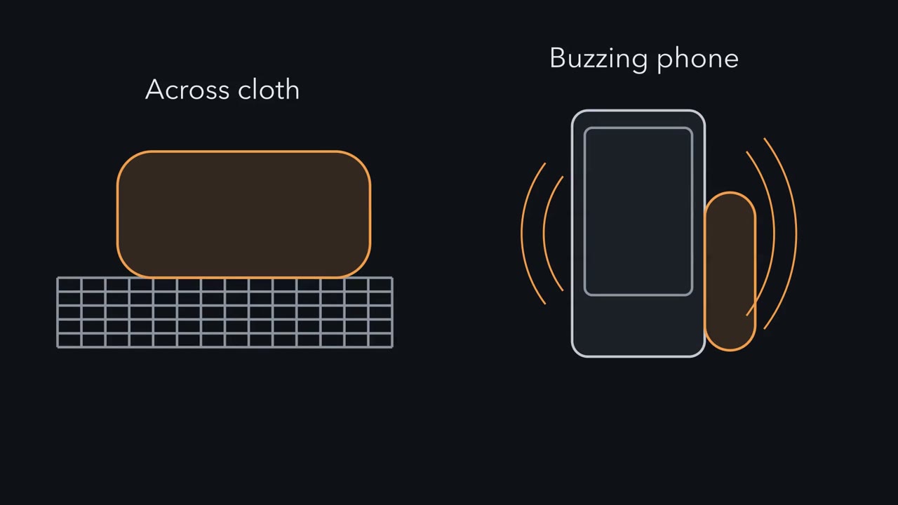
Slide a fingertip across cloth and Meissner corpuscles report the movement; hold a buzzing phone and Pacinian corpuscles report the hum. Each is a nerve ending wrapped in cells that help it feel. How do the two split the work of sensing change?
Pause when the reading reaches a film, and offer to play it. Saved on this device.
Watch the film now, or keep listening
Builds on:Piezo Channels and the Physics of ForceThe Low-Threshold Mechanoreceptors: One Touch, Several SignalsFive Ways to Turn a Stimulus into a CurrentWhy these?
 ▶ Watch Reporters of Change · 2:18
Read the transcript of “Reporters of Change”
Film transcript: Reporters of Change
Slide a fingertip across cloth, or hold a buzzing phone. Two kinds of touch organ answer. What are they reporting? They are the Meissner and Pacinian corpuscles. Each is a nerve ending and the cells that help it feel. Push a probe into the skin, hold it still, then lift it. One kind of fibre, called slowly adapting, keeps firing through the whole press. Predict. What does a Meissner or Pacinian fibre do while the press is held? And when the probe lifts?
Typically, it fires a burst as the probe pushes in. It goes quiet during the hold. Then it often fires again as the probe lifts. That is rapidly adapting. The firing dies away while the press is held. A burst at the release is common, not required.
So these fibres report change, not level. They fire when the skin starts to move, stops, or keeps moving. Vibration is change that repeats. So it drives these fibres strongly. In 1968, Talbot and colleagues vibrated people's hands. From about 5 to 40 Hz, it felt like a light flutter. From around 60 to 80 Hz upward, it felt like a deeper hum. The same study recorded single fibres in monkeys. Flutter matched fibres now tied to Meissner corpuscles. The hum matched Pacinian fibres. The best frequency depends on species. Behavioural sensitivity peaked near 240 Hz in people, and near 1000 Hz in mice. Here is the idea to carry. Compare pushes of the same size.
The faster the response dies away in a hold, the more it discounts slow change. That dying-away can happen in three places. The capsule, the ending's channels, and the spike generator. Keep this. These corpuscles report change, not level.
From the film
- Meissner and Pacinian corpuscles report change, not level: their afferents fire when the skin starts to move, stops or keeps moving, and go quiet while a press is held.
- Meissner afferents serve low-frequency flutter and Pacinian afferents high-frequency vibration, and the best frequency differs between humans and mice.
- The brevity of the response can arise in three places: the capsule, the terminal's channels and the spike generator.
Press a fingertip on a surface and hold it still. Many nerve fibres from that skin fire as the press begins, go quiet while you hold, and often fire again as you let go. Where in the sense organ does a held press become two brief events?
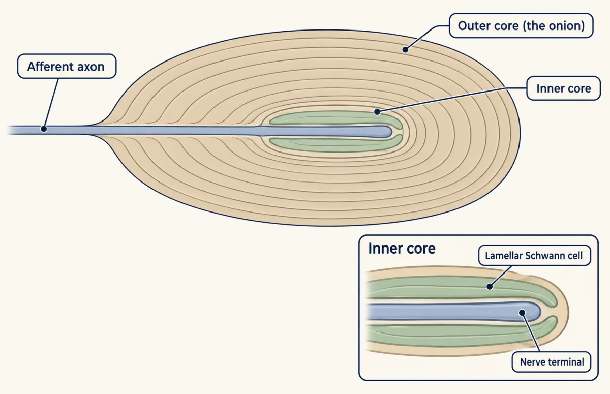
Those fibres are afferents, sensory neurons whose axons carry the signal inward, and they end in Meissner and Pacinian corpuscles, the skin’s two detectors of change. Slide a fingertip across cloth and Meissner afferents report the movement; hold a buzzing phone and Pacinian afferents report the hum. Neither organ is a bare nerve ending. Each is a nerve ending and the cells that help it feel. A lamellar cell is a flattened, Schwann-cell-like cell wrapped around the nerve terminal inside a corpuscle. In the Pacinian corpuscle the lamellar cells form the inner core, and many concentric layers outside them, the onion or outer core, form the capsule.
The touch and pain lesson introduced Meissner and Pacinian afferents as rapidly adapting, with small and large receptive fields, and the LTMR lesson set them in the full class table. This lesson takes the response itself, then the one settled step, where force becomes current, then the central question: why the response is so brief, and what the cells around the ending add. Last come the two afferents of the Meissner corpuscle and what the brain and the animal do with the signal.
The typical response: a burst at each end
Push a probe into the skin, hold it still for a second, then lift it, and record from one afferent at a time.
Predict. A slowly adapting afferent, such as the Merkel cell afferent (SA I), keeps firing while the press is held. What should a Meissner or Pacinian afferent do during the hold, and what should it do when the probe lifts?
Check your prediction
It typically falls silent during the hold and fires again at the release.
| Phase of the press | Typical Meissner or Pacinian afferent (rapidly adapting) | Slowly adapting afferent, for comparison |
|---|---|---|
| Probe pushes in | a burst of spikes | starts firing |
| Probe held still | typically silent | keeps firing |
| Probe lifts | often a second burst | firing ends |
Rapidly adapting means that the firing dies away while a press is held. The release burst is common in classical units but is not part of the definition. The names label afferent classes, not organs. Meissner corpuscles carry the rapidly adapting type I (RA I) afferents, which detect movement across the skin and low-frequency vibration. Pacinian corpuscles carry the rapidly adapting type II (RA II) afferents, which respond to higher-frequency vibration.
Flutter and hum
Vibration is change that repeats, so it drives these afferents strongly. Put a vibrating probe on your fingertip and turn up the frequency, and the percept changes kind. Talbot, Darian-Smith, Kornhuber and Mountcastle (1968, Journal of Neurophysiology) asked healthy adults to describe sinusoidal stimulation of the hand. At about 5 to 40 Hz they reported a light flutter. From around 60 to 80 Hz upward the sensation became a deeper, diffuse hum of vibration.
The same paper recorded single afferents from the median nerve of anaesthetized rhesus monkeys, a second preparation, not a measurement of the human subjects. The comparison tied flutter to the rapidly adapting afferents now linked to Meissner corpuscles and the hum to Pacinian afferents. Read the boundary between them as a description of two percepts, not a sharp receptor cut-off. Why the two organs prefer different frequencies is thought to come from their different structures (Handler et al. 2023), which is why this lesson looks inside them.
Frequency preferences also depend on species. Prsa and colleagues (2021, Nature Communications) found that behavioural vibration sensitivity peaked near 240 Hz at the human index fingertip and near 1000 Hz in their mouse forepaw preparation. “Pacinian tuning around 250 Hz” is therefore a human figure. Behavioural sensitivity is also not identical to the tuning of a single isolated afferent.
The one idea to carry through this lesson. A corpuscle reports change, not level. Its afferent fires when the skin starts to move, stops or keeps moving, and goes quiet while a press is held. Compare two pushes of the same size: the faster its response dies away during a hold, the more it discounts slow change, so a push that builds over seconds gives a smaller response than one that arrives in milliseconds. That dying-away can happen in three places: the capsule shapes the force that reaches the ending, the terminal’s channels set how fast the current falls, and the spike generator sets how many impulses follow. Because more than one place can make the response brief, removing one need not abolish it, and an unchanged response does not prove the removed part did nothing.
The idea covers rapidly adapting afferents: Meissner and Pacinian corpuscles in mammals and their counterparts in the duck bill. It does not cover slowly adapting afferents, or the minority of Ret-positive Meissner afferents that also fired during maintained indentation (later in this lesson). The rule’s push comparison is a simple linear filter model; real firing also depends on amplitude, threshold and recovery. It explains the low-frequency side of an afferent’s band, a graded roll-off rather than a hard detection limit, not its best frequency, which also depends on what fails at high frequencies. And it does not settle how lamellar cells act in mammals: in mice, manipulating them changes detection and the afferent’s vibration responses, but under artificial drive, so their normal coupling is unresolved.
Where force becomes current: Piezo2 in the axon
A corpuscle is built from three elements: the terminal of an Aβ afferent (a large, fast-conducting, myelinated fibre), the lamellar cells (non-myelinating glia), and connective-tissue sheaths that form the capsule. Start with the step that is settled. Force opens mechanically gated channels in the afferent terminal, and the current that flows, the transduction current, is what drives spikes. In mammals the established channel is Piezo2. Mice lacking Piezo2 have an almost complete loss of light touch, and people with PIEZO2 mutations lose touch perception selectively (reviewed by Cobo and colleagues 2020 and Suazo and colleagues 2022). In mouse Meissner and Pacinian corpuscles, Piezo2 sits in the sensory axon membrane, including many small protrusions that tether the axon to the surrounding non-neuronal cells, and it is needed for their light-touch responses, while deleting it from lamellar cells left those responses intact (Handler et al. 2023). The authors’ model is that a stimulus stretches these protrusions across the end organ and opens axonal Piezo2. The mechanotransduction channels lesson sets out what proof a transducer claim needs.
Why the response is brief: capsule, terminal and spike
Read the transcript of “Peel the Onion”
Film transcript: Peel the Onion
A Pacinian fibre typically fires at the press and at the release. In between, it is silent. Where does that brevity come from? There are three suspects. First, the capsule. Its onion of layers could absorb a held press before it reaches the ending. Second, the ending's channels. They could open at the press, then close while the press is still held. Third, the spike generator. It could stop firing even while the ending stays depolarized. Two terms. The generator potential is the ending's graded depolarization, driven by the transduction current. Impulses are the all-or-none action potentials it triggers further along the axon. The classic suspect was the capsule.
In 1966, Loewenstein and Skalak modelled it as fluid between elastic layers. Follow one press. At the instant of the press, the fluid cannot move fast enough. The ending is squeezed. During the hold, the fluid flows and the stretched layers take up the load. The core relaxes. At the release, the layers spring back and jolt the core a second time. So, in this model, the capsule strongly damps steady pressure and passes quick change. That is a high-pass filter. A year earlier, in 1965, Loewenstein and Mendelson peeled most layers from cat corpuscles. Then they pressed and held. Predict. If the capsule did all the work, what should now last through the hold?
Only half of it. The generator potential decayed much more slowly, mainly under intense compression. But the impulses did not persist. The ending typically still fired a single spike. So brevity had two stages. One is the mechanics around the ending. The other is the spike-generating membrane. In 2025, researchers recorded at the ending itself. They used bill skin from duck embryos. They removed the onion, and kept the inner cells that hug the ending. Predict. If the onion sets the brief response, what should happen to the ending's current?
It stayed fast. And the fibre's frequency tuning did not change significantly. The current did grow larger. So the onion may still damp the force that arrives. How do the two results fit? In the duck, currents slowed only as stripped endings deteriorated. So a damaged, dying ending can look like one that has lost its filter. That is one reading. Keep this. In this duck preparation, the ending stayed fast without the outer core. So fast adaptation does not need the onion there.
From the film
- Brevity can arise in the capsule, the terminal's channels or the spike generator, and the classical account put the filtering in the capsule.
- In cat corpuscles with most layers peeled, the generator potential decayed more slowly but the impulses did not persist, so brevity had two stages.
- In the duck preparation, the terminal stayed fast without the outer core, so fast adaptation does not need the onion there.
Something between the skin and the axon turns a held Pacinian press into two brief events. There are three candidates, and this section tests them.
- The capsule. The onion of concentric layers could absorb a held press before it reaches the nerve ending.
- The terminal’s channels. The mechanically gated channels in the ending could open at the press and then close, or inactivate, even while the press is held.
- The spike generator. The membrane where impulses start could stop firing even while the ending stays depolarized.
Two terms come first. The generator potential is the graded depolarization of the nerve ending driven by the transduction current. The impulses are the all-or-none action potentials it triggers further along the axon.
The classical answer: the capsule
Loewenstein and Mendelson (1965, Journal of Physiology) dissected Pacinian corpuscles from the cat mesentery, removed most of the capsule’s layers (the lamellae) and pressed on what remained.
Predict. If the capsule alone made the response brief, what should the bare ending do during a held press? What happens to the generator potential, and what happens to the impulses?
Check your prediction
If the capsule did all the work, both should now last through the hold. Half of that happened: after decapsulation the generator potential decayed much more slowly, an effect that later reviewers note appeared mainly under intense compression. The impulses did not persist: the ending typically still fired a single spike. So adaptation had two stages. One lay in the mechanics around the terminal; another lay in the terminal’s spike-generating membrane, which stopped firing despite a lasting depolarization.
Loewenstein and Skalak (1966) then modelled the capsule. In their model, fluid between the lamellae supplies viscosity and the lamellae supply elasticity. Follow one press through time. At the instant of the press the fluid cannot move fast enough, so the force passes straight to the core and the ending is squeezed. During the hold the fluid flows and the stretched layers take up the load, so the core relaxes although the skin is still pressed. At the release the layers spring back and jolt the core a second time. The capsule thus suppresses slow or steady input and passes transients, at both ends of a press. For decades that high-pass filter was the textbook explanation of rapid adaptation.
Testing the filter at the terminal itself
A filter is best tested by recording at the place it is supposed to protect. Ziolkowski and colleagues (2025, Science Advances) patch-clamped the afferent terminal inside Herbst corpuscles, the bird counterpart of the Pacinian corpuscle, in bill skin from late-stage duck embryos, ex vivo. They then removed the outer core and kept the inner core.
Predict. If the onion sets the brief response and the frequency tuning, what should happen to the terminal’s transduction current, and to the afferent’s tuning, once the outer core is gone?
Check your prediction
The current should become slow and sustained, and the tuning should change. Neither happened. Removing the outer core left the rapid kinetics of the mechanotransduction current intact and did not significantly change the afferent’s frequency tuning. In this preparation, fast adaptation and tuning do not need the onion. It is one bird preparation, not a cat or a human hand. Decapsulation also increased the terminal’s current amplitude, so the outer core may still damp the stimulus.
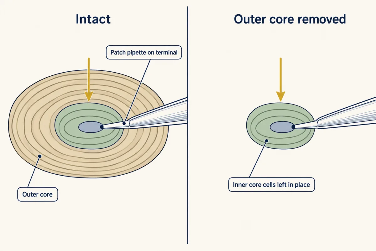
So in the duck, the terminal can adapt without the onion. An earlier duck study had already shown that the terminal inside a Grandry corpuscle, the bird counterpart of the Meissner corpuscle, is a transducer in its own right, with transduction currents at both the onset and the offset of indentation (Ziolkowski, Gracheva and Bagriantsev 2023).
How do 1965 and 2025 fit together? The duck recordings offer one reading. Slower inactivation appeared only as decapsulated terminals deteriorated, so a damaged, slowly dying terminal can look like a terminal that has lost its filter.
| Experiment | What was removed | Signal at the terminal during a held press | Afferent output |
|---|---|---|---|
| Cat Pacinian, 1965 | most of the capsule | generator potential decayed much more slowly, mainly under intense compression | typically still one spike |
| Duck Herbst, 2025 | outer core; inner core kept | transduction current kept its rapid kinetics, and grew larger | frequency tuning not significantly changed |
The verdict on the three suspects, so far. The capsule can shape the signal (the cat generator potential; the duck current’s size), but the duck terminal did not need it to adapt. In the duck, fast current decay persisted without the outer core, which supports a contribution from the terminal’s channels, though the retained inner-core cells and mechanics may also shape that decay. The spike generator adds its own brevity (the cat’s single spike). Adaptation is set jointly, the one idea of this lesson at work.
Adaptation explains only one side of the frequency band. A stage that discards slow change sets a graded low-frequency edge: well below it, the response largely dies away during each slow push. On its own it has no best frequency. A peak, such as the human peak sensitivity near 240 Hz, also needs something that fails at high frequencies, such as the mechanics, the channel kinetics or the timing of spikes, and that side of the band is less well understood. So, on a simple filter model, neither the capsule nor the terminal “makes” the peak by adapting.
That leaves one more candidate still wrapped around the ending once the onion has gone: the inner-core lamellar cells.
What is left once the onion is gone: the lamellar cells
Read the transcript of “The Cells That Help It Feel”
Film transcript: The Cells That Help It Feel
Peel away the onion, and cells still hug the nerve ending. What do they add? First, the settled step. In mice, force opens a channel called Piezo2, in the sensory axon itself. Delete it from the wrapping cells, and touch responses stay.
But the wrapping cells feel force too. In duck bill corpuscles, they carry their own mechanically activated currents. In duck Grandry corpuscles, the Meissner type, depolarizing one wrapping cell could make its fibre fire, after a variable delay.
In mice, switching Pacinian wrapping cells on with light drove the fibre. Switching them off meant a vibration had to be about five times larger to be detected. So these cells contribute to detection. How they normally do it is not yet known. A 200 Hz vibration repeats every 5 milliseconds. In duck Grandry corpuscles, delays ran from 20 milliseconds to about 9 seconds. Predict. Could those cells be passing on each cycle?
A steady delay could shift every cycle, so delay alone cannot rule it out. But the slow, scattered firing suggests a supporting role. Those delays followed electrical depolarization of a wrapping cell, not vibration. Passing each cycle needs a direct test. For Pacinian wrapping cells, the question is open. Meissner corpuscles hold a surprise too. In mice, most hold two molecularly distinct nerve fibres, told apart by genetic labels. Mice lacking TrkB in sensory neurons from development grew up without Meissner corpuscles. They needed about twice the push to detect a touch. They still detected stronger pushes. So these corpuscles help set sensitivity, but in this experiment detection survived without them.
In freely moving mice, Pacinian neurons fired during the mouse's own movements. The most sensitive also answered vibrations from a tap on the branch 2.5 metres away. Two detectors of change, each a nerve ending and the cells that help it feel.
From the film
- In mice, force opens Piezo2 in the sensory axon, but the wrapping cells also feel force and contribute to detection; how they normally do so is not yet known.
- Switching off Pacinian wrapping cells in mice meant a vibration had to be about five times larger to be detected.
- Mice without Meissner corpuscles needed about twice the push to detect a touch yet still detected stronger pushes, so these corpuscles help set sensitivity.
The older picture made the cells around the ending passive padding. The current evidence is that they help set what the afferent feels. Three findings build the case.
- They feel force. In duck bill skin, the Bagriantsev and Gracheva laboratories showed that lamellar cells of Grandry and Herbst corpuscles carry mechanically activated currents (in Herbst corpuscles these recordings were from outer-core cells).
- They can make the afferent fire. Depolarizing one Grandry lamellar cell could make its afferent fire, with calcium influx required, and in Herbst corpuscles depolarizing an inner-core cell by current injection lowered the afferent’s mechanical threshold (Ziolkowski et al. 2025). So in the authors’ sense the corpuscle is bicellular: two kinds of cell, the afferent and the lamellar cells, each able to feel force and to influence the spike.
- In mice they matter for detection. Chen and colleagues (2025, Science Advances; Huber laboratory) expressed optogenetic tools in mouse Pacinian lamellar Schwann cells. Activating them drove time-locked afferent spiking, and inhibiting them raised the hindpaw vibrotactile detection threshold of awake mice about fivefold: a vibration had to be about five times larger to be detected. Inhibition also lowered the afferent’s spike rate and phase locking and shifted its preferred frequency downward. The genetic line used also reaches some other cell types, and the authors note that optogenetic drive may not reproduce normal physiology, so the result shows that lamellar cells contribute to detection, not how they normally do it.
Meissner lamellar cells matter in mice too: without USH2A, a large membrane protein they carry, Meissner (RA I) afferents lose much of their vibration sensitivity (Schwaller et al. 2021, reviewed by Suazo and colleagues 2022).
What remains open is the coupling. Does a lamellar cell pass on each cycle of a vibration, or does it set how sensitive the terminal is?
Predict. To pass on each cycle, a lamellar cell’s signal would have to keep time to within a few milliseconds. A 200 Hz vibration repeats every 5 ms (1 ÷ 200 Hz = 0.005 s). In duck Grandry corpuscles, the delay between depolarizing one lamellar cell and afferent firing ran from 20 ms to about 9 s. Could the lamellar cells there be carrying each cycle?
Check your prediction
These data cannot settle it. The shortest delay, 20 ms, spans 20 ms ÷ 5 ms = 4 cycles, but a steady delay would only shift every cycle by the same amount. Nor do first-spike delays measure cycle-to-cycle precision: they followed electrical depolarization of one lamellar cell, not vibration. In duck Grandry corpuscles, the Meissner-type organ, the delayed, irregular firing suggests a complementary, modulating role rather than the fast primary response; testing cycle following would need repeated mechanical stimulation and timing measured in the same cell–afferent pair. In mouse Pacinian corpuscles the shortest latencies under intense light were very short, which the authors read as tight coupling. Whether Pacinian lamellar cells signal fast enough to follow each cycle of a 200 Hz vibration, or instead set the terminal’s sensitivity, is open.
The channel behind the duck terminal currents, the lamellar-cell transducer and the signal from lamellar cell to terminal remain unidentified. Merkel cells give a parallel case of a non-neuronal partner cell in touch.
Two afferents in one Meissner corpuscle
The Meissner corpuscle sits in a dermal papilla of glabrous skin, a stack of lamellar cells with Aβ axons coiled between them. The usual picture has one physiological type of afferent per corpuscle, although several distinct endings per corpuscle had been described before. Neubarth, Emanuel, Ginty and colleagues (2020, Science) found that most corpuscles in adult mouse paw pads hold two genetically distinct Aβ afferents, intermingled among the lamellar cells.
The two kinds were marked by developmental genetic labels, a Ret label and a TrkB lineage label. TrkB protein was undetectable in adult terminals, so the TrkB label marks a developmental lineage, not a current receptor. Receptive fields of the same kind fitted together across the skin like floor tiles, and the two kinds’ tilings were offset from each other.
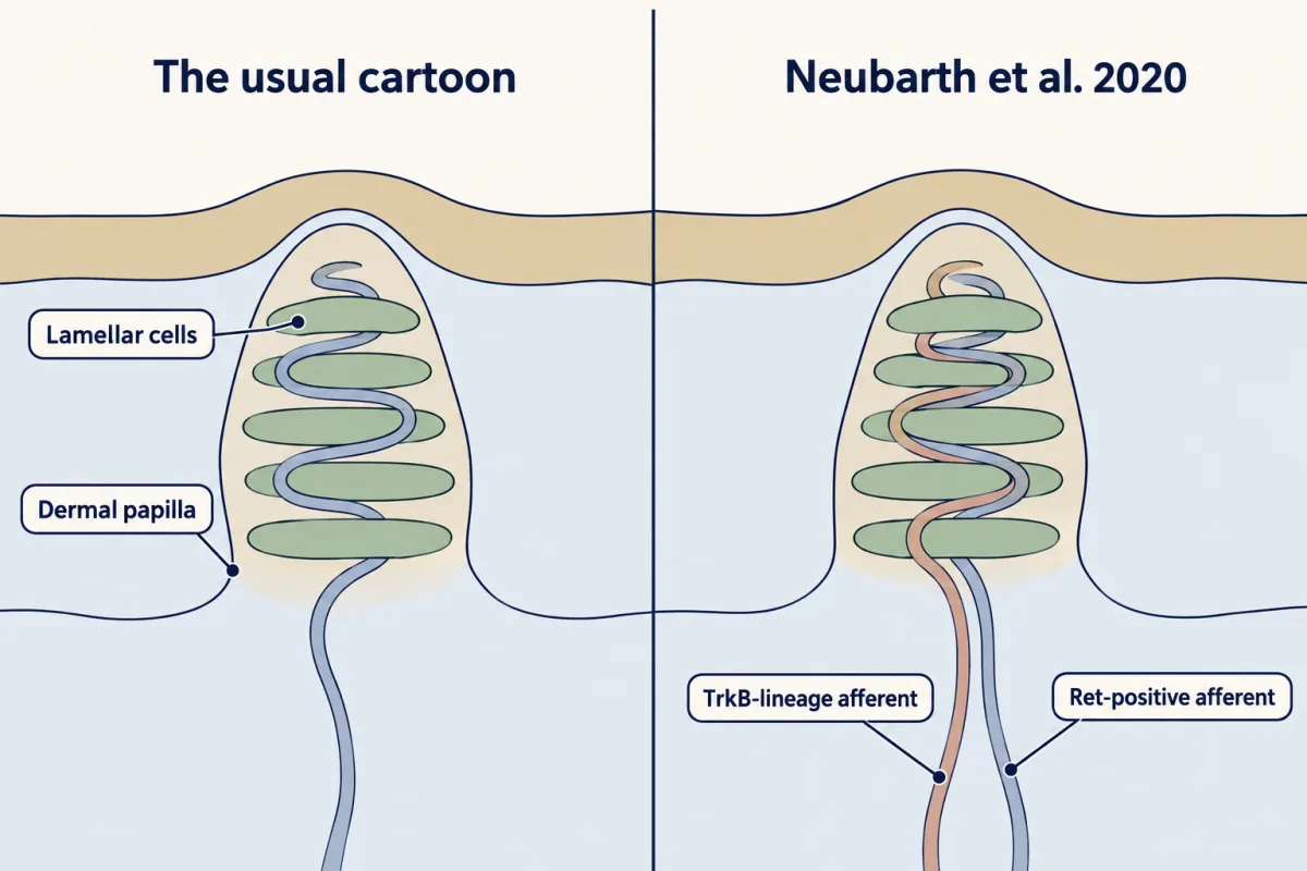
The two differed in physiology. TrkB-lineage afferents were the more sensitive. Ret-positive afferents were less sensitive and rarely fired at the release, and 4 of 10 recorded also fired during maintained indentation, so “two types of rapidly adapting afferent” overstates the result: one subtype included units with a sustained response.
Then the behaviour. Deleting TrkB from sensory neurons during development produced adult mice without the Meissner system. Mice were trained in an operant task to report a step indentation of the forepaw.
Worked problem: a doubled threshold
Adult control mice detected the push at a threshold of about 10 mN; mice lacking Meissner corpuscles, at about 20 mN (Neubarth et al. 2020).
Predict. If Meissner afferents were required for detecting a gentle push, what would the mutants’ threshold look like? What does a measurable but higher threshold say instead?
Check your prediction
If no detection were possible without Meissner afferents, the mutants would never reach the detection criterion over the forces tested, and no threshold could be measured.
- Ratio: 20 mN ÷ 10 mN = 2.0, a twofold rise.
- The mutants still had a measurable threshold, so they still detected larger pushes; their normal sensitivity was impaired.
- Conclusion: Meissner afferents contribute to gentle-touch detection, lowering the threshold by about half, but other afferents can support detection without them.
Two limits apply. The loss was developmental and TrkB was deleted in all sensory neurons, not only Meissner afferents, so other afferents may have compensated or contributed, and an acute silencing could give a larger or smaller effect. And the task measured detection only, not discrimination of flutter or slip.
Vibration in the brain and in the wild
What does cortex do with a Pacinian signal? Prsa, Morandell, Cuenu and Huber (2019, Nature) delivered vibrations to the mouse forelimb and recorded neurons in forelimb primary somatosensory cortex (S1). The responses depended on deep Pacinian input. The neurons did not fire in step with each cycle; their firing rates were tuned to a feature combining frequency and amplitude. That is a rate code for a composite feature. The authors likened this to pitch coding in auditory cortex and contrasted it with primate S1, where low-frequency flutter entrains spiking cycle by cycle. The afferents themselves do phase-lock, much as auditory nerve fibres do in hearing and balance; the loss of entrainment is a cortical result.
Prsa and colleagues (2021) then measured the percept. Trained mice and adult human volunteers compared vibrations, and in both species a change in amplitude pulled perceived pitch toward the frequency of best sensitivity (about 1000 Hz in mice, about 240 Hz in humans). Near 440 Hz, below the mouse peak but above the human one, the two species therefore went opposite ways: in mice a louder vibration felt higher, and in humans it felt lower. For mice near a 450 Hz reference the equal-pitch data were fitted with a power law, which the matcher below uses: in that fit, a 360 Hz vibration at double amplitude matched the 450 Hz reference. It is a local mouse fit and does not apply to humans at this frequency, where the fitted exponent is about −0.24, so the sign reverses.
- equal-pitch curve f* (in this sketch)
- reference, 450 Hz at 5.6 µm
- your comparison vibration
- Exponent used
- 0.32
- (A/5.6)0.32
- 1.000
- P = f × (A/5.6)0.32
- 450.0 Hz
- Against the reference (450)
- matched
- Frequency that matches the reference at this A
- f* = 450 ÷ 1.000 ≈ 450.0 Hz
- Check
- 450.0 × 1.000 = 450.0 Hz
Reference point: the lesson's measured reference, 450 Hz at 5.6 µm.
In this sketch: the power-law fit P = f × (A/5.6 µm)0.32 near a 450 Hz reference in head-fixed C57BL/6 mice (a fit to four mice), assumed to hold from the reference amplitude up to double it (5.6 to 11.2 µm). Frequencies far from 450 Hz are extrapolation. The 1% band is a display tolerance in this sketch, not a perceptual threshold.
Rule to keep: the model is a local fit, so any amplitude other than 5.6 µm gives a prediction, not a measurement. Humans also showed amplitude-dependent pitch, but no human exponent is given, so none is applied here.
Finally, natural behaviour. Turecek and Ginty (2024, Neuron) recorded single Pacinian neurons in the L4 dorsal root ganglion of awake, freely moving mice. The neurons fired during the mouse’s own movements as well as during external vibration, and sensitivity varied widely between neurons. The most sensitive responded to substrate vibrations generated 2.5 m away, the furthest point tested on that surface, not a known limit. Whether the brain separates self-generated from external vibration was not tested. The dorsal horn lesson follows these signals into the spinal cord and brainstem, and prediction returns to how the brain discounts its own movements.
Design problem
A mouse Pacinian afferent fires briefly at the onset and offset of a press and is most sensitive to high-frequency vibration. Design an experiment that decides how much of that behaviour comes from the outer core, from the inner-core lamellar cells and from the terminal itself.
Saved in this browser only.
One way to do it
- Measure the right signal. Voltage-clamp the terminal inside one corpuscle and record the transduction current, and record spikes from the same fibre separately, so generator current and spiking are not confused.
- Remove the outer core in the same corpuscle. Record before and after. Deliver step indentations and sinusoids from 10 to 1000 Hz at matched displacement at the terminal (measured, for example, by imaging the inner core), so that any change in tuning can be assigned to mechanics or to the membrane.
- Control for damage. Track time after dissection and the terminal’s leak current. If adaptation slows only as leak rises, deterioration, not loss of a filter, explains the change. Include sham-handled corpuscles kept for the same time.
- Test the lamellar cells. With the outer core on and off, optogenetically inhibit the lamellar Schwann cells (as Chen and colleagues did in 2025) during the same stimuli, and measure the afferent’s threshold and the animal’s behavioural detection threshold. Allow for the genetic line also reaching other cell types.
- Interpret. If adaptation and tuning survive outer-core removal but change when lamellar cells are inhibited, the inner core shapes them; if neither manipulation measurably changes them, the terminal’s own mechanisms become the leading candidate, once incomplete inhibition, retained mechanics, redundant contributions and assay sensitivity are excluded; if they change only in healthy decapsulated terminals, the outer core shapes adaptation, and the displacement measurements in step 2 say whether it does so mechanically. Duck results do not transfer automatically, and human corpuscles have not been patch-clamped this way, so human answers would need other methods, such as microneurography and psychophysics.
Going deeper
Anatomy, terms and framing
The outer-core layers of the Pacinian corpuscle are made by a different, non-glial cell type from the inner-core lamellar Schwann cells, though papers sometimes call them lamellar cells too. Reviews now treat the afferent terminal, the lamellar cells and the capsule as one sensor.
The 1965 and 2025 decapsulation experiments in detail
The 2025 duck recordings used bill skin from embryonic day 25 to 27 duck embryos, ex vivo. Decapsulation also slightly lowered the terminal’s current threshold, and the authors propose that the outer core protects the inner core.
A simple filter model of adaptation
This is an illustrative model, not a measurement. Treat adaptation as one first-order high-pass stage with time constant τ, the Loewenstein and Skalak idea in its simplest form.
- A press, hold and release. Run a step of force through the stage. For a stage normalized to pass fast change fully, the output jumps to +1 of the step just after the onset, decays toward zero during the hold (as e−t/τ), and jumps the opposite way at the release, to e−T/τ − 1 for a hold of length T: about −0.99 for a hold of 5τ. A transducer that responds to deformation in either direction then fires at both ends, much as the duck Grandry terminal shows onset and offset currents; the model is an analogy, not an account of how those currents arise.
- Two stages in series. Put a capsule stage and a terminal stage one after the other. With equal time constants the step response is e−t/τ(1 − t/τ): it starts at +1, crosses zero at τ, dips to about −0.14 at 2τ and settles to zero during a long hold. Remove either stage and the output is still transient, though its shape changes: the guiding rule in miniature.
- No peak on its own. The gain of a high-pass stage, |H| = ωτ/√(1 + (ωτ)²), rises with frequency all the way up; it sets a corner frequency of 1/(2πτ), a graded low edge rather than a cut-off (at a tenth of the corner the gain is still about 0.0995), and has no best frequency. For stages with the same high-frequency gain and inputs of the same amplitude, a shorter τ attenuates slow change more strongly. With τ = 9 ms, the onset inactivation time constant of the duck Grandry terminal current, the corner is 1/(2π × 0.009 s) ≈ 17.7 Hz. That falls inside the human flutter range, but it is a cross-species coincidence of order of magnitude, not evidence.
- A peak needs a second, low-pass element. For example (illustrative values, not measured), a high-pass time constant of 2.5 ms combined with a low-pass time constant of 0.25 ms peaks near 200 Hz, at 1/(2π × √(τhigh × τlow)).
Duck bill recordings, step by step
Three papers built the lamellar-cell case in steps, all in duck bill skin ex vivo.
- 2020 (Nikolaev and colleagues, Science Advances; embryonic day 24 to 26). Grandry lamellar cells fired action potentials, predominantly through R-type calcium channels. Herbst outer-core cells were mechanosensitive but not excitable.
- January 2023 (Ziolkowski, Gracheva and Bagriantsev, eLife). The onset current in the Grandry terminal inactivated in 8.95 ± 1.82 ms (mean ± SEM), and tetrodotoxin blocked the afferent’s action potentials.
- Later 2023 (Nikolaev, Ziolkowski and colleagues, Science Advances). Latencies from lamellar-cell depolarization to afferent firing ran from 20 ms to about 9 s. Chemical transmission was proposed but not shown.
Grandry lamellar cells lack functional gap junctions with surrounding cells. In duck Herbst corpuscles, the Pacinian type, the inner-core lamellar Schwann cells are coupled to one another by gap junctions, and depolarizing one evoked an inward current in the terminal (Ziolkowski et al. 2025), although dye did not pass from the terminal into them.
Mouse lamellar cells: optogenetics
Chen and colleagues activated the lamellar Schwann cells in anaesthetized adult mice and saw first-spike latencies up to about 40 ms. Electron microscopy showed lamellar cells linked to one another by many desmosomes and gap junctions and anchored to the axon by desmosomes. The Etv1-CreER line marks inner-core lamellar cells but is also expressed in other cell types, so the manipulation is not perfectly cell-specific.
Meissner afferent labels, tallies and wrapping
The Ret label used Ret-CreER given tamoxifen in early embryos. The TrkB lineage label marked cells that expressed TrkB during development: an inducible Cre driver, TrkB-CreER, given tamoxifen, switched on a permanent reporter in them. Both afferent types conducted in the Aβ range. By electron microscopy, TrkB-lineage endings had more lamellar-cell wrappings than Ret-positive endings, and the authors proposed that the extent of wrapping helps set an afferent’s force threshold and kinetics. The behavioural deletion used Advillin-Cre.
Worked problem: the equal-pitch fit
The 2021 mice were C57BL/6, awake and head-fixed, and the equal-pitch fit near 450 Hz came from four mice:
P = f × (A / Aref)0.32, with Aref = 5.6 µm
The frequency that keeps pitch constant. Reference: 450 Hz at 5.6 µm. Amplitude doubles to 11.2 µm. What frequency feels the same?
- Set pitches equal: 450 Hz × (5.6/5.6)0.32 = f2 × (11.2/5.6)0.32.
- Left side: 450 Hz × 1 = 450 Hz. Right side: f2 × 20.32.
- 20.32 = e0.32 × 0.693 = e0.2218 = 1.248.
- f2 = 450 Hz ÷ 1.248 = 360.5 Hz.
- Change: (450 − 360.5) ÷ 450 = 0.199, a 19.9% decrease. Check: 360.5 × 1.248 = 449.9 Hz.
The amplitude ratio has no units, so the answer stays in hertz. This is a prediction of a local mouse fit, assumed to hold at double amplitude; it is not a new measurement.
Your notes
Select any text in the lesson to highlight it or add a note to it. Your highlights and notes are saved in this browser and collected in your notebook.
This browser is not letting the site save anything (a private window or strict privacy settings), so highlights and notes will be lost when you leave the page.
How we measure it
Terminal patch clamp with single-fibre recording
A pipette is sealed onto the afferent terminal inside an intact corpuscle while a second electrode records spikes from the same fibre. You see the transduction current itself and can ask whether its timing, rather than the capsule's, sets adaptation. So far this has been done in ex vivo duck bill skin.
Cre and lineage genetics with operant psychophysics
Genetic labels (Ret, a TrkB lineage tracer) mark afferent subtypes, a conditional deletion removes an end organ, and trained mice report whether they felt a calibrated push. The result shows what that end organ contributes to detection, within the limits of a developmental rather than acute loss.
Optogenetics in non-neuronal cells
A light-gated channel or pump expressed in lamellar Schwann cells lets you activate or inhibit them while recording the afferent or testing behaviour. Activation shows what the cells can do; inhibition tests how much the afferent and the behaviour depend on them, both under artificial drive.
DRG recording in freely moving mice
A stabilized electrode in the lumbar dorsal root ganglion records single sensory neurons for a few hours after the mouse wakes from anaesthesia, while the mouse moves about freely. It asks what an afferent reports in natural life instead of during imposed sine waves.
Practice questions on this lesson (3) → MCAT-style, original, each with an explanation
Where this lesson fits
Before thisReview first
- Piezo Channels and the Physics of ForceThis course · Lesson 2
You need Piezo2 as the force-gated channel and the proof a transducer claim requires, which this lesson takes as settled.
- The Low-Threshold Mechanoreceptors: One Touch, Several SignalsThis course · Lesson 4
This lesson builds on the class table that places Meissner and Pacinian afferents among the touch neurons.
- Five Ways to Turn a Stimulus into a CurrentThis course · Lesson 1
The split between generator potential and spike adaptation here rests on how a stimulus becomes a current.
After thisStudy next
- Where Touch Is First Processed: Dorsal Horn and Dorsal Column NucleiThis course · Lesson 8
It follows these rapidly adapting afferent signals into the spinal cord and brainstem.
- Hairy Skin, Stroking and Affective TouchThis course · Lesson 7
It applies the same afferent-response ideas to other touch fibres, comparing them with the corpuscle afferents.
- Touch Over-reactivity in Autism ModelsThis course · Lesson 9
It tests how changes in these touch afferents alter behaviour in mice.
Elsewhere on the siteFilms, simulations and method films in other courses
- The Spike and Its PuzzlesFilm · Neuroscience
It shows spike generation, one of the three places where brevity can arise.
- What a Spike MeansFilm · Neuroscience
It shows that receptor spikes carry stimulus change as rate, the basis for flutter and vibration coding.
Suggested by an AI model from the lessons themselves, then checked. How we use AI
Finish line
Test yourself
8 questions
Practice and MCAT-style, with explained answers.
Next · Lesson 7
Hairy Skin, Stroking and Affective Touch
Brush a forearm at a few centimetres per second and one slow, unmyelinated fibre fires hardest. Is that why it feels nice?
Continue →← Lesson 5 · Merkel Cells: Touch Receptors That Are Not Neurons