Sensory Systems · Unit 2: Touch
The Low-Threshold Mechanoreceptors: One Touch, Several Signals
Rewritten on : new text, flashcards and film. Your notes and highlights stay, but the text around them may have moved.
A light stroke on your skin is not one signal. Different kinds of touch neuron are most sensitive to different parts of it: the motion, the steady press, the vibration, the slow stroke. On a mouse's back, three of these kinds can even share a single hair. Why build a sense that way?
Pause when the reading reaches a film, and offer to play it. Saved on this device.
Watch the film now, or keep listening
Builds on:Five Ways to Turn a Stimulus into a CurrentPiezo Channels and the Physics of ForceSensory Neurons by Their Genes: the Atlas EraWhy these?
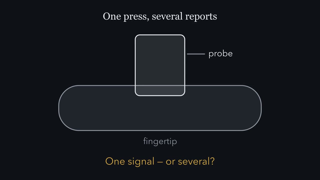 ▶ Watch One Press, Several Reports · 2:50
Read the transcript of “One Press, Several Reports”
Film transcript: One Press, Several Reports
Press a fingertip and hold it. Is that one signal reaching your brain, or several? In Touch and pain, you sorted four touch afferents of the hand into a two-by-two table. You used firing alone. So what lies behind each box? And why split one touch into several signals at all? Start simple. A smooth probe presses into a fingertip, holds still for a second, then lifts. Record from each kind of touch afferent in turn, and line up its spikes under the probe. Which afferent keeps firing while the probe is held still?
The slowly adapting afferent that ends at Merkel cells. It fires going in, and keeps firing through the hold. It reports that something is there, and where its edges are. The rapidly adapting afferent in a Meissner corpuscle fires going in, and again on lifting. During the hold, it is quiet. The Pacinian afferent is silent during the hold. At most, it gives a brief burst as the probe goes in and lifts. Now make the probe vibrate fast, with tiny movements. Which afferent detects the smallest vibration?
The Pacinian afferent. It is the most sensitive to fast vibration. The slowly adapting afferent hardly responds. Each class has a band where it is most sensitive. A band of best sensitivity, not a cutoff. Same skin, same probe, different records. Each is one part of the touch. Here is the one idea to carry. Each class of low-threshold mechanoreceptor is most sensitive to a different part of a touch. Like overlapping filters, each class favours its own part. A held press. Skin motion. Fast vibration. The slightest bend of a hair. A slow stroke. The ending, with the neuron's own properties, shapes what a class responds to best. Axon size and myelin set how fast the report travels.
Why split at all? The recorded classes are complementary. One keeps reporting a held press. Another catches fast vibrations too small for the rest. No class reports the whole touch. So the nervous system reads the pattern across the classes that share the skin. This extends the Touch and pain rule. Endings help decide what a line can report, even within one sense. One press, several reports. Next, mouse hairy skin, where three classes can share a single hair.
From the film
- Each class of low-threshold mechanoreceptor is most sensitive to a different part of a touch.
- The ending and the neuron's own properties shape what a class responds to best, and axon size and myelin set how fast the report travels.
- No class reports the whole touch, so the nervous system reads the pattern across classes.
Bend one awl/auchene hair on a mouse’s back and three classes of touch neuron can answer, because all three have endings along its follicle. Each class is most sensitive to a different part of the movement, and their reports travel at different speeds.
Touch and pain sorted the four touch afferents of the human hand into a two-by-two table, by how fast they adapt and how big their receptive field is, and sorted axons into Aβ, Aδ and C by speed using a latency problem. That table was built from firing alone. This lesson asks what lies behind each box, which ending and which neuron, and what hairy skin adds. It also answers the question the table raises: why split one touch into several signals at all?
One press, several reports
Start with the simplest touch there is. A smooth probe presses into a fingertip, holds still for a second, then lifts. Record from each kind of touch afferent in turn, and line up its spikes under the probe’s position.
- A slowly adapting afferent ending at Merkel cells (SA I) fires as the probe goes in and keeps firing while it is held. It reports that something is there, and where its edges are.
- A rapidly adapting afferent ending in a Meissner corpuscle (RA) fires while the skin moves, as the probe goes in and again as it lifts, and is quiet during the hold. It reports that something is changing.
- A Pacinian afferent (PC) is silent during the hold. It gives at most a brief burst as the probe goes in and as it lifts, and with a smooth, slow ramp even that may be weak.
Predict. Now make the probe vibrate fast, at 250 Hz, with tiny movements. Which afferent detects the smallest such vibration?
Check your prediction
The Pacinian afferent. It is the most sensitive of the classes to fast vibration, best near 100 to 300 Hz, while the slowly adapting afferent hardly responds. Each class has a band in which it is most sensitive. These are bands of best sensitivity, not cutoffs: a strong enough vibration drives other classes too.
Same skin, same probe, different records; each is one part of the touch.
Why not one all-purpose ending? The experiments here do not test that. They show that the recorded classes are complementary: SA I afferents keep signalling a held press, while Pacinian afferents detect fast vibrations too small to drive the others much. Together they give the nervous system several views of the same contact.
The one idea to carry through this lesson. Each class of low-threshold mechanoreceptor is most sensitive to a different part of a touch, much as overlapping filters each favour one band of a sound: a held press, skin motion, fast vibration, a stroke over a wide area, the slightest bend of a hair, a slow stroke. Its end organ, together with the neuron’s own channels and excitability, shapes which part. Its axon’s diameter and myelin set how fast the report travels, and the fibre groups Aβ, Aδ and C summarize those speeds. No class reports the whole touch, so the nervous system reads the pattern across the classes that share the skin.
This extends a rule from Touch and pain, that the ending helps decide what can activate a line, to the parts of one sense. It has limits, and the end of the lesson lists them.
Two terms, now that you have seen them work. A low-threshold mechanoreceptor (LTMR) is a cutaneous sensory neuron activated by touch too light to hurt (Abraira and Ginty, 2013). “Low threshold” describes the bottom of its range, not the top. In mice, Aβ field-LTMRs respond vigorously to a gentle stroke, and their firing kept increasing as indentation entered the noxious range (Bai and colleagues, 2015). An LTMR can fire to a pinch; what makes it an LTMR is that it also fires to a touch too light to hurt.
RA (rapidly adapting) and SA (slowly adapting) are names for the response to one test, the held step you just watched: RA afferents fire at onset and sometimes at offset, SA afferents keep firing (Handler and Ginty, 2021). A step response does not tell you everything a neuron reports during natural movement. Sensory coding treats adaptation in general.
A note on names. This lesson uses Roman numerals for human microneurography classes (SA I, SA II, with RA and PC) and Arabic numerals for mouse genetic classes (Aβ SA1-LTMR, Aβ RA1); the literature mixes both. SA I, RA and PC match SA1, RA1 and RA2 by firing; whether SA II matches a mouse SA2 class is open.
Hairy skin: two endings and the class table
Two kinds of ending recur in hairy skin. A lanceolate ending is an axon branch that runs lengthwise along a hair follicle, parallel to the shaft, like one picket in a fence around the hair. A circumferential ending wraps around the follicle instead. A terminal Schwann cell is a non-neuronal cell whose processes interleave with lanceolate endings.
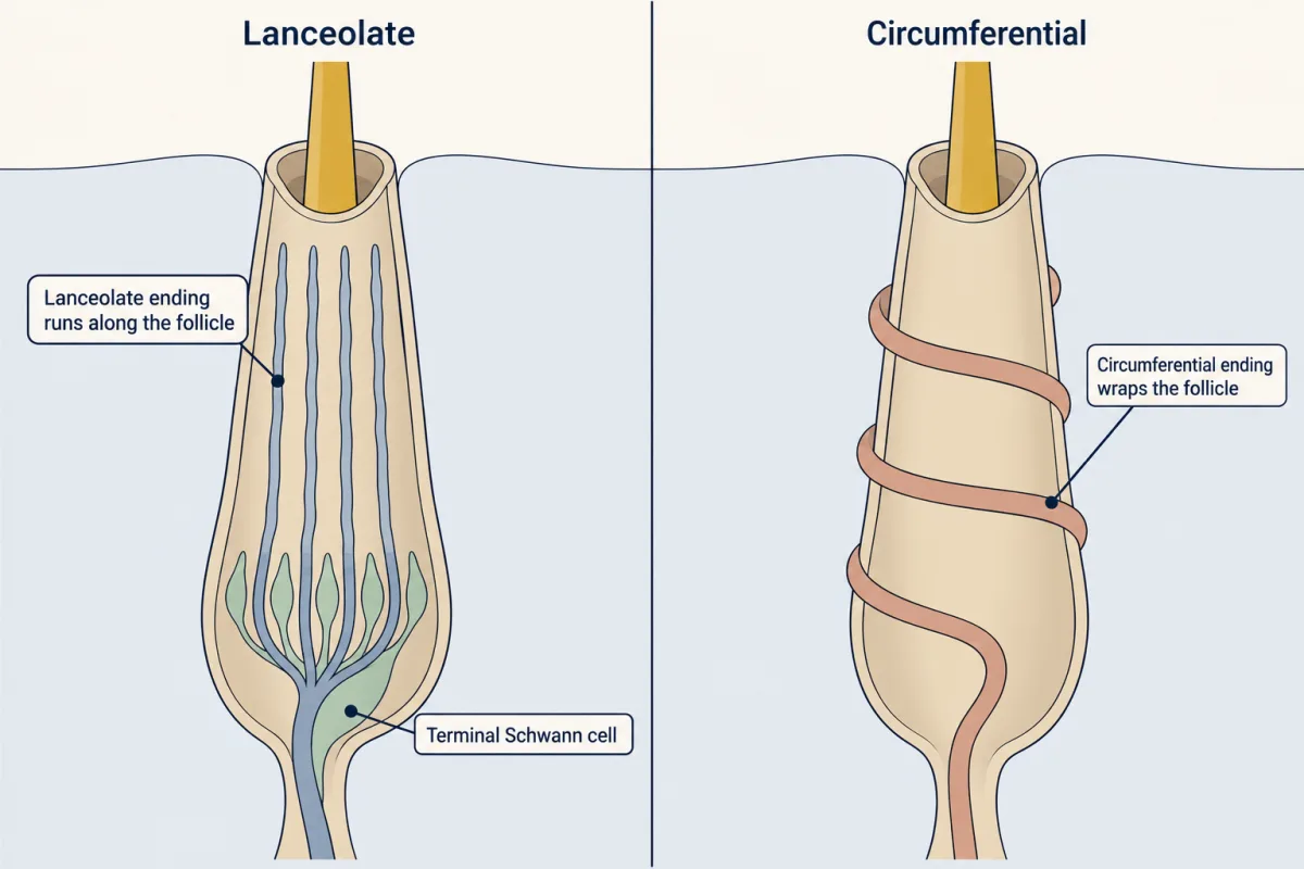
Now the six well-characterized classes, glabrous and hairy skin together. Read the last column first: it says what each class is most sensitive to.
| Class | End organ | Mouse conduction velocity | Response to a step | Best stimulus |
|---|---|---|---|---|
| Aβ RA1 | Glabrous: Meissner corpuscle. Hairy: lanceolate endings at guard and awl/auchene hairs | 13.6 ± 2.7 m/s, hairy skin (Li 2011) | Onset, sometimes offset | Skin motion; Meissner best near 40 to 60 Hz |
| Aβ RA2 | Pacinian corpuscle; in mice mostly in periosteum of digits and limbs | No mouse value here | Onset and offset | Vibration, most sensitive near 100 to 300 Hz |
| Aβ SA1 | Merkel cell-neurite complex; at guard hairs, a touch dome (a cluster of Merkel cells beside the hair) | No mouse value here | Sustained | Indentation, points and edges |
| Aβ field | Circumferential endings around many follicles | 17.0 ± 2.0 m/s (Bai 2015) | Rapid near threshold, intermediate at higher force | Stroke over a wide area |
| Aδ-LTMR | Lanceolate endings at awl/auchene and zigzag hairs (TrkB) | 5.8 ± 0.9 m/s (Li 2011) | Rapid | Tiny hair deflection, below 0.07 mN von Frey force |
| C-LTMR | Lanceolate endings at awl/auchene and zigzag hairs (TH) | 0.58 ± 0.02 m/s (Li 2011) | Intermediate | Gentle deformation of hairy skin |
The speeds are means from one preparation in one study each, not universal boundaries for Aβ, Aδ and C fibres; the frequency bands come from reviews. The Aδ threshold is a von Frey force, set by a calibrated filament pressed until it bends.
Now read the velocity column. The two Aβ classes with mouse values, Aβ RA1 and field, have means only 3.4 m/s apart, so speed alone is a poor guide to which of them a single neuron belongs to. The large separations are between fibre groups: in the mouse, C-LTMRs conduct about ten times more slowly than Aδ-LTMRs. That is why the one idea gives speed to the fibre group, not to each class.
Predict. A soft brush is drawn lightly across a mouse’s back. It bends the hairs but barely presses into the skin. Using the last column, which rows would you expect to answer, and which to stay nearly quiet?
Check your prediction
The hair rows answer: Aβ RA lanceolate endings, Aδ-LTMRs and C-LTMRs. Field-LTMRs, tuned to a stroke over a wide area, should answer too. The SA1 afferents of the touch domes should respond little: they respond to indentation of the dome, not to deflection of the hair alone, and this brush barely indents the skin. A brush that also deformed the dome could recruit them. A held poke aimed at a guard hair’s touch dome would reverse the picture, driving its SA1 afferents through the hold, while other classes still fire as the skin moves in and out. That is the model of Abraira and Ginty (2013): which classes fire depends on the stimulus, and touch is read from these ensembles of impulses from physiologically distinct LTMRs, not from one labelled line per sensation.
Their impulses also reach the spinal cord at different times. The next section shows by how much, and how anyone learned which endings belong to which neurons.
The key experiment: label, record, map
Read the transcript of “One Hair, Three Classes”
Film transcript: One Hair, Three Classes
Bend one hair on a mouse's back. How many kinds of touch neuron can answer? It depends on the hair. Mouse trunk skin has three main hair types: long guard hairs, awl/auchene hairs, and short, bent zigzag hairs. For decades, touch classes in hairy skin were defined by recording. The ending behind each was only inferred. What changed was genetic access. A Cre driver mouse, crossed to a reporter, marks cells that expressed one chosen gene. But a label is only a handle, until recording confirms what the labelled neurons do. Li and colleagues, in 2011, did that check. Neurons labelled by TrkB behaved as Aδ-LTMRs. They answer the tiniest hair deflections.
Neurons with C-LTMR properties stained for TH. Slow, with intermediate adaptation. A third label, for some early Ret-expressing neurons, picked out Aβ beta R -A -L -T -M -R -S, Fast, and rapidly adapting. Then they imaged whole sheets of trunk skin, to see which follicles each label surrounded. Now bend an awl/auchene hair and hold it. Which labelled classes answer?
Three classes can: Aβ beta RA, Aδ delta, and C. Their lanceolate endings fence the same follicle. Each ending runs lengthwise along the follicle, like one picket in a fence. Most awl/auchene and zigzag follicles also have circumferential endings from field-LTMRs, which Li did not label. That makes four classes at an awl/auchene follicle, and three at a zigzag one. Guard hairs carry Aβ beta RA endings, and a touch dome beside the hair. Zigzag hairs carry Aδ delta and C. These combinations repeat across the skin. One movement of an awl/auchene hair can be reported by three classes in parallel. Assume their spikes start together and travel equal paths of 2 centimetres.
Conduction alone gives these arrival times. The Aβ beta and Aδ delta reports arrive about 2 milliseconds apart. The C report arrives about 30 milliseconds later. During the hold, the two rapid classes fall quiet. The C unit keeps firing for part of it. On longer paths, every gap grows. What fires, and when its spikes arrive, both shape what the spinal cord receives. One caution. A single neuron branches to many hairs. So endings at a hair are not a count of neurons. One hair, three classes, three speeds. The skin splits even a single hair movement into parallel reports.
From the film
- One movement of an awl/auchene hair can be reported in parallel by three classes, at three speeds.
- The combinations of endings repeat across the skin and differ between hair types.
- A single neuron branches to many hairs, so endings at a hair are not a count of neurons.
For decades, classes in hairy skin were defined by recording, and the ending behind each was inferred. What changed was genetic access. A Cre driver is a mouse line that expresses Cre recombinase from one gene’s regulatory DNA, so that a reporter marks cells that expressed that gene; CreER adds a tamoxifen switch so the experimenter picks the labelling window (tools for development covers how). A label is a handle, not a class, until physiology confirms it. Transcriptomic atlases, covered in the atlas lesson, now offer many more handles, and each still needs this step.
Mouse trunk skin has three main hair types: long guard hairs, awl/auchene hairs, and the short, bent zigzag hairs. Li and colleagues (2011) labelled the C, Aδ and Aβ RA classes genetically and checked each label by recording. They did not label field-LTMRs, which return below. No mouse line for SA1 neurons existed yet, so they marked SA1 endings through the Merkel cells they contact. They then imaged fixed whole mounts of trunk skin to see which follicles each label surrounded. Here is what each handle gave them.
| Genetic handle | Recording showed | Ending | Follicles |
|---|---|---|---|
| TrkB | Aδ-LTMR: 5.8 m/s, rapidly adapting, threshold below 0.07 mN | Lanceolate | Awl/auchene, zigzag |
| TH | C-LTMR: 0.58 m/s, intermediate adaptation | Lanceolate | Awl/auchene, zigzag |
| Npy2r-GFP, a subset of the early Ret-expressing neurons | Aβ RA-LTMR: 13.6 m/s, rapidly adapting | Lanceolate | Guard, awl/auchene |
| None for SA1 neurons; Merkel cells stained instead | Not recorded | Touch dome | Guard |
In their recordings, adult TrkB-expressing dorsal root ganglion neurons behaved as Aδ-LTMRs, and the cutaneous C-fibre neurons with C-LTMR properties stained TH-positive. That is the check that turns a handle into a class.
Predict. An awl/auchene hair is bent and held. Which labelled classes answer? If you record at a ganglion 2 cm away, in what order do their spikes arrive, and which keep firing through the hold?
Check your prediction
Three classes can answer, because three classes’ lanceolate endings fence that follicle: Aβ RA, Aδ and C. Assume the bend drives all three and their first spikes start together.
Worked problem: the arrival race. The bottom line: the C report arrives about ten times later than the Aδ report, and the two fast reports about 2 ms apart. Take an illustrative 2.0 cm path, Li’s mean velocities, constant speed and conduction alone.
In words The travel time is the path length divided by the conduction velocity.
- t“t”
- the conduction times
- L“L”
- the length of the path along the axonm
- v“v”
- the conduction velocitym/s
Sanity check Double the path and the time doubles; double the speed and it halves. Units: metres divided by metres per second leaves seconds.
Try one number
A shortcut for the units: 1 m/s is the same as 1 mm/ms, because metres and seconds both shrink by a factor of 1000.
So 2.0 cm = 20 mm at 13.6 mm/ms takes 20 ÷ 13.6 ≈ 1.5 ms. Step 1 below does the same in SI units.
Need a refresher? units and prefixes · rates and “per”
- Aβ RA at 13.6 m/s: 0.020 m ÷ 13.6 m/s = 0.0015 s, about 1.5 ms.
- Aδ at 5.8 m/s: 0.020 m ÷ 5.8 m/s = 0.0034 s, about 3.4 ms.
- C at 0.58 m/s: 0.020 m ÷ 0.58 m/s = 0.034 s, about 34 ms.
- Check: Aδ is ten times faster than C (5.8 ÷ 0.58 = 10), so the C time should be ten times the Aδ time: 3.4 ms × 10 = 34 ms.
These are conduction times, not measured response latencies. Both gaps grow with path length, and whether a 2 ms gap matters is a question for the circuits that receive these signals. The rapidly adapting Aβ RA and Aδ units fire as the hair moves and fall quiet in the hold; the C unit, with intermediate adaptation, keeps firing for part of it.
The full map gave each hair type its own combination:
- Guard: Aβ RA lanceolate endings, plus Merkel-associated Aβ SA1 endings in the touch dome beside the hair.
- Awl/auchene: Aβ RA, Aδ and C lanceolate endings, interdigitated.
- Zigzag: Aδ and C lanceolate endings, in virtually all follicles examined.
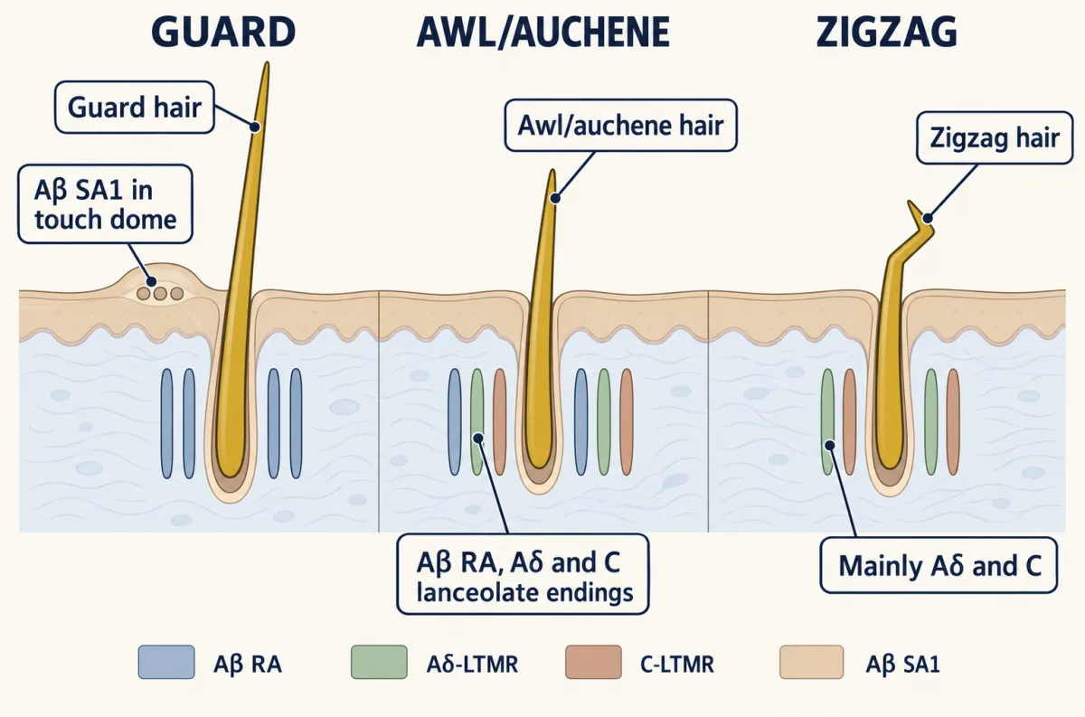
“Stereotyped” keeps its exceptions: the paper reports very few Aβ RA associations with zigzag follicles. The three hair types occurred in an approximate 1:23:76 ratio (guard to awl/auchene to zigzag), and recurring clusters of them led the authors to describe peripheral low-threshold mechanoreceptor (LTMR) units. The field-LTMRs Li did not label add circumferential endings around most awl/auchene and zigzag follicles (Bai and colleagues, 2015), making four LTMR classes at an awl/auchene follicle and three at a zigzag follicle. Li and Ginty (2014) later found that a single terminal Schwann cell can host endings of more than one LTMR type.
This is what the map means for coding. One movement of an awl/auchene hair can be reported in parallel by three classes, at three speeds and with three time courses. One warning when you count: a single afferent branches to many follicles, so a count of endings at hairs is not a count of neurons. The same handles let later studies ablate, silence or activate one class, as in the C-LTMR experiment under “Where the idea stops”.
Your own skin: human touch classes and C-tactile afferents
Read the transcript of “Your Own Skin: The Slow Stroke”
Film transcript: Your Own Skin: The Slow Stroke
A soft brush strokes your forearm. Does some nerve fibre care how fast it moves? Mouse genetics cannot be done in people. So human touch classes rest on another method, microneurography. A fine tungsten electrode goes through the skin into a nerve, in a conscious person. It records single afferents. Recordings from the hand sorted human afferents into four classes, by firing. The ending is never seen. Hairy skin adds another class. Slow, unmyelinated afferents that respond to gentle deformation. These are the C-tactile afferents, physiologically similar to mouse C-LTMRs. The brush strokes from very slow to fast. Which speed drives a C-tactile afferent hardest?
Not the fastest. C-tactile firing rises, then falls: an inverted U. It peaks in the slow-stroke range. Pleasantness ratings correlated with that firing. Fast myelinated Aβ afferents behaved differently. Their firing kept rising with speed. Reviews conclude that these afferents support gentle, affective touch, the social side of touch. But not every affective touch relies on them. And affective touch need not be pleasant. The human C-tactile ending is still unknown. The mouse hair map cannot be transplanted onto human hair. So the idea carries over. Each class is most sensitive to its own part of a touch. The nervous system reads the pattern.
From the film
- In people, C-tactile afferents fire most in the slow stroke range, in an inverted U across stroking speeds.
- These afferents support gentle, affective touch, but not every affective touch relies on them.
- The human C-tactile ending is still unknown, so the mouse hair map cannot be transplanted onto human hair.
Mouse genetics cannot be done in people, so the human classes rest on another method. In microneurography, a tungsten microelectrode inserted through the skin into a peripheral nerve records single afferents in a conscious person. Recordings from the glabrous skin of the hand sorted human low-threshold mechanoreceptors (LTMRs) into four classes, RA, PC, SA I and SA II (Johansson and Vallbo, 1979). By their firing, RA, PC and SA I line up with the Aβ RA1, RA2 and SA1 rows of the table. The human classes rest on firing and receptive fields; no ending was seen. That is why SA II, slowly adapting units with large receptive fields that fire to skin stretch, is not yet tied to an ending in either species.
Hairy skin adds another class. Vallbo and colleagues (1999) recorded unmyelinated afferents in human forearm hairy skin that respond to gentle deformation, the C-tactile (CT) afferents, physiologically similar to mouse C-LTMRs.
Predict. A soft brush strokes the forearm at speeds from very slow to fast. Which speed drives a CT afferent hardest? Will a fast myelinated Aβ afferent follow the same curve?
Check your prediction
Not the fastest. In microneurography by Löken and colleagues (2009), CT firing followed an inverted U across brushing velocities, peaking at about 1 to 10 cm/s, the slow, gentle stroking commonly felt as pleasant. The volunteers’ pleasantness ratings correlated with CT firing. Aβ afferents behaved differently: their firing kept rising with velocity. This inverted U is the CT’s characteristic response, as the held step is for SA and RA afferents.
Reviews of the field conclude that CTs support gentle, affective touch, the social side of touch, while fast myelinated Aβ afferents serve discriminative touch (Schirmer and colleagues, 2023; McGlone and colleagues, 2026). The link has a limit: not every affective touch relies on CTs, and affective touch need not be pleasant (Schirmer and colleagues, 2023). The human CT ending is unknown, and the awl/auchene and zigzag map cannot be transplanted onto human hair. Hairy skin and affective touch takes CTs further.
How a tuning is built: direction
One class shows that what a neuron is most sensitive to can be built into its wiring during development. Aδ-LTMRs are among the most sensitive hair afferents in the mouse, and in adult mouse trunk skin they prefer one direction: they fire more to hair deflection toward the head (caudal-to-rostral) than to an equal deflection the other way (Rutlin and colleagues, 2014). Their lanceolate endings are enriched on the caudal, tail side of each follicle, where brain-derived neurotrophic factor (BDNF) in the follicle epithelium is also enriched.
Two things here are easy to confuse. Think of a weathervane: where a vane is mounted is not the way it points. The endings are mounted on the tail side; the neuron’s preferred movement is the hair tip swinging toward the head. How one leads to the other mechanically is unknown. BDNF signalling through TrkB is covered in Trk and p75.
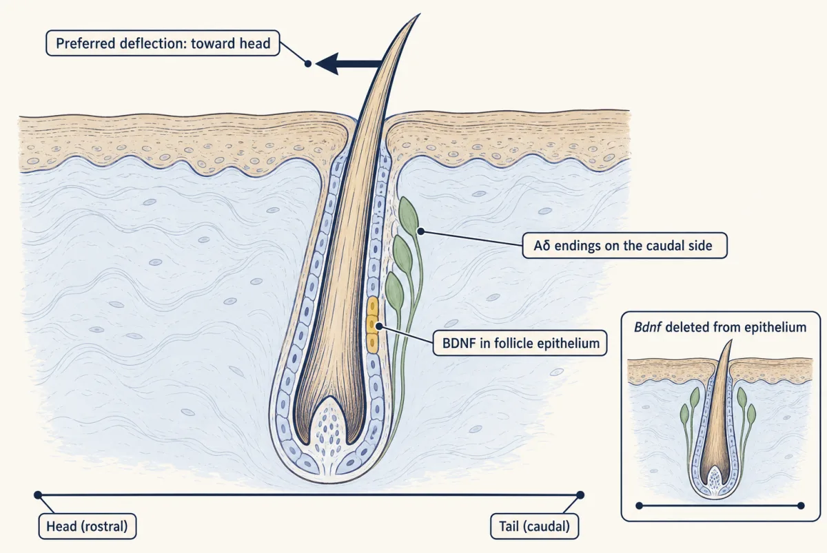
Rutlin and colleagues deleted Bdnf from the epithelium with K5Cre and compared these mutants with littermate controls on three outcomes.
| Mice | Aδ endings on the caudal side? | Tuning lined up with the body axis? | Aδ-LTMRs survive? |
|---|---|---|---|
| Littermate controls | Yes | Yes, toward the head | Yes |
| K5Cre;Bdnf (epithelial deletion) | ? | ? | ? |
Predict. If epithelial BDNF organizes the endings during development, fill in the mutant row. Why does the last column matter?
Check your prediction
The caudal polarity of the endings was disrupted, and the neurons’ preferred direction was randomized relative to the body axis. Aδ-LTMR survival and general maturation were retained. The last column rules out the dull explanation: tuning that no longer lines up with the body axis is not explained by lost neurons. Because the deletion acted through development, the result shows that epithelial BDNF is required to organize the endings, not that BDNF is released during touch.
A direction index puts a number on the preference, on a scale from −1 to +1.
In words The difference between the two spike counts, divided by their sum.
- H“H”
- the spike count for deflection toward the head
- T“T”
- the spike count for an equal deflection toward the tail
Sanity check Equal nonzero counts give 0: no preference along this axis. No spikes toward the tail gives +1; no spikes toward the head gives −1. If both counts are zero the index is undefined and the trial is unresponsive.
Try one number
Take round illustrative counts: 6 spikes toward the head (H = 6) and 2 toward the tail (T = 2).
Difference: 6 − 2 = 4. Sum: 6 + 2 = 8.
Index: 4 ÷ 8 = 0.50. A threefold preference for the head gives an index of one half.
Need a refresher? rates and “per”
The simulation below lets you compute it.
Up is rostral (toward the head) in both pictures. In each dial the thin line is the neuron’s preferred direction; the bold arrow’s length is its direction index. The number under a dial is that neuron’s body-axis index, DI × cos θ. The ringed dial is the neuron drawn on the follicle.
- Ratio P / O
- Direction index, DI = (P − O) / (P + O)
- Mean body-axis index, expected
- Mean body-axis index, observed (12 sketch neurons)
In this sketch: spike counts are illustrative, not Rutlin’s data. Mutant angles are drawn uniformly at random, which is why the expected mean is 0. Two assumptions are mine, not findings: the body-axis index is cosine tuning, DI × cos θ, and every neuron has the same DI, which each mutant neuron keeps while only its angle is randomized (the lesson does not say whether single-neuron tuning strength survived). The deletion shows a developmental role for epithelial BDNF, not BDNF release during touch. The rule to keep is DI = (P − O) / (P + O): 0 is no preference, 1 is no response to the opposite direction.
Design problem
A transcriptomic atlas offers a new marker gene for mouse afferents that may be the missing SA2-like class. Design the experiments that would establish these neurons as a low-threshold mechanoreceptor (LTMR) class: what they respond to, how fast they conduct, and where they end. State what the design could and could not conclude.
One way to do it
Get genetic access. Make a Cre or CreER driver from the marker gene and cross it to a reporter. CreER lets you choose the labelling window, which matters if the gene is also expressed by other neurons at other times.
Check the label with physiology. Record labelled neurons in an ex vivo skin, nerve, ganglion and spinal cord preparation. Measure conduction velocity, threshold, the response to a held step indentation and the response to skin stretch. An SA2-like class should fire to innocuous force, keep firing during a held stimulus and respond to stretch.
Find the ending. Image whole mounts of labelled skin, and confirm the ending of recorded neurons afterwards, to see whether the endings are Ruffini-like or something else.
Test what the class does. With genetic access you can ablate, silence or activate the class and ask which behaviour changes, as was done for C-LTMRs and the wet dog shake. A partial loss would show that the class contributes, not that it is the only route.
Conclude, at most, that mice have a genetically defined SA2-like class with a known response and ending. The design cannot show what human SA II units end in, because microneurography does not see the ending.
Where the idea stops
The one idea says “most sensitive to”, not “reports only”, and gives the ending a share in tuning, not all of it. Here is why.
- Classes overlap. In a small human study that changed surface friction during touch, frictional transients evoked time-locked spikes across the sampled afferent classes (Dione and colleagues, 2021).
- Same ending, different tuning. Aβ RA, Aδ and C-LTMRs all make lanceolate endings at the same awl/auchene follicle, yet differ in threshold, adaptation and speed. The ending type alone does not set the tuning.
- A C-LTMR is not only a line for gentle stroking. In mice, optogenetic activation of C-LTMRs evoked wet dog shakes, and adult ablation cut oil-droplet-evoked shakes by about half (Zhang and colleagues, 2024).
- Some endings are unknown. SA II physiology is secure, but its ending is unidentified in both species, and the human C-tactile (CT) ending is unknown too, so the end organ’s share in their tuning cannot yet be checked.
- The idea stops at the skin. How the spinal cord combines these reports is the subject of dorsal horn touch.
Going deeper
The details below support the main line without changing it.
Where the class-table numbers come from
The Li velocities come from intracellular recordings in an ex vivo skin, nerve, ganglion and spinal cord preparation from 2 to 3 month old mice, with five C-LTMRs, 19 Aδ-LTMRs from seven animals and 18 Aβ RA-LTMRs from five animals. The often-quoted 15 to 100 m/s for Aβ is a review range pooled across species; the mouse hairy-skin Aβ RA mean sits below it. The Li whole mounts were of trunk skin from 3 to 10 week old mice, and the SA1 endings were found through their Merkel cells with anti-Troma1 staining.
What the ± values can tell you. The main text of Li et al. gives them without saying whether they are standard deviations or standard errors, and the size of a value is no guide to which. Either way, treat the values as summaries of these samples, not as boundaries between classes. If both Aβ values are standard deviations, the mean ± 1 SD intervals, 10.9 to 16.3 m/s for Aβ RA and 15.0 to 19.0 m/s for field-LTMRs, already overlap; if they are standard errors, single neurons spread more widely still.
The Meissner band in the table is a review value; Touch and pain describes the same afferents as most sensitive to low-frequency vibration, the range of flutter and slip.
The dorsal horn columns
The dorsal horn columns came from separate experiments in which cholera toxin B (CTB), a tracer taken up by axons, was injected into small patches of back skin at postnatal day 14 to 25. They showed that the central endings of afferents serving one patch line up in columns in the dorsal horn. That alignment is anatomy; how the columns combine their inputs is the subject of dorsal horn touch.
Multi-afferent end organs
The awl/auchene follicle is not the only end organ with more than one afferent type. The 2021 review by Handler and Ginty adds that mouse Meissner corpuscles receive two afferent populations, some of them more slowly adapting, a result from Neubarth and colleagues (2020) that the corpuscle lesson takes up. Handler and colleagues (2023) located tagged Piezo2 in the sensory axon membrane of lanceolate, Meissner and Pacinian endings (Piezo channels covers the channel). Merkel cells get their own lesson, and field-LTMRs and C-LTMRs return in hairy skin and affective touch.
Counting follicle associations is not counting neurons
Worked problem: associations versus neurons. Take an idealized patch of exactly 100 mouse trunk follicles in Li’s ratio: 1 guard, 23 awl/auchene, 76 zigzag. Use only the dominant map above, count each class once per follicle it innervates, and leave out field-LTMRs and the rare exceptions.
- Aβ RA: guard plus awl/auchene = 1 + 23 = 24 associations.
- Aβ SA1: guard only = 1.
- Aδ-LTMR: awl/auchene plus zigzag = 23 + 76 = 99.
- C-LTMR: awl/auchene plus zigzag = 23 + 76 = 99.
- Total: 24 + 1 + 99 + 99 = 223.
- Check by follicle: (1 × 2) + (23 × 3) + (76 × 2) = 2 + 69 + 152 = 223.
associations = sum over follicle types of (number of follicles × classes per follicle)
Is 223 a neuron count? No. One afferent contacts many follicles; a single mouse field-LTMR arbor reached 20 to 180 follicles (Bai and colleagues, 2015). And 1:23:76 is a ratio of hairs, not of neurons. The calculation tells you how often each class has endings at a hair.
SA II and the Ruffini question
Johansson and Vallbo (1979) recorded 334 low-threshold units through the median nerve; their SA II class is defined by firing, so its Ruffini pairing is an inference. The 2021 review by Handler and Ginty keeps that pairing controversial, and SA2-like responses have been recorded in mice without a corresponding Ruffini ending being identified. Three claims go too far. “Mice lack SA2” goes beyond the evidence, whether mice have a distinct SA2 class is still open, and “SA2 is the Ruffini afferent” is unproven in both species. The experiment that would settle it in mice is the one in the design problem.
Small human samples: palm CTs and friction
C-tactiles (CTs), long thought to be confined to hairy skin, have been found, sparsely, in the glabrous skin of the hand. Watkins and colleagues (2021) reported three CT afferents there, at roughly one per 80 myelinated afferents encountered. The authors used that ratio to estimate a CT density about seven times lower than in hairy skin, an estimate that rests on electrode sampling. Dione and colleagues (2021) changed surface friction with an ultrasonic device while recording from eight afferents in five women aged 21 to 28.
Direction tuning in detail
In the ex vivo recordings, genetically identified Aδ-LTMRs fired, on average, more than three times as many spikes to caudal-to-rostral deflection as to an equal deflection the other way. The caudal brain-derived neurotrophic factor (BDNF) was shown by reporter histology. The mutants were K5Cre;Bdnf conditional mutants: an epithelial driver avoids the problem that a germline Bdnf knockout would affect many neurons and the animal’s health. Rutlin and colleagues recorded from TrkB-GFP neurons in these mutants. A TrkB-GFP knock-in labels Aδ-LTMRs without recombining Bdnf; a tamoxifen-induced TrkB-CreER line would also excise the floxed Bdnf allele in the labelled neurons. TrkB is expressed by other neurons too, so identity should be confirmed by conduction velocity and threshold. Separating development from adult function would need an adult, inducible deletion, and touch-evoked release would still have to be measured. The experiment says nothing directly about human hair afferents.
How to test it
The three columns of the prediction table map onto three measurements in mutants and littermate controls: ending polarity (the fraction of Aδ endings on the caudal side of each follicle, in whole mounts), tuning (spikes to equal rostral and caudal deflections, as a direction index, in ex vivo recordings) and survival (counts of labelled dorsal root ganglion (DRG) neurons).
Worked problem: a direction index. Suppose, as illustration, an Aδ-LTMR fires 12 spikes to a caudal-to-rostral deflection (H = 12) and 3 to an equal rostral-to-caudal one (T = 3).
direction index = (H − T) / (H + T), for H + T > 0
- Ratio: 12 / 3 = 4, in the range of the paper’s over-3-fold average.
- Index: (12 − 3) / (12 + 3) = 9 / 15 = 0.60.
- Check: the head direction is fixed by the body axis, so a neuron tuned the other way gets a negative index; 1 means no response to the opposite direction. A ratio of 3 gives (3 − 1) / (3 + 1) = 0.50, so 0.60 is a plausible illustrative value; the paper reports an average over tested spots, not a per-neuron floor.
Your notes
Select any text in the lesson to highlight it or add a note to it. Your highlights and notes are saved in this browser and collected in your notebook.
This browser is not letting the site save anything (a private window or strict privacy settings), so highlights and notes will be lost when you leave the page.
How we measure it
Genetic labelling with Cre and CreER drivers
A Cre driver is a mouse line that expresses Cre recombinase under one gene's control, so a reporter marks the cells that expressed that gene at some time. CreER adds a tamoxifen switch, so the induction window can be chosen. The label is a handle on a population, not proof of a pure type until physiology checks it.
Intracellular recording in an ex vivo skin-nerve-DRG-spinal cord preparation
Skin, nerve, dorsal root ganglion and cord are dissected out together, and a sharp electrode records a labelled cell body while the skin is touched or the nerve stimulated. It gives conduction velocity, threshold and adaptation for a neuron whose genetic identity and ending can be confirmed afterwards.
Whole-mount immunostaining and confocal microscopy
A sheet of fixed skin is stained intact and imaged in three dimensions, so each labelled ending can be assigned to the follicle and hair type it surrounds.
Human microneurography
A tungsten microelectrode is inserted through the skin into a peripheral nerve of a conscious person to record single afferents. Units are classified by firing and receptive field; their endings are not seen.
Practice questions on this lesson (3) → MCAT-style, original, each with an explanation
Where this lesson fits
Before thisReview first
- Five Ways to Turn a Stimulus into a CurrentThis course · Lesson 1
You need the ways a stimulus becomes a current to understand how each ending shapes its class's response.
- Piezo Channels and the Physics of ForceThis course · Lesson 2
The Piezo channel in the ending is what turns skin force into current in each class.
- Sensory Neurons by Their Genes: the Atlas EraThis course · Lesson 3
Genetic handles from the atlas are what this lesson checks by recording to turn them into classes.
After thisStudy next
- Merkel Cells: Touch Receptors That Are Not NeuronsThis course · Lesson 5
The Aβ SA1 class and its Merkel ending, only briefly met here, get a full lesson.
- Meissner and Pacinian Corpuscles: Flutter and VibrationThis course · Lesson 6
The RA1 and Pacinian classes, tuned to flutter and vibration, are worked through in detail.
- Hairy Skin, Stroking and Affective TouchThis course · Lesson 7
It takes the C-LTMR and human C-tactile slow-stroke tuning further, including the affective touch link.
Elsewhere on the siteFilms, simulations and method films in other courses
- Lateral inhibition at an edgeSimulation · Neuroscience
It shows how a sensory neuron's spikes carry meaning from its tuning, as each LTMR class is tuned to part of a touch.
- What a Spike MeansFilm · Neuroscience
It shows that a spike means what its neuron is tuned to, the principle behind reading touch across classes.
- Why Capsaicin Feels HotFilm · Neuroscience
It covers the skin touch receptors and the pathways that carry their signals to the spinal cord.
Suggested by an AI model from the lessons themselves, then checked. How we use AI
Finish line
Test yourself
8 questions
Practice and MCAT-style, with explained answers.
Next · Lesson 5
Merkel Cells: Touch Receptors That Are Not Neurons
A skin cell that is not a neuron helps a nerve keep firing for as long as you press. How does it hold the press, and what does it tell the nerve?
Continue →