Sensory Systems · Unit 2: Touch
Touch Over-reactivity in Autism Models
In one experiment, deleting a gene from peripheral sensory neurons, but not from the brain, was enough to change a mouse's behaviour in anxiety-like and social assays.
Pause when the reading reaches a film, and offer to play it. Saved on this device.
Watch the film now, or keep listening
Builds on:The Low-Threshold Mechanoreceptors: One Touch, Several SignalsWhere Touch Is First Processed: Dorsal Horn and Dorsal Column NucleiFive Ways to Turn a Stimulus into a CurrentWhy these?
 ▶ Watch Delete Here, Learn Where · 3:15
Read the transcript of “Delete Here, Learn Where”
Film transcript: Delete Here, Learn Where
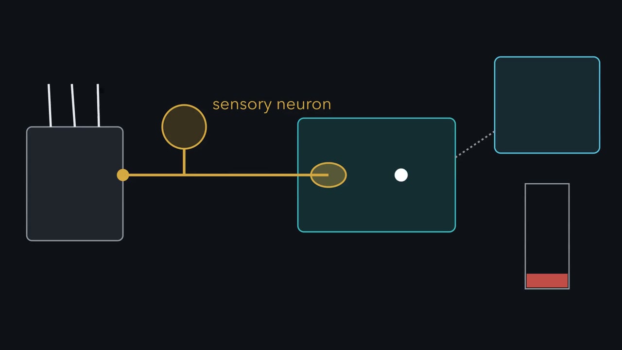
If the brain keeps its gene, can changing the first touch neuron still change a mouse's response? A gentle air puff bends hairs on the back. A gold sensory neuron carries the signal through a teal spinal cord toward a blue brain. The mouse's movement becomes our readout. In these mouse experiments, a fixed air puff measured movement, not perception. The puff stays constant while the movement gauge changes. That differs from a detection threshold. It also cannot tell us how the animal experiences touch. Now place a gene badge in every cell. Advillin-Cre removes a floxed gene from dorsal-root and trigeminal sensory neurons. The driver also reaches sympathetic ganglion neurons.
Resident spinal and brain cells keep their badges. The sensory neuron's central terminal does not. Deleting Mecp2 or Gabrb3 occurs only in that targeted population. The resident central nervous system copies remain intact. If the first neuron contributes to the measured outcome, what should happen after the restricted deletion?
The response became stronger. Texture discrimination and tactile prepulse inhibition also changed. Developmental deletion shifted selected anxiety-like and social-interaction assays. Gene loss in the Advillin-targeted population was sufficient for selected mouse outcomes. Sufficient does not mean exclusive. The reverse test started with an otherwise null mouse.
Researchers restored Mecp2 mainly in the Advillin-targeted population. Tactile sensitivity and selected anxiety-like and social measures shifted toward control. Lethality, memory deficits and motor deficits did not. Restoring Mecp2 mainly in that population rescued selected outcomes but left others unchanged. The rescue therefore draws a boundary around the claim. A peripheral sensory neuron can contribute to part of this mouse phenotype. It does not explain every feature, or establish the same mechanism in people. Now gather what those restricted tests earned. A restricted manipulation can locate one contribution without explaining the whole phenotype. Keep the full pathway in view, and ask exactly which cells each experiment changed.
This logic does not ask whether the brain matters. It asks whether one targeted population can move one measured outcome while other cells remain unchanged. The answer is bounded by the cells, assay, species and developmental schedule.
From the film
- In these mouse experiments, a fixed air puff measured movement, not perception.
- Gene loss in the Advillin-targeted population was sufficient for selected mouse outcomes.
- Restoring Mecp2 mainly in that population rescued selected outcomes but left others unchanged.
- A restricted manipulation can locate one contribution without explaining the whole phenotype.
With the restricted-manipulation logic from the first film in hand, the next task is to stop three unlike readouts from sliding into one another.
The one idea. Within these mouse models, the same-looking tactile over-reactivity can arise from different defects along the early touch pathway; locate each claim by the cells manipulated, the mechanism measured and the developmental window tested.
Scope: this rule applies to the mouse lines and assays here. It does not identify one mechanism for autism, turn a mouse movement into a reported percept or establish a human developmental window.
Touch and pain introduced the afferent classes, and dorsal horn touch introduced presynaptic control of their terminals. The genetics of Rett syndrome, fragile X and autism-associated synapse genes belong to synapse disorders. Here the problem is experimental: what can each manipulation actually locate?
First separate the readouts
Sensory features are part of the diagnostic criteria for autism. Criterion B4 of the DSM-5 covers hyperreactivity, hyporeactivity and unusual interest in sensory aspects of the environment. It is one of four restricted and repetitive behaviour criteria, of which at least two must be met. Sensory difference therefore runs in both directions, and no one sensory feature is required for a diagnosis. Not every autistic person is over-reactive to touch.
Three measurements that sound similar ask different questions:
| Readout | What is held fixed? | What is measured? | What it cannot report by itself |
|---|---|---|---|
| Detection threshold | A response rule | The smallest stimulus a person reports detecting | Everyday comfort or distress |
| Mouse air-puff reactivity | One puff: 0.9 psi for 50 ms on back hairy skin | Size of a startle-like movement | Detection threshold or experience |
| Daily-life sensory report | Neither laboratory stimulus nor response | A person’s response in context | A single receptor or circuit locus |
Puts and colleagues (2014) tested 32 autistic and 67 typically developing children aged 8 to 12. In the static finger-vibration task, the autistic cohort’s mean detection threshold was 9.13 micrometres, against 6.59 micrometres. That is a 2.54-micrometre difference, or 38.5 per cent relative to 6.59.
Predict. Does the higher mean threshold show better or poorer detection in this task?
Check your prediction
Poorer detection. The stimulus had to become larger before it was reported. That result neither contradicts nor measures everyday over-reactivity: the quantities differ.
The group distributions overlap heavily, so the means do not set any one child’s threshold or say how a touch feels. Use the interactive to see why the difference between means and the fraction beyond a chosen threshold are not the same quantity.
- Difference
- 2.54 µm
- Ratio
- 1.385
- Relative increase
- 38.5%
| Expected (means: before clipping at 0) | Drawn | |
|---|---|---|
| Controls at or below cursor | ||
| Autistic at or below cursor | ||
| Control sample mean | ||
| Autistic sample mean | ||
| Difference of sample means |
In this sketch: normal shapes drawn from the mean and SD in Puts and colleagues 2014 (an assumption), clipped at 0 µm because a threshold cannot be negative, and children drawn independently; the paper’s individual data are not shown. Rule to keep: a group mean difference is not a statement about one child.
Locate a contribution with conditional genetics
Orefice and colleagues (2016, Cell, Ginty laboratory) began with four mouse lines carrying mutations in genes associated with autism in people: Mecp2-null, Mecp2R306C, Shank3B heterozygous and Fmr1-null. At 6 to 8 weeks, all four reacted more strongly than controls to the fixed air puff.
A primary somatosensory neuron reaches from skin into the central nervous system: its cell body lies in a dorsal-root or trigeminal ganglion, and its central terminal lies inside the spinal cord or brainstem. Advillin-Cre supplies Cre recombinase to the whole population of dorsal-root and trigeminal neurons that have expressed Advillin at any time, plus sympathetic ganglion neurons, not to one mechanoreceptor subtype. Crossing it to a floxed allele removes the gene from the targeted neuron, including its central terminal, while resident brain and spinal-cord cells retain their copies.
The two directions of the experiment support different inferences:
| Manipulation | Genetic background | Outcomes in the assays tested | Inference |
|---|---|---|---|
| Delete Mecp2 or Gabrb3 in the Advillin-targeted population | Gene retained in resident CNS cells | Stronger air-puff responses, altered tactile prepulse inhibition, impaired texture discrimination in a textured novel-object task, and selected anxiety-like and social-interaction differences | Gene loss in this population is sufficient to produce those changes under this manipulation |
| Restore Mecp2 mainly in the Advillin-targeted population | Otherwise Mecp2-null | Tactile sensitivity and selected anxiety-like and social measures moved toward control; lethality, memory and motor deficits did not | This population can rescue part, not all, of the phenotype |
Here sufficient means that the restricted manipulation can produce the outcome. It does not mean that the targeted population is the only route to it.
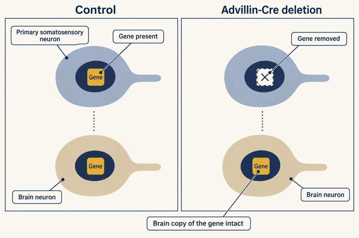
Timing supplies another control. An inducible Advillin-CreERT2 version began deletion at P28 rather than during development.
Predict. If the tactile and anxiety-like outcomes have the same developmental requirement, what should P28-induced deletion do to both?
Check your prediction
They should change together. Instead, the tactile deficits still appeared while the anxiety-like phenotype did not. The paper calls this “adult deletion,” but P28 is the induction age to keep. The two outcomes therefore have different timing requirements in this experiment.
Build the terminal mechanism as an evidence ladder
The deletion locates a contribution; it does not yet say what changed inside the neuron. The authors tested a candidate control point at the neuron’s central ending. A low-threshold mechanoreceptor, or LTMR, is an afferent that responds to innocuous mechanical stimulation.
First came anatomy. GABRB3-containing GABA-A receptors localise to presynaptic compartments of A-beta- and A-delta-LTMR terminals in mouse dorsal horn. In sensory-neuron Mecp2 mutants, GABRB3 puncta associated with vGlut1-positive terminals fell by about 70 per cent. A punctum is an immunolabelled cluster in fixed tissue. The count supports loss of receptor marker near terminals; it is not a conductance measurement and does not say that every terminal lost every receptor.
Predict. Does about 70 per cent fewer receptor-associated puncta establish about 70 per cent lower GABA-A conductance?
Check your prediction
No. Puncta are counted marker clusters in fixed tissue; conductance is an electrical measurement. The imaging result does not provide a conversion between them.
Separate physiology then tested release. In P14-to-P19 slices, dorsal-root stimulation evoked more asynchronous quantal excitatory postsynaptic currents (EPSCs) in postsynaptic dorsal column neurons after sensory-neuron deletion of Mecp2 or Gabrb3. Frequency rose without a corresponding amplitude change, supporting a presynaptic change.
The second physiological readout tested the GABA-A pathway. Because these afferents hold chloride high, GABA-A receptor activation depolarises them (synaptic integration explains why the gradient sets the sign). That depolarisation can travel backward into a dorsal root as a dorsal root potential, a readout of primary afferent depolarisation. Evoked dorsal root potentials were greatly diminished in isolated cords after sensory-neuron deletion of Mecp2 or Gabrb3.
Put the observations together: less receptor-associated marker, more afferent-evoked quantal release and less GABA-A-dependent primary afferent depolarisation. The authors attributed the touch phenotype to deficient GABA-A-mediated presynaptic inhibition.
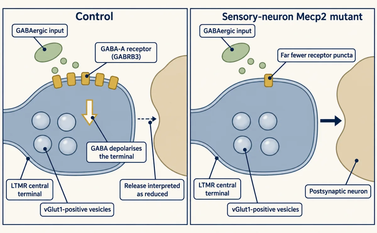
Now refine that useful first model. Depolarising is not identical to exciting. The 2016 study interprets GABA-A depolarisation as reducing action-potential-evoked release. Later work in proprioceptive afferents instead places axonal GABA-A receptors mainly near nodes and branch points, where they aid spike propagation, and assigns inhibition of release to GABA-B receptors (Hari and colleagues, 2022). Keep the presynaptic-inhibition account attached to these LTMR experiments rather than treating it as settled physiology for every sensory afferent.
The 2019 follow-up supplied a counterexample. Large-diameter dorsal-root-ganglion neurons cultured for one to two days from 6- to 8-week-old Shank3 mutants had reduced Ih, the hyperpolarisation-activated current, and greater excitability. No altered GABRB3 expression was detected at Shank3 LTMR central terminals. Two peripheral routes already reach one endpoint; the second film adds the developmental and spinal map.
Read the transcript of “One Touch Behaviour, Several Routes”
Film transcript: One Touch Behaviour, Several Routes
Chapter one located one contribution in the first touch neuron without making it the whole story. If several mice over-react to the same puff, did the same circuit defect cause it? Keep the colour map: gold sensory neuron, teal dorsal horn, blue brain. Start at the gold neuron's terminal inside the teal dorsal horn. In Mecp2 and Gabrb3 models, evidence supported reduced GABA-A-receptor-dependent presynaptic inhibition. Receptor-associated puncta fell, afferent-evoked quantal release rose, and dorsal root potentials shrank. Reset the pathway. The Shank3 model did not show that terminal GABRB3 change. Cultured large sensory neurons instead had reduced Ih and greater excitability.
The Shank3 model therefore reached a similar tactile endpoint through a different measured defect. The same tactile endpoint can arise from different defects in these mouse models. Mecp2 and Gabrb3 models support a reduced presynaptic GABA-A inhibition account. The Shank3 model instead showed reduced Ih and greater sensory-neuron excitability. Mechanism is only one axis.
Slide a timeline beneath four mouse-model lanes, from postnatal day four to adult testing. Every lane later shows a tactile abnormality. Must every adult tactile phenotype already be present at postnatal day four? No. Gabrb3 and Mecp2 loss-of-function pups were already more reactive at P4. Nlgn2 and Rorb mutants were not, although both showed tactile abnormalities as adults. Two sampled ages separate early from later onset, but do not identify the onset day. Nlgn2 also changed the locus. Deleting it from dorsal-horn neurons was sufficient for adult tactile alterations.
Emphasise the teal spinal block, not the gold afferent. Across these models, early reactivity tracked selected later behaviours, but that association did not prove causation. Locus, mechanism and developmental timing must be tested separately. A shared mouse movement cannot choose among them. Conditional genetics locates a sufficient contribution. Physiology tests a mechanism. Measurements at different ages map a schedule without turning association into a human developmental rule. The map explains why one endpoint cannot name its cause. It does not turn air-puff movement into perception, or mouse development into a human window. Those translation questions need their own measurements.
From the film
- The same tactile endpoint can arise from different defects in these mouse models.
- Mecp2 and Gabrb3 models support a reduced presynaptic GABA-A inhibition account.
- The Shank3 model instead showed reduced Ih and greater sensory-neuron excitability.
- Locus, mechanism and developmental timing must be tested separately.
What the route map did not show: the causal control
Orefice and colleagues also asked whether activating peripheral GABA-A receptors could change the measured response. Isoguvacine is a GABA-A agonist. At the assayed high dose and sampling conditions, it was detected in blood and liver but not in brain or cerebrospinal fluid. That is non-detection at stated limits, not proof that no molecule can ever cross. Low acute doses reduced tactile over-reactivity in five genetic models and one maternal-immune-activation model, without the sedation reported for the brain-penetrating comparator midazolam in those assays.
The decisive control was genetic. In anaesthetised mice, subcutaneous isoguvacine reduced stimulus-evoked A-beta-LTMR firing recorded from the L4 ganglion.
Predict. What should happen to this drug effect after homozygous Gabrb3 deletion in sensory neurons?
Check your prediction
It should disappear if the drug acts through GABA-A receptors in those neurons. It did: isoguvacine no longer changed touch-evoked spiking. A receptor deletion in the neuron removed the drug’s effect on that neuron.
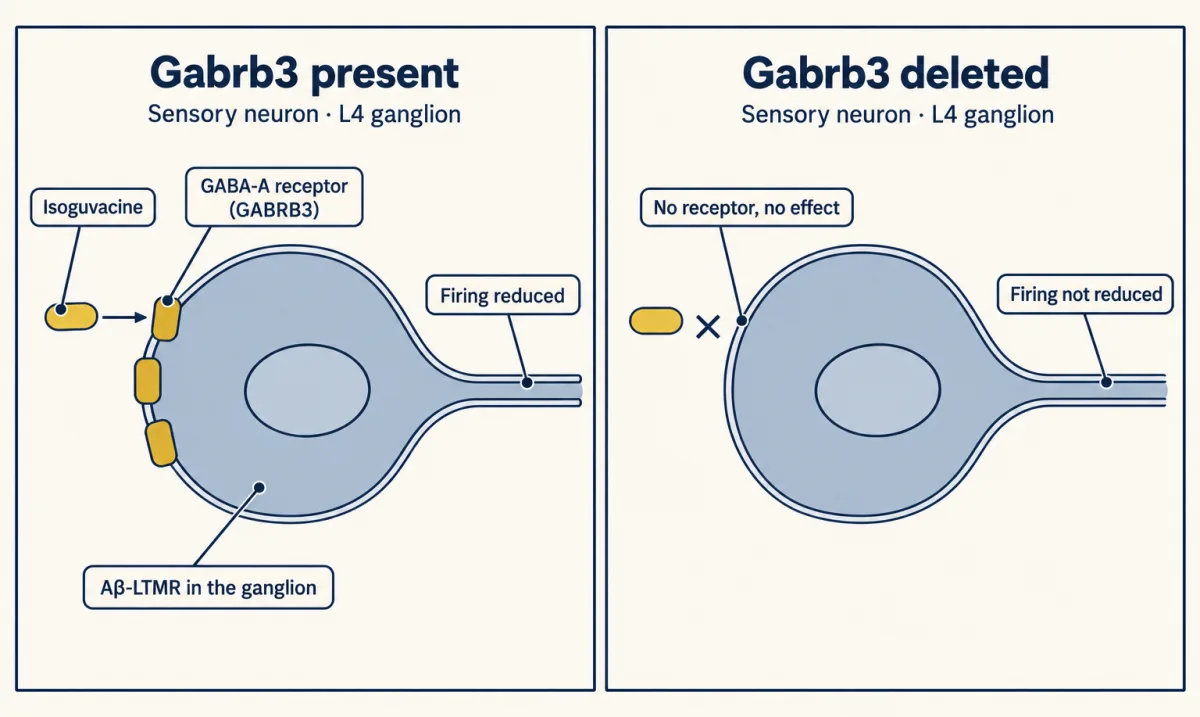
Chronic dosing drew a second boundary. Litters from Mecp2R306C or Shank3B heterozygous dams received 2 mg/kg daily from P1 to P42, with testing from P42. Each litter contained mutant and control pups, and the whole litter received the same treatment. Body condition, some brain abnormalities, anxiety-like behaviours and some social impairments improved. Texture discrimination and memory deficits persisted, as did Mecp2 motor impairment and Shank3 overgrooming. These are selective effects on mouse endpoints, not a general rescue.
Timing requires two different designs
The P28 experiment intervened on when gene loss began. Tasnim and colleagues (2024, Nature Neuroscience, Ginty corresponding author, Orefice a coauthor) asked a different question: which models already show the tactile phenotype at P4?
Gabrb3 and Mecp2 loss-of-function pups were more reactive to an air puff at P4, measured as maximal body displacement within 500 milliseconds. Nlgn2 and Rorb mutants were not, although both showed tactile abnormalities as adults.
Nlgn2 sits in a later spinal system. Deleting it from dorsal-horn neurons with Lbx1-Cre was sufficient for adult tactile alterations.
Predict. If Nlgn2 loss weakens dorsal-horn synaptic inhibition, what should miniature inhibitory currents show at P19 to P21?
Check your prediction
Their frequency and amplitude should fall. In P19-to-P21 Cdx2-Cre Nlgn2 mutants, deleted in all cells below cervical level 2, both measures were reduced in the dorsal horn.
Sampling two ages does not identify the onset day, although it does separate early-onset from later-onset models. Across the models tested, neonatal over-reactivity was associated with adult anxiety-like and social differences, while later-emerging over-reactivity was not, and the authors treat that dissociation as their central result. That is an association across genotypes, not proof that the early tactile state causes the later behaviours. These mouse results do not establish corresponding human windows.
Candidate control sites are compatible with the timing split. GABRB3 immunoreactivity was detectable near VGLUT1-positive sensory terminals at P0 to P1. Gabrb3 deletion virtually abolished GABA-evoked depolarisations in medium- and large-diameter dorsal-root-ganglion neurons at P4 and P18 to P30. Deep dorsal-horn miniature inhibitory currents were smaller and rarer at P4 than at P19 to P21, and feedforward inhibition in the LTMR-recipient zone was predominantly glycinergic by P19. These observations do not by themselves prove why each model starts when it does.
Why timing can matter is developed in sensitive periods in behaviour and critical period control.
What this does and does not mean for people
The short reminder is: test locus, mechanism and timing separately. In these models, part of a tactile phenotype and selected accompanying behavioural measures can begin outside the brain. That does not make autism a peripheral condition. The manipulations are mouse sufficiency tests, the behaviours are operational assays rather than experiences, and the rescue experiments left major features unchanged.
Human measurable differences are task-specific, run in both directions and vary between people more than a group mean conveys. Human peripheral data are not absent, but they do not bridge the mechanism. In skin biopsies from 32 autistic men, intraepidermal nerve-fibre density was lower than in controls, 5.53 against 11.13 fibres per millimetre, and about half met a criterion for skin denervation (Chien and colleagues, 2020). Those are small fibres, not the A-beta- and A-delta-LTMRs central to the mouse work.
No peripheral GABA-A therapy for tactile over-reactivity in autism has been approved. Isoguvacine remains experimental: a 2025 review reported no documented human administration, and a registry search found no registered studies, though that cannot exclude other registries or unregistered work. The 2019 authors described peripheral GABA-A intervention as a strategy requiring tests in patients and disclosed pending patents on peripherally restricted GABA-A drugs for that use.
What would move the field is a human measurement with some of the cellular specificity supplied by the mouse genetics. Chronic pain and spinal pain circuits face the same gap from the other side.
Design problem
Test whether early tactile over-reactivity causes later anxiety-like behaviour in mice, or merely predicts it.
Saved in this browser only.
One way to do it
Use a tamoxifen-inducible sensory-neuron driver to delete Gabrb3 in defined windows: before P4, from P28 and in adulthood. In a parallel arm, restore it in the same windows on a mutant background. Include tamoxifen-treated Cre-negative and untreated Cre-positive littermates, because inducible drivers can recombine without ligand to an extent that varies with driver, tissue and age. Tools for development covers the required Cre controls.
Measure air-puff reactivity at P4 and the later anxiety-like and social assays in the same animals. Each animal then supplies both ends of the association. Add dorsal-horn Nlgn2 deletion as a later-locus contrast: if the adult outcome follows the early tactile state rather than the gene, that arm should separate them.
Genotype and induction window are assigned per animal, so analyse per animal with litter as a clustering factor. Keep litter as the treatment unit only where whole litters are dosed, as in the chronic isoguvacine experiments. Report cells and animals separately wherever physiology enters. State that this mouse design does not establish a human counterpart of either window.
Going deeper
Human psychophysics beyond the mean
Puts and colleagues delivered vibration to the second and third digits of the left hand. The dynamic ramp used 25 hertz rising at 2 micrometres per second. They also measured amplitude and frequency discrimination. Baseline amplitude discrimination was worse in the autistic cohort, while frequency discrimination showed no significant diagnosis effect.
The static thresholds were 6.59 plus or minus 2.81 micrometres in the typically developing cohort and 9.13 plus or minus 4.81 micrometres in the autistic cohort, reported as mean plus or minus standard deviation. The standard deviations are large beside the 2.54-micrometre difference.
difference = 9.13 micrometres minus 6.59 micrometres = 2.54 micrometres
ratio = 9.13 divided by 6.59 = 1.385
relative increase = 2.54 divided by 6.59 times 100 = 38.5 per cent
Other studies point in other directions: lower thresholds at 200 hertz but not 30 hertz in adults with Asperger syndrome (Blakemore and colleagues, 2006), and heightened forearm vibration and thermal-pain sensitivity with normal light-touch thresholds (Cascio and colleagues, 2008). Threshold findings vary with frequency, body site and age, so the Puts result is one result among inconsistent ones.
Recording details behind the timing map
The GABA-evoked depolarisations at P4 and P18 to P30 were recorded with a high-chloride internal solution, which sets the driving force the experimenter wanted to see. In the P19-to-P21 dorsal-horn miniature-current comparison, seven cells per genotype came from two mice per genotype. Cells from one mouse share genotype, litter, preparation and day, so seven cells are not seven independent animals.
Isoguvacine pharmacology and dosing
The half-maximal effective concentrations of isoguvacine were 19.7 micromolar at recombinant human alpha-one beta-three gamma-two receptors and 18.7 micromolar at alpha-two beta-three gamma-two receptors expressed in HEK293T cells. These are in-vitro receptor measurements, not tissue exposure targets.
After 20 mg/kg intraperitoneally in male mice aged 8 to 12 weeks, liquid chromatography–mass spectrometry detected isoguvacine in blood and liver but not brain or cerebrospinal fluid. Detection limits were 640 picomolar in fluids and 16 nanomolar in tissue. The acute behavioural dose was 2 mg/kg. A ClinicalTrials.gov search for isoguvacine on 27 October 2026 returned no registered studies.
The embryo preparation
In E18.5 exteriorised embryos, optogenetic activation of forepaw Ret-positive A-beta-LTMRs evoked greater movement in Gabrb3 heterozygotes than in controls: 11 control and 9 heterozygous embryos. That is evoked movement in an embryo preparation, not a prenatal test of anything else.
Your notes
Select any text in the lesson to highlight it or add a note to it. Your highlights and notes are saved in this browser and collected in your notebook.
This browser is not letting the site save anything (a private window or strict privacy settings), so highlights and notes will be lost when you leave the page.
How we measure it
Vibrotactile psychophysics
A controlled vibrating probe on the finger, with a staircase or ramp, estimates a detection threshold in micrometres and discrimination performance. It measures what a participant can report in a laboratory task, which is a different quantity from how a stimulus feels in daily life.
Conditional deletion and selective restoration
A floxed allele plus a sensory-neuron Cre driver removes a gene from primary somatosensory neurons; a tamoxifen-inducible CreERT2 version sets when. The reverse design restores a gene only in those cells on an otherwise null background, which tests sufficiency of that cell population for a given outcome.
In vivo dorsal root ganglion recording
Multi-unit recording from a ganglion in an anaesthetised mouse reports how many spikes mechanical stimulation of the skin evokes in peripheral afferents. Combined with a conditional receptor deletion, it tests whether a drug's effect runs through the receptor in those neurons.
Ex vivo spinal cord electrophysiology
Dorsal root potentials in an isolated cord measure back-propagating primary afferent depolarisation, and whole-cell recordings in slices measure miniature inhibitory currents at a chosen age. Both report synaptic function in tissue, not perception.
Practice questions on this lesson (3) → MCAT-style, original, each with an explanation
Where this lesson fits
Before thisReview first
- The Low-Threshold Mechanoreceptors: One Touch, Several SignalsThis course · Lesson 4
The lesson works with A-beta and A-delta low-threshold mechanoreceptors, whose classes and responses are introduced there.
- Where Touch Is First Processed: Dorsal Horn and Dorsal Column NucleiThis course · Lesson 8
You need its account of presynaptic control of touch afferent terminals to follow the GABA-A inhibition mechanism here.
- Five Ways to Turn a Stimulus into a CurrentThis course · Lesson 1
The lesson assumes you know how a sensory neuron turns a stimulus into a current.
After thisStudy next
- When Pain PersistsThis course · Lesson 14
It applies the same locus, mechanism and timing questions to touch that turns painful after nerve injury.
- Spinal Circuits That Gate PainThis course · Lesson 12
It uses spinal inhibitory gate failure, the same kind of defect that lets touch signals escape control here.
Elsewhere on the siteFilms, simulations and method films in other courses
- What Can a Label Prove?Film · Developmental Neuroscience
It shows what a conditional Cre-based label or deletion can and cannot prove about where a gene acts.
- Conditional mouse builder: where does Cre act?Simulation · Developmental Neuroscience
It lets you see where Cre acts in a conditional mouse, the logic behind the peripheral-versus-brain deletion here.
Suggested by an AI model from the lessons themselves, then checked. How we use AI
Finish line
Test yourself
8 questions
Practice and MCAT-style, with explained answers.
Next · Lesson 10
Hot, Warm, Cool and Cold
Mice lacking the cold-and-menthol receptor TRPM8 failed completely to learn to report forepaw warming in a 10-day task. Why would a cold receptor matter for warmth?
Continue →← Lesson 8 · Where Touch Is First Processed: Dorsal Horn and Dorsal Column Nuclei