Sensory Systems · Unit 2: Touch
Where Touch Is First Processed: Dorsal Horn and Dorsal Column Nuclei
Rewritten on : new text, flashcards and film. Your notes and highlights stay, but the text around them may have moved.
The brainstem can hear about one touch of the paw twice: directly, and again by a route through the spinal cord that, in a simple conduction model, arrives a few milliseconds later. What does the second report add?
Pause when the reading reaches a film, and offer to play it. Saved on this device.
Watch the film now, or keep listening
Builds on:The Low-Threshold Mechanoreceptors: One Touch, Several SignalsSensory Neurons by Their Genes: the Atlas EraWhy these?
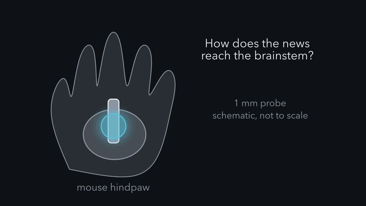 ▶ Watch Two Routes, One Patch of Skin · 2:19
Read the transcript of “Two Routes, One Patch of Skin”
Film transcript: Two Routes, One Patch of Skin
Press the skin of a mouse's hindpaw. How does the news reach the brainstem? The textbook draws one route. There are two. Start with the textbook drawing. A touch afferent, an Aβ low-threshold mechanoreceptor, enters the spinal cord. Its main branch climbs the dorsal column on the same side. It reaches the gracile nucleus in the brainstem with no synapse on the way. Call this the direct route. Now look below that drawing. The same afferent also sends branches into the dorsal horn, a layered circuit. Those branches drive local interneurons. Through them, and in places directly, they drive a spinal neuron whose own axon climbs the same dorsal column.
This is a postsynaptic dorsal column neuron, or PSDC neuron. It is driven in the dorsal horn and projects to the brainstem. Recordings in cats described these neurons by the early 1980s. Mouse work adds genetic access, and a test of each route. So draw a second line, in gold, from the afferent through the circuit and up to the gracile nucleus. Pause and predict. Does the gold line end on the same brainstem neurons as the blue line?
For thalamus-projecting gracile neurons, yes. The two inputs had highly aligned receptive fields. Both reported the same patch of skin. But the two reports are not the same. The direct route keeps precise timing. The indirect route reports sustained pressure, up to high forces. That is the idea for this lesson. Two routes, one patch of skin. One report keeps the timing. The other is rebuilt in the cord, not copied.
The limits: these are mouse studies, mostly in anaesthetized adults, and the dorsal column route only. The takeaway: one touch, two routes, two different reports on the same skin.
From the film
- A touch on the mouse hindpaw reaches the gracile nucleus by two routes: the afferent's own axon, and a route through the dorsal horn.
- For thalamus-projecting gracile neurons, the two inputs had highly aligned receptive fields, so both report the same patch of skin.
- The direct route keeps precise timing, while the indirect route reports sustained pressure, up to high forces.
When the skin of a mouse’s hindpaw is pressed, the brainstem can hear about it twice. Once from the touch afferent’s own axon, which climbs the spinal cord without a synapse. And once from a neuron in the spinal cord that has already worked on the signal.
What does the second report add? Three mouse studies, from 2017 and 2022, answer the question.
Two routes on one drawing
Touch and pain draws the dorsal column-medial lemniscus pathway as a blue line. The first-order axon ascends on the same side and synapses in the gracile or cuneate nucleus (together, the dorsal column nuclei, DCN). The second-order axon crosses in the medulla and reaches the ventral posterolateral thalamus (VPL). That route is correct. This lesson starts below the drawing, in the cord, and follows touch from the hindlimb to the gracile nucleus.
Picture the cross-section of the cord again: the grey butterfly, with the dorsal horn at the top and the white-matter dorsal column above it. The touch afferents that matter here are Aβ low-threshold mechanoreceptors (Aβ-LTMRs; the LTMR classes are sorted in the LTMR lesson). As each one enters, it does two things.
- The direct route. Its main branch climbs the dorsal column to the gracile nucleus with no synapse on the way. Keep drawing this in blue.
- The indirect route. Other branches end in the dorsal horn, a layered region where what happens is a circuit, not a handoff. There the afferent drives local interneurons and, through them and in places directly, a spinal neuron whose own axon climbs the same dorsal column to the same nucleus. Draw this in gold.
Other LTMR classes have no direct branch to the gracile nucleus; they can contribute through spinal circuits.
That spinal neuron is a postsynaptic dorsal column (PSDC) neuron: a spinal neuron that receives input in the dorsal horn and sends an axon up the dorsal column to the DCN. PSDC neurons are not a new discovery. Recordings in cats described their complex receptive fields by the early 1980s. What the mouse work adds is genetic access, and a test of what each route carries.
In Touch and Pain the blue line was one axon to the medulla. Below that drawing there are two.
The one idea to carry through this lesson. Two routes, one patch of skin. A touch reaches the gracile nucleus by two routes. The direct route is the afferent’s own axon, and it keeps precise timing. The indirect route runs through the dorsal horn, where local circuits rebuild the signal; in the illustrative model below (equal cord-to-brainstem paths, matched final synapses, a PSDC axon no faster than the afferent) it arrives a few milliseconds later, and it reports sustained pressure, up to high forces. The two reports describe the same patch of skin and meet on single brainstem neurons.
The lesson builds it in three steps, then marks its limits: the experiment that showed what each route carries, the arithmetic of what a slower route costs, and what the dorsal horn does to the signal on the way.
Predict. You can record from one gracile neuron while you touch the paw, and both routes feed it. How would you find out what each route contributes?
Check your prediction
Take one route away at a time and see what the neuron loses: silence the afferent terminals in the gracile nucleus, or block the spinal synapses. The next section shows how that was done.
The key experiment: silence one route at a time
Read the transcript of “Silence One Route”
Film transcript: Silence One Route
Two routes feed one gracile neuron. How can you tell what each one carries? Take one route away at a time, and watch what the neuron loses. Turecek, Lehnert and Ginty recorded single gracile neurons in the hindlimb region of anaesthetized adult mice. They identified neurons that project to the thalamus. Stimulating their axons there sent spikes backward, checked with a collision test.
Switch one removes the direct route. Light on the gracile nucleus silences the afferent terminals there, through a light-gated chloride channel. Switch two removes the indirect route. Drugs on the lumbar cord block its spinal synapses. The afferents’ direct branches climb past without a spinal synapse, so they keep conducting. Then three stimuli on the hindpaw. A vibration. A gentle displacement of the skin. And a sustained press with a blunt probe. Pause and predict. Which route carries vibration? Which carries a hard, sustained press? And gentle displacement?
Vibration first. The direct input carried vibration with high temporal precision, up to high frequencies. The indirect route can add to slow, 10 hertz vibration. Now the press. Blocking the spinal synapses almost eliminated sustained firing in most neurons. The block also raised the force needed to make the neuron fire at all. So the indirect route is not just a late add-on.
Gentle displacement used both routes. So direct for vibration, indirect for light touch, is too simple. Now map each input's receptive field on one neuron. The direct and indirect fields were highly aligned. So the two routes are not two maps. They are two reports on one spot, meeting on one neuron.
One limit. The light silencer was validated only for gentle forces. So the high-force result rests on the drug. And that drug is not specific to PSDC neurons. And strong firing at high force is not by itself evidence of pain. In awake mice, the high-force press evoked no pain-related behaviour. The takeaway: fast timing rides the direct route. Sustained pressure rides the indirect one. Gentle touch uses both, and both report the same skin.
From the film
- Silencing one route at a time showed that the direct input carries vibration with high temporal precision.
- The indirect route carries sustained pressure, and gentle displacement uses both routes.
- The high-force result rests on a drug that is not specific to PSDC neurons, so it carries that limit.
Turecek, Lehnert and Ginty (Nature, 2022) recorded single neurons in the hindlimb part of the gracile nucleus of adult, urethane-anaesthetized mice. They picked out the neurons that project to the thalamus (VPL) by antidromic activation: stimulating their axons there and testing for collision with a spontaneous spike, which proves the projection.
Then they took one route away at a time.
- To remove the direct route, they silenced afferent terminals reversibly with a light-gated chloride channel, ACR1. The light was aimed at the gracile nucleus, sparing the afferents’ branches in the cord.
- To remove the indirect route, they applied glutamate receptor antagonists to the lumbar cord. That blocks the spinal synapses that drive PSDC neurons, while the afferents’ ascending branches, which have no synapse on their way up the dorsal column, keep conducting.
They tested three kinds of stimulus on the hindpaw: vibration, gentle displacement of the skin, and sustained indentation with a blunt probe 1 mm across, at forces from 1 to 300 mN.
Predict. For each stimulus, which route do you expect to carry it? Use what you know: the direct route has no synapse before the brainstem, and the indirect route passes at least one synapse in the cord (two, through an interneuron).
Check your prediction
| Stimulus | Direct route (afferent axon) | Indirect route (through PSDC neurons) |
|---|---|---|
| Vibration | Carried, with high temporal precision; it underlies the high-frequency tuning | Can contribute to low-frequency (10 Hz) vibration responses |
| Gentle displacement | Contributes | Contributes |
| Sustained indentation, up to high force | Not enough on its own (see the next column) | Carried: with the lumbar synapses blocked, leaving only the direct route, sustained-indentation firing almost disappeared in most units |
If you gave fast vibration to the direct route, you were right, and the next section shows why a relay could struggle to keep that timing. If you gave all light touch to the direct route, the data say otherwise: both routes contributed to gentle displacement, so “direct means vibration and indirect means light touch” is too simple. Blocking the cord also raised the onset threshold, the force needed to make the neuron fire at all, so the indirect route is not only a late add-on.
Now the result that gives the lesson its idea. On single VPL-projecting neurons, the direct and indirect inputs had highly aligned receptive fields (a receptive field is the patch of skin whose touch drives the neuron). These neurons had a small excitatory field surrounded by a larger suppressive region, where touch reduced their firing. So the two routes are not two maps but two reports on one spot, meeting on one neuron.
One limit, said once. ACR1 silencing was validated only for gentle stimuli, about 20 to 30 mN or less, so the high-force conclusion rests on the lumbar drug, and that drug is not specific to PSDC neurons.
What a slower route costs
Read the transcript of “Why Later Is Fine, and What the Cord Does”
Film transcript: Why Later Is Fine, and What the Cord Does
In a simple model, the indirect route arrives later. What does a slower report cost, and what does it add? Take illustrative numbers for a mouse hindpaw. In this model, the indirect input arrives about 4.5 milliseconds later. That lag is a model, not a measurement, and its size is uncertain. With equal paths from cord to brainstem, matched final synapses and a PSDC axon no faster, the spinal synapses make it later. Pause and predict. Does a lag of a few milliseconds matter more for a long press, or for fast vibration?
For the steady firing during a 300 millisecond press, it may matter little. The lag is a sliver at the start, though onset timing is a separate question. At 500 hertz, one cycle lasts 2 milliseconds. The lag spans more than two whole cycles. A fixed delay does not blur locking on its own route. Every spike is equally late, so the spikes still share one phase. That phase shifts, by a quarter cycle here. What blurs timing is jitter. In this toy model, each synapse adds random scatter to when the next spike fires. With assumed jitter, two synapses nearly erase locking at 500 hertz. At 50 hertz, locking barely changes. That model is not the paper's explanation.
But it shows how relay jitter could explain why fast timing rides the route with no relay. Now a second question. What does the cord do to the signal? PSDC neurons get most of their excitation from local interneurons. single dorsal horn neurons received input from both low-threshold and high-threshold mechanoreceptors. Two kinds of inhibition shape the output. In feedforward inhibition, the afferent excites the PSDC neuron and an inhibitory interneuron. That interneuron then inhibits the PSDC neuron. Here, Rorβ interneurons supplied it, using glycine. Presynaptic inhibition lands on the afferent terminal itself, and scales down the transmitter it releases.
Pause and predict. Remove either kind of inhibition. Does the PSDC receptive field shrink, or grow? It grows. And losing presynaptic inhibition left neurons firing after the indentation ended. These were chronic knockouts, recorded under anaesthesia. And no study here recorded these neurons in an awake animal. The takeaway: two routes, one patch of skin. One keeps the timing. The other, rebuilt in the cord, reports the pressure.
From the film
- In a simple model the indirect route arrives a few milliseconds later, but that lag is illustrative, not a measurement.
- A fixed delay does not blur a route's own timing; random jitter at each synapse could, which would cost fast vibration more than a sustained press.
- Losing feedforward or presynaptic inhibition enlarged the receptive fields of PSDC neurons in anaesthetized mice.
What does a later report cost? Start by estimating how much later.
Worked problem: how much later? Suppose a gracile neuron receives both inputs from a mouse hindpaw. These are illustrative numbers, not measurements. The model assumes constant velocity, conduction plus synaptic delay only (see the action potential lesson), one shared start at the paw, equal path lengths from cord to brainstem, and the same delay at each route’s final synapse in the gracile nucleus, which then cancels.
t = L / v
- Direct: 0.08 m at 16 m/s = 0.005 s = 5.0 ms.
- Indirect, paw to lumbar cord: 0.04 m at 16 m/s = 2.5 ms.
- Indirect, two spinal synapses at an assumed 1 ms each = 2.0 ms.
- Indirect, PSDC axon: 0.04 m at an assumed 8 m/s = 5.0 ms.
- Indirect total: 2.5 + 2.0 + 5.0 = 9.5 ms. Difference: 9.5 − 5.0 = 4.5 ms.
The indirect input arrives about 4.5 ms later in this model. Its size is not a measurement: other plausible path lengths, speeds and delays give lags from under 1 ms to about 12 ms (Going deeper). Its sign is firmer: the two routes make separate synapses onto the same gracile neuron, and under the stated assumptions, with a PSDC axon no faster than the afferent, the spinal synapses alone make the indirect input later. Whether two components that far apart can be told apart in a real recording depends on how much their timing varies and overlaps.
Predict. Take the 4.5 ms lag. Does it matter more for a 300 ms press, or for a 500 Hz vibration?
Check your prediction
For a readout of average firing during the plateau of a 300 ms press, probably little: 4.5 ms is 1.5 percent of the press, though that fraction says nothing about onset detection, integration with other inputs or perception. For vibration at 500 Hz, the highest frequency tested, one cycle lasts 1/500 s = 2.0 ms, so the lag spans more than two whole cycles (2.25).
A fixed delay does not blur one route’s own locking: if every spike is late by the same amount, the spikes still share one phase. That phase moves, though. At 500 Hz, 2.25 cycles shifts the locked phase by a quarter cycle (90°), which matters where the two routes meet on one neuron. What blurs locking is variability: in the model below, each synapse adds a little random jitter to when the next spike fires.
A toy model shows how much jitter could matter. Vector strength is a locking score: how tightly spikes lock to one phase of a vibration cycle, from 1 (every spike at the same phase) to 0 (no net phase preference, as when spikes spread evenly over the cycle, or split equally between opposite phases). If spike times scatter around one phase with a normal spread σ, vector strength = exp(−(2πfσ)²/2) at frequency f (f in Hz, σ in seconds). Assume the direct route has σ = 0.2 ms, and that each of two spinal synapses adds independent random jitter of σ = 0.5 ms. Independent spreads add as squares, so the indirect route has σ = √(0.2² + 0.5² + 0.5²) ≈ 0.73 ms.
| Vibration | One cycle | Vector strength, direct (σ 0.2 ms) | Vector strength, indirect (σ 0.73 ms) |
|---|---|---|---|
| 50 Hz | 20 ms | 1.00 | 0.97 |
| 300 Hz | 3.3 ms | 0.93 | 0.38 |
| 500 Hz | 2.0 ms | 0.82 | 0.07 |
The jitter values are assumptions, not Turecek and colleagues’ measurements, and relays need not add this much, so the model shows a possibility, not the paper’s explanation: if each spinal synapse adds even half a millisecond of independent jitter, locking at hundreds of hertz would be mostly lost on the relayed route, while locking at 50 Hz survives. It also fits some gracile neurons better than others. Most thalamus-projecting neurons entrained to vibration (fired in time with each cycle) only below 150 Hz, where the model predicts little cost; it was most inferior colliculus-projecting neurons, identified the same antidromic way, that phase-locked from 10 to 500 Hz.
Against a 300 ms press, the same 0.73 ms of scatter is small (0.25 percent), though small is not proven harmless. So for a plateau-rate readout the lag is probably cheap, and what the indirect route adds is what it carries. Whether onset timing or the relative arrival times matter is open.
Turecek and colleagues did not measure the lag; they measured something more telling, what each route carries (the table in the last section). Try the arithmetic in the sketch below.
Left and right are the mouse’s own. Everything below the medulla is on the stimulated paw’s side; nothing crosses in the cord. Only the second-order axon crosses, in the medulla. The direct and indirect afferents are drawn as separate lines for clarity.
- Direct, t = L / v
- Indirect: afferent + synapses + PSDC axon
- Indirect minus direct
In this sketch: illustrative lengths and velocities, not measurements; constant speed; conduction plus synaptic delay only; anaesthetized adult mouse logic. The rule to keep is t = L / v for each stage, then add the synaptic delays.
What the dorsal horn does to the signal
Start with one anatomical fact from the atlas in the next section: PSDC neurons received their excitatory input mainly from local interneurons of the zone where touch afferents end, rather than straight from the afferents. So most of what a PSDC neuron sends up is the output of a circuit, rebuilt rather than passed on.
Chirila and colleagues (Cell, 2022) asked what that circuit does to a touch signal. They recorded from the dorsal horn of adult, urethane-anaesthetized mice while the skin was indented, and sorted the responses into six functional profiles. Those are response clusters, not six cell types. They found convergence, one neuron receiving input from more than one kind of afferent: individual neurons received input from both low-threshold and high-threshold mechanoreceptors. High-threshold mechanoreceptors are afferents that respond only to stronger, often damaging, stimuli.
Two inhibitory motifs shaped the output of PSDC neurons.
- Feedforward inhibition. The same afferent input that excites a PSDC neuron also excites an interneuron that inhibits it, so excitation is followed by inhibition. Rorβ interneurons supplied glycinergic feedforward inhibition onto PSDC neurons.
- Presynaptic inhibition. Inhibition is applied to the afferent terminal, scaling down the transmitter it releases. PVi neurons, the parvalbumin-expressing inhibitory population, form axo-axonic contacts on afferent terminals and take part in this GABA-A receptor dependent inhibition, but they are not its only source. Chirila and colleagues removed it in all sensory neurons, not by targeting PVi neurons.
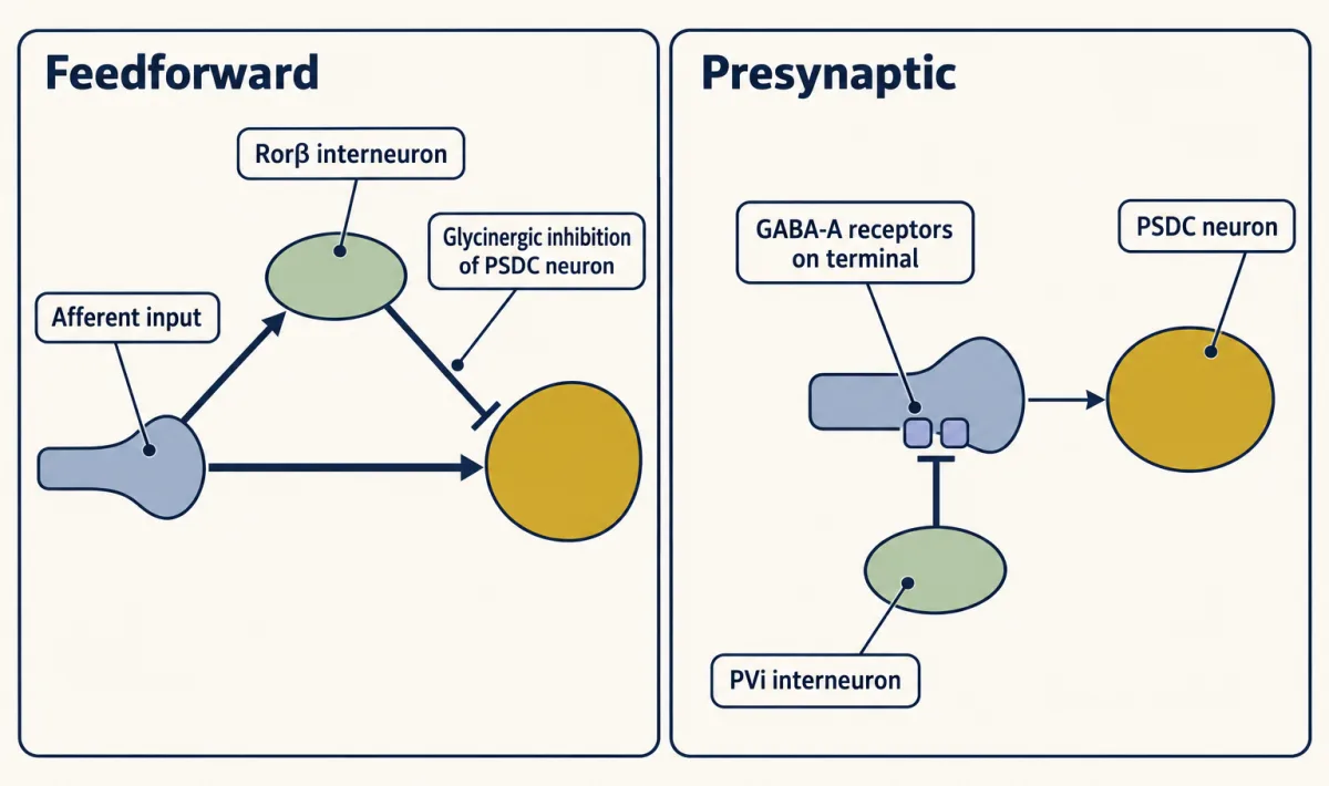
Predict. Remove either kind of inhibition. Will the PSDC neuron’s receptive field shrink or grow? And what might happen at the end of a step indentation?
Check your prediction
It grows. Disrupting either motif enlarged PSDC receptive fields: a larger field means touches farther from its centre now drive the neuron. Loss of presynaptic inhibition also produced after-discharge, continued firing after the end of a step indentation.
Disrupting feedforward inhibition also changed touch responses in primary somatosensory cortex (S1) of awake mice, and those mice showed tactile over-reactivity and a texture discrimination deficit. The study did not show that the dorsal column carried the cortical change. The limits are plain: the dorsal horn and PSDC recordings were made in anaesthetized mice, and both disruptions were chronic conditional knockouts (Vgat deleted in Rorβ neurons for feedforward inhibition, and the GABA-A β3 subunit gene deleted in sensory neurons for presynaptic inhibition), which cannot be assumed to isolate acute circuit function. The dorsal horn is therefore not a simple relay on the way to the dorsal column.
Who is in the zone: an atlas
Abraira and colleagues (Cell, 2017) asked an earlier question: which cells sit where touch afferents end? In the mouse lumbar cord, low-threshold mechanoreceptors (LTMRs) end in a band in the middle of the dorsal horn, the LTMR recipient zone (LTMR-RZ). It begins just below the band labelled by IB4, a marker of a set of nociceptor terminals.
They described seven excitatory and four inhibitory interneuron populations there. A reporter-defined population is one marked in a mouse line by a gene it expresses, which is a molecular label, not proof of a single cell type (genetic access is covered in tools for development). Two have already appeared, Rorβ and PVi; cholecystokinin (CCK) and Rorβ return in the texture test below.
Three facts set the scale. About 70 percent of the neurons intrinsic to the zone were excitatory and about 30 percent inhibitory. The reporter lines together covered an estimated 71 to 82 percent of LTMR-RZ neurons, so the atlas is incomplete. And the populations were not private lines. By counting pre- and postsynaptic marker apposition, each LTMR subtype assayed contacted four to eleven interneuron subtypes, and each interneuron subtype sampled one to three of the LTMR classes assayed (which does not exclude classes not assayed), so the zone mixes its inputs. The interneurons also received descending corticospinal input.
PVi and a second inhibitory population, Cdh3, formed axo-axonic contacts (synapses onto another neuron’s axon terminal) on sensory terminals, consistent with presynaptic inhibition of the afferents (see synaptic integration).
Did any of it matter for behaviour? The authors silenced spinal CCK or Rorβ lineages and tested a texture version of the novel object recognition test, in which a mouse that remembers an object explores a new one more. Both silenced groups lost the preference for the newly textured object.
Predict. A mouse that no longer prefers a novel texture might have lost touch. What else could it have lost, and what control would rule that out?
Check your prediction
It might have stopped exploring, or lost memory, motivation or movement. The control is the same recognition task with objects that differ in colour and shape rather than texture. The silenced mice kept the preference, so the deficit is not a general failure to explore or recognize objects.
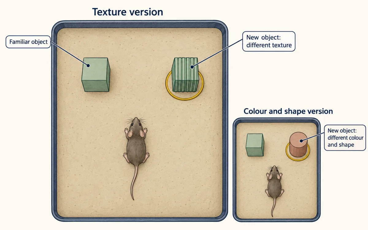
One limit to keep in mind: a lineage includes every cell that ever expressed the marker, so the silencing is not acute and not one pure type. It shows these lineages contribute to the task, not that each subtype is needed.
When inhibition fails
If inhibition holds back touch from other circuits, loss of inhibition could let it reach them. Duan and colleagues (Cell, 2014) tested that idea for spinal pain circuits in mice and found that dynorphin-lineage inhibitory neurons gated Aβ input away from somatostatin-lineage neurons needed for mechanical pain behaviours; spinal circuits that gate pain covers the work in full. This is a separate circuit from the PSDC experiments. An enlarged PSDC receptive field is not allodynia (pain from normally non-painful touch), and the blunt 300 mN indentations did not evoke pain behaviour in awake mice. A later lesson takes touch over-reactivity in autism models further.
Where the idea stops
Here is how far “two routes, one patch of skin” holds.
- Mouse hindlimb only. The evidence is from the mouse lumbar dorsal horn and gracile nucleus, recorded mostly in urethane-anaesthetized adults. There are no human data.
- The hindlimb and gracile example only. The cuneate nucleus also belongs to the dorsal column pathway but is not analysed here; the anterolateral and trigeminal systems are separate pathways.
- Rebuilt is not copied. The indirect route is not a delayed duplicate: it supports sustained-intensity coding into the high-force range that direct input alone did not reproduce: blocking lumbar transmission nearly abolished sustained responses in most recorded units. Both routes contribute to gentle displacement, so neither is a single-submodality channel.
- The direct route skips the dorsal horn, not processing. It still ends at a synapse in the gracile nucleus, and the thalamus-projecting neurons there have suppressive surrounds. Silencing the direct afferent terminals reduced that surround suppression, which points to direct input acting mainly through local inhibitory neurons of the nucleus; the full microcircuit was not resolved.
- What reaches the brainstem, not what the animal perceives. No study here recorded PSDC or gracile neurons in awake animals.
Design problem
Chirila and colleagues found that PSDC receptive fields grow when presynaptic inhibition is lost. Design an experiment to show that this matters for perception, and say what would be needed in people.
One way to do it
- Make the change acute. Remove presynaptic inhibition of the afferents acutely in adult mice, not by a developmental or germline deletion, which can change the cord’s wiring. Silencing PVi neurons alone would not do this, because they are one of several sources of axo-axonic contacts and also make ordinary synapses.
- Tie it to PSDC output. Add a perturbation or rescue restricted to PSDC output. A spinal drug blockade is not PSDC-specific, so a drug result must not be read as one.
- Record awake. Record PSDC neurons in awake mice, not under urethane, to check that the field enlargement survives without anaesthesia.
- Measure perception with a control. Measure tactile acuity behaviourally, for example discrimination of closely spaced textures, paired with a control that does not depend on texture, like Abraira’s colour and shape object test, so a general change in exploration is not mistaken for a tactile one.
- People. Mouse data do not transfer by default. In people, you would need psychophysical acuity measures and a way to change spinal inhibition safely, and a result would still be limited to that preparation.
Going deeper
Ages and preparations
The 2017 study used fixed tissue mostly from mice aged 30 to 35 days, spinal slices from mice aged 13 to 21 days, and behaviour in males aged 7 to 12 weeks. In the two 2022 studies the dorsal horn and gracile nucleus were recorded in adult mice under urethane anaesthesia: older than 6 weeks for the dorsal horn recordings of Chirila and colleagues, and older than 5 weeks, of both sexes, for the gracile recordings of Turecek and colleagues.
Where touch afferents end
Low-threshold mechanoreceptor (LTMR) terminals overlap in laminae IIiv to IV of the dorsal horn (laminae are the layers numbered from the surface; IIiv is the inner, ventral part of lamina II). In the mouse lumbar cord, the LTMR-RZ was sampled from the IB4 border to about 250 µm below it.
The interneuron populations and their firing patterns
The excitatory populations were PKCγ, Cbln2, NeuroD4, PVe, cholecystokinin (CCK), 5HTr6 and Igfbp5. The inhibitory ones were PVi, Kcnip2, Rorβ and Cdh3. Parvalbumin is not an inhibitory marker by default here: PVe is excitatory and PVi is inhibitory.
In spinal slices, the populations fired differently when current was injected. Cbln2 neurons burst at the start and PKCγ neurons spiked after a delay. Among inhibitory neurons, PVi cells fired tonically, Kcnip2 and Rorβ cells after a delay, and Cdh3 cells with gaps.
Counting inhibitory contacts
Abraira counted 2.9 ± 0.1 (mean ± SEM) axo-axonic inhibitory contacts per sensory terminal in fixed mouse tissue, of which 1.5 ± 0.1 were from PVi neurons. 1.5 / 2.9 = 0.52, so about 52 percent of the contacts. These are contact counts, not conductances, so they do not say PVi neurons supply half of the inhibition.
No core-shell map
Turecek and colleagues could not detect a clear core-shell organization in the mouse gracile recordings, so the split in this lesson is direct versus indirect route, not a map of submodalities across the nucleus.
How big is the lag?
Under the worked problem’s assumptions, the lag is L(cord to DCN) × (1/v(PSDC) − 1/v(afferent)) plus the number of spinal synapses times the synaptic delay. A sweep over illustrative mouse ranges (paw to cord 3 to 5 cm, cord to gracile nucleus 2.5 to 4.5 cm, afferent 10 to 25 m/s, PSDC axon 5 to 16 m/s and never faster than the afferent, one to three spinal synapses at 0.5 to 1.5 ms each), sampled on the grid paw to cord 5 cm, cord to gracile 4.5 cm, afferent 25 m/s, PSDC 16 m/s, synapses 3 at 1.5 ms, with every admissible combination weighted equally (810 combinations), gave lags from 0.5 to 11.7 ms, with a median of about 4.0 ms. The median is a property of that grid, not a biological estimate. Every lag was positive. These ranges are assumptions for illustration, not measurements.
The Duan experiment
Ablating somatostatin-lineage excitatory spinal neurons impaired mechanical pain behaviours, and dynorphin-lineage inhibitory neurons gated Aβ input from activating the somatostatin neurons (ablation in vivo; slice recordings from mice aged 23 to 30 days).
Your notes
Select any text in the lesson to highlight it or add a note to it. Your highlights and notes are saved in this browser and collected in your notebook.
This browser is not letting the site save anything (a private window or strict privacy settings), so highlights and notes will be lost when you leave the page.
How we measure it
Reporter lines and intersectional silencing
Mouse lines in which a genetic driver switches on a fluorescent reporter label cells that express a marker gene. Combining two drivers restricts the label, or a silencing tool such as tetanus toxin, to cells in the spinal cord. The result tells you which cells have the marker and, for silencing, which behaviours need that lineage, not that the marked cells are one pure cell type.
Whole-cell patch clamp in spinal slices
A glass pipette records voltage or current from a single labelled neuron in a thin slice of mouse spinal cord. Injecting current shows the cell's firing pattern, such as an initial burst, delayed spiking, tonic firing or gaps. The preparation is from young animals and cut off from most of its inputs, although some slices keep dorsal roots attached so afferents can be stimulated.
In vivo multielectrode recording with antidromic identification
Electrode arrays record spikes in anaesthetized mice while a motorized probe indents or vibrates the skin. Stimulating a neuron's output axon at a candidate target and checking that the evoked spike collides with a spontaneous one proves that the neuron projects to that target. This labels neurons by target, such as thalamus or inferior colliculus.
Reversible input silencing
A light-gated silencer (ACR1) in afferent terminals, or a glutamate receptor antagonist applied to the lumbar cord, removes one route's contribution to a recorded neuron while leaving the other route in place. Each tool has limits of efficacy and specificity that must be checked before reading the result.
Practice questions on this lesson (3) → MCAT-style, original, each with an explanation
Where this lesson fits
Before thisReview first
- The Low-Threshold Mechanoreceptors: One Touch, Several SignalsThis course · Lesson 4
This lesson starts from the Aβ low-threshold mechanoreceptor classes whose branches split into the direct and dorsal horn routes.
- Sensory Neurons by Their Genes: the Atlas EraThis course · Lesson 3
The reporter-defined interneuron populations and genetic access used here build on how sensory neurons are sorted by their genes.
After thisStudy next
- Spinal Circuits That Gate PainThis course · Lesson 12
It takes the dorsal horn inhibitory gating of Aβ input further, covering the Duan experiment this lesson only sketches.
- Touch Over-reactivity in Autism ModelsThis course · Lesson 9
It follows the loss of feedforward and presynaptic inhibition into tactile over-reactivity, which this lesson introduces.
Elsewhere on the siteFilms, simulations and method films in other courses
- Where Every Command EndsFilm · Neuroscience
It shows a sensory axon branching to act on several targets at once, as the touch afferent does here.
- Why Capsaicin Feels HotFilm · Neuroscience
It introduces touch pathways and the spinal cord, which this lesson works out in detail.
Suggested by an AI model from the lessons themselves, then checked. How we use AI
Finish line
Test yourself
8 questions
Practice and MCAT-style, with explained answers.
Next · Lesson 9
Touch Over-reactivity in Autism Models
In one experiment, deleting a gene from peripheral sensory neurons, but not from the brain, was enough to change a mouse's behaviour in anxiety-like and social assays.
Continue →