Genetics and Molecular Biology · Unit 2: Mapping and gene function
Complementation and Gene Function: From Beadle and Tatum to Benzer
Rewritten on : new text, flashcards and film. Your notes and highlights stay, but the text around them may have moved.
Two pure-breeding white-eyed flies can have red-eyed offspring. What does that tell you about their genes?
Pause when the reading reaches a film, and offer to play it. Saved on this device.
Watch the film now, or keep listening
Builds on:Mendel's Peas: Segregation and Independent AssortmentExtending Mendel: Dominance, Multiple Alleles and Gene InteractionLinkage and Recombination MappingWhy these?
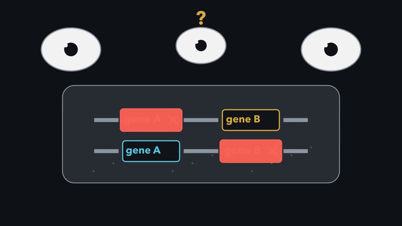 ▶ Watch When Two Mutants Rescue Each Other · 3:28
Read the transcript of “When Two Mutants Rescue Each Other”
Film transcript: When Two Mutants Rescue Each Other
How can two pure-breeding white-eyed flies have red-eyed offspring? Watch their chromosome copies enter one shared cell. Each parent carries a different hidden loss. The offspring eye begins white, waiting to see whether normal function returns. A red eye needs several genes working together. Some genes make pigments. Others move pigments into granules. A missing shared transporter leaves the eye white. So does blocking both pigment pathways.
Appearance alone cannot tell which gene broke. An allele is one version of a gene. A diploid cell carries paired chromosome copies, so it can carry paired alleles. For a recessive loss, one working allele usually makes enough product. That product works at its normal place in the cell. First check each mutation against wild type. If a working copy restores the ordinary phenotype, the mutation is recessive. That check matters because complementation reads rescue. A dominant mutation can keep the cell mutant beside any partner. Now combine the checked recessive mutations on opposite chromosome copies in one diploid cell. This arrangement is trans.
One chromosome has broken gene A and working gene B. The other has working A and broken B. Will this cell recover its red eye, or stay white? Follow the working copies, not the broken ones. Decide whether the shared cell now has every function it needs.
The working A copy supplies the blue function. The working B copy supplies the gold function. With both functions present in the cell, red pigment appears. Normal function returns, so the mutations complement. Reset the cell. Now both chromosomes carry broken alleles of gene A, while gene B works. No working A product appears. The eye stays white, so these mutations fail to complement under the simple model. Mutations that fail with one another form a complementation group. In the simple model, a group usually marks one gene. Testing many pairs turns one shared phenotype into groups, revealing how many functions the screen happened to hit. The rule has boundaries.
Intragenic complementation can rescue different alleles of one gene. Mutations in different genes can sometimes fail together. Those exceptions refine the test. They do not change what a clean recessive result usually means. Always name what the test measured. A red eye is observed rescue, not direct proof of chromosome position. Mapping asks where mutations sit. Complementation asks whether their functions can supply each other in one cell.
From the film
- For recessive mutants, rescue in trans usually means different genes.
- Check each mutation against wild type before interpreting a complementation test.
- Mutations that fail to complement form a group that usually marks one gene.
- Dominant mutations make the test uninformative, and known recessive exceptions can mislead it.
The first film built the test: check that each mutation is recessive, combine two in trans in one cell, read rescue. Here: the notation, sorting many mutants, and the limits.
The one idea. Rescue tells you whether two mutations share a function, and brackets a pathway block between supplied compounds.
Scope: complementation uses recessive loss-of-function mutations; pathway rescue assumes one linear route whose supplied compounds enter the cell and can be used. Rescue narrows where a defect lies; it does not name the broken reaction or map a chromosome position.
A recessive allele (an allele is one version of a gene) shows its effect only when no working allele is present, so a diploid is mutant only when both copies are defective: homozygous for one mutant allele, or a compound heterozygote carrying two different mutant alleles of one gene. An inactivating mutation is usually recessive because one working copy makes enough product. A gene’s place on a chromosome is its locus; linkage and mapping and three-point mapping locate loci.
The complementation test: put two mutations in one cell
In symbols, with recessive mutations in genes a and b, the trans diploid is a/+ ; b/+ (for linked genes, a + / + b). It is wild type: the mutations complement. Two alleles of one gene give a1/a2, with no working copy, so the diploid stays mutant. The white sweet peas in extending Mendel that produced purple hybrids show the same rescue. Mutations that fail to complement form a complementation group, which usually marks one gene.
Predict. Mutant 1 fails to complement mutant 3, and mutant 3 fails with mutant 5. If mutant 1 complements mutant 5, can the three results form one clean complementation group under the simple model?
Check your prediction
No. In a simple complementation group, every inside pair should fail. Recheck the crosses and recessiveness before invoking an exception.
Worked problem: how many genes? Six white-eyed mutants of an idealized fly (1 to 6) are crossed in all fifteen pairs, and the F1 is scored (+ is wild-type F1, − is white). Assume each mutation is recessive, autosomal and fully penetrant.
1x2 +, 1x3 −, 1x4 +, 1x5 −, 1x6 +, 2x3 +, 2x4 +, 2x5 +, 2x6 −, 3x4 +, 3x5 −, 3x6 +, 4x5 +, 4x6 +, 5x6 +.
- List the failures only: 1x3, 1x5, 2x6, 3x5.
- Start with mutant 1. It fails with 3 and 5, so {1, 3, 5} is a group. Check the inside pair: 3x5 fails, as it must if all three are in one gene.
- Mutant 2 fails only with 6: group {2, 6}. Check that neither 2 nor 6 fails with 1, 3 or 5. Neither does.
- Mutant 4 complements all five others: group {4}.
Three complementation groups, so at least three genes. “At least”, because other genes needed for red pigment may not have been hit in these six mutants.
Six recessive, autosomal, fully penetrant white-eyed mutants of an idealized fly, 1 to 6; the scores are the lesson’s worked problem. Click a cell to cross two of them and score the F1 diploid.
Mutations that fail to complement form a complementation group, which in the simple model is one gene; “at least” that many genes, because other genes needed for red pigment may not have been hit. Exceptions exist: intragenic complementation can make same-gene mutants look wild type, and non-allelic non-complementation can make different-gene mutants look white. This is a sketch of the logic, not to scale.
Now the limits the film only named. A dominant allele stays mutant opposite any partner, so its trans result is uninformative, and a dominant-negative product that spoils the wild-type product can falsely look like “same gene.” Recessive exceptions mislead rather than say nothing. In a protein built from several identical subunits, differently damaged subunits can sometimes rebuild a working enzyme: intragenic complementation. In human argininosuccinate lyase each active site holds residues from three subunits, which explains how. Conversely, mutations in different genes sometimes fail to complement.
Beadle and Tatum: one mutant, one blocked step
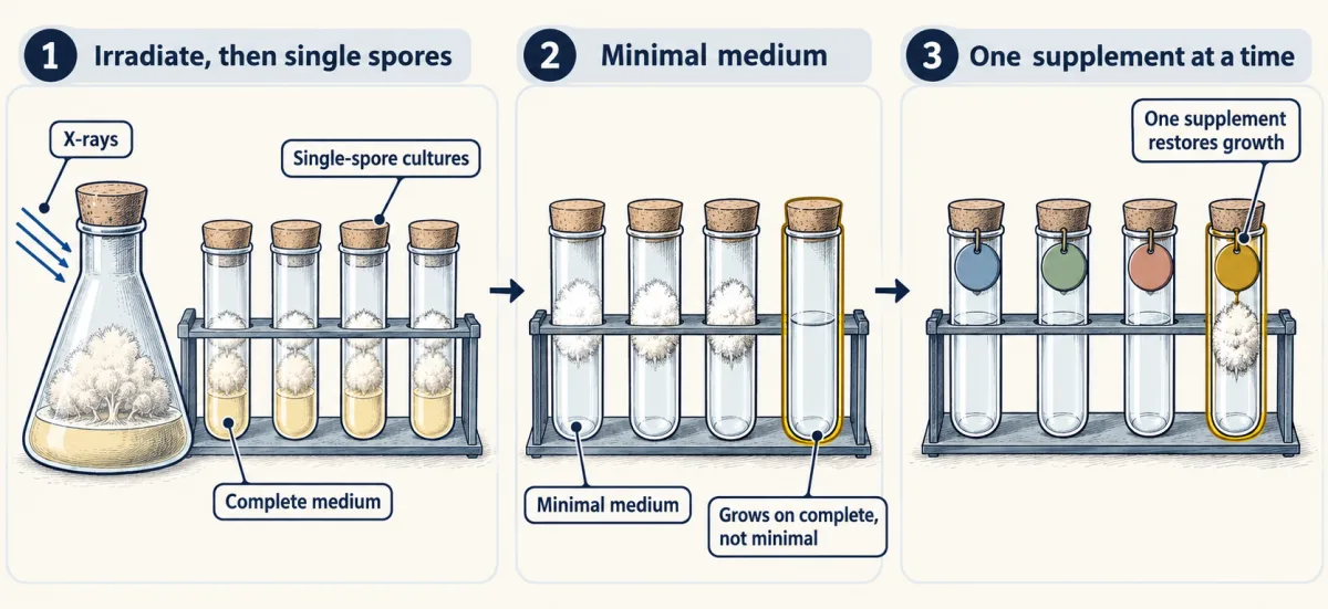
George Beadle and Edward Tatum turned the question around at Stanford: start with known chemistry, then find mutants that have lost part of it. The bread mold Neurospora grows on a simple minimal medium and makes its amino acids and most vitamins itself. Its growing phase is haploid, with one chromosome copy, so a recessive mutation shows at once.
- Irradiate the mold with X-rays. Beadle and Tatum irradiated fruiting bodies before meiosis, so each spore taken was haploid.
- Grow single-spore cultures on a complete medium, rich in amino acids and vitamins, so that a mutant unable to make one of them stays alive.
- Test each culture on minimal medium. Keep the ones that grow on complete medium but not on minimal.
- Add supplements one at a time to minimal medium. The single compound that restores growth names the synthesis that was lost. Then cross the mutant to wild type and check that the defect segregates as one gene.
Predict. A supplement restores one culture’s growth. Why must the researchers still cross that culture to wild type before calling the defect a single-gene mutation?
Check your prediction
Growth rescue identifies a missing synthesis, not its genetic basis. Segregation tests whether the inherited difference behaves as one gene.
Their 1941 paper reported three mutants among about 2,000 strains, needing pyridoxine (vitamin B6), the thiazole half of thiamine or para-aminobenzoic acid. The pyridoxine and thiamine mutants were Neurospora sitophila, and the para-aminobenzoic acid mutant N. crassa. Crosses of the pyridoxine mutant fit a single-gene difference, on what the authors called limited data. Beadle and Tatum shared the 1958 Nobel Prize with Joshua Lederberg.
A single supplement names one missing synthesis. The second film asks what the pattern of rescues across many mutants reveals: the order of the steps.
Read the transcript of “Reading a Pathway Backward”
Film transcript: Reading a Pathway Backward
The first film used one genome to supply what another lacked. Can a supplied compound reveal where a pathway is blocked? Watch one cell build a needed product through a chain of steps. Each arrow needs a gene-controlled function. A damaging mutation can remove one function and stop the flow. The cell then lacks the final product, even though every step after the block may still work. A nutritional screen makes that hidden block visible. Keep each mutant alive on complete medium, then test minimal medium. Adding candidate compounds separately asks which supplied material can restore the missing synthesis and growth. Add an upstream intermediate.
It enters before the broken arrow and reaches the same barrier. Nothing arrives downstream. The growth gauge stays dark. Failure to rescue tells us only that this supply did not bypass the block. Now add a downstream intermediate. It enters beyond the broken arrow. The later functions convert it into the needed product, and growth returns.
Rescue places the block somewhere before that entry point. A new mutant blocks the last arrow. Which supply can restore growth: the early intermediate, or the final product? Trace each route before choosing. The early intermediate still meets the last block, so it cannot rescue. The final product needs no remaining conversion. Supplying it restores growth. The useful supply entered after every blocked step. Compare several mutants. An early block is rescued by many later supplies. A middle block has fewer options. A late block may accept only the product. Nested rescue patterns let us read pathway order backward. This reading needs assumptions.
The supplied compound must enter the cell and join the pathway. The pathway must behave like the tested chain. A rescue class can contain several genes, because different functions may act between the same supplies. A strain can carry more than one block. An early supply may bypass the first barrier yet stop at a later one.
Growth returns only when the supplied material enters after every remaining block. Complementation and feeding experiments ask related rescue questions, but their groups differ. Complementation groups sort mutations by shared function. Rescue classes sort blocks by pathway position. Keeping those readouts separate prepares us to compare function with recombination. Rescue narrows the possibilities; it does not expose the broken reaction directly. Measure accumulated compounds or test more intermediates when neighboring steps remain possible.
From the film
- A usable supplement rescues only by entering after the blocked step.
- More rescuing intermediates place a block earlier in a simple linear pathway.
- A rescue class can contain several genes that act between the same supplied compounds.
- Growth rescue requires the supplied compound to enter the cell and join the pathway.
Ordering a pathway from rescue data
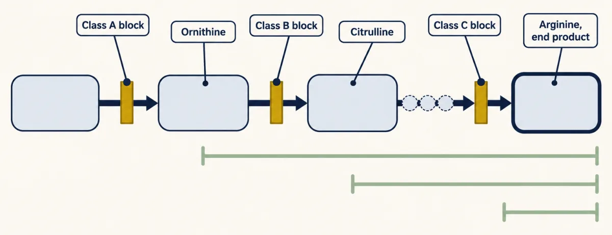
The film read a schematic chain backward. Adrian Srb and Norman Horowitz read the real arginine pathway in 1944. Its compounds also appear in the urea cycle in amino acid catabolism: ornithine gains a carbamoyl group to become citrulline, then two further reactions pass through argininosuccinate to arginine. Argininosuccinate lyase carries out the last reaction. As the one idea says, rescue brackets the block between supplied compounds, because the enzymes after it still work (see enzyme action).
Worked problem: put the steps in order. Three classes of arginine-requiring Neurospora mutants are tested on minimal medium plus one supplement. Assume each mutant blocks one step and that uptake of every supplement is normal.
- Class A grows with ornithine, citrulline or arginine.
- Class B grows with citrulline or arginine.
- Class C grows only with arginine.
- Arginine rescues every class, so it is the end product.
- Citrulline rescues A and B but not C, so C’s block lies after citrulline. Ornithine rescues only A, so ornithine comes earlier than citrulline.
- The order is precursor → ornithine → citrulline → arginine.
- A is blocked before ornithine, B between ornithine and citrulline, and C between citrulline and arginine.
- A block often makes the compound just before it build up. Class C’s stretch, citrulline to arginine, holds two reactions, so C may build up citrulline or argininosuccinate. Rescue data alone cannot say which, and feedback changes how much builds up, so accumulation has to be measured.
The real data were less tidy. Srb and Horowitz had fifteen arginine-requiring strains. Heterokaryon tests and crosses showed at least seven were genetically different, yet those seven fell into the same three classes: one rescue class need not be one gene. Under their one-gene, one-step model they inferred at least seven steps from sugar and ammonia to arginine. In double mutants, the block nearer arginine set the requirement, because a supplement must enter after every remaining block.
Benzer: function versus fine structure
A complementation test asks whether two mutations damage the same function. A recombination test asks whether the two sites can be separated by crossing over. Seymour Benzer, working at Purdue in the 1950s, showed how differently the two can answer.
His system was the rII region of the phage T4. rII mutants form large plaques on E. coli strain B but cannot grow on K12S(λ), a K-12 strain that carries the phage lambda. That host difference gives two tests.
Predict. Two rII mutants fail to complement, yet crossing them produces wild-type recombinants. Do those results contradict each other?
Check your prediction
No. Failure places the mutations in one functional unit; wild-type recombinants show separable sites inside it. Function and position differ.
- Function. Coinfect K12S(λ) with two rII mutants. If they lie in different functional units, each phage supplies what the other lacks and the cells release progeny. Benzer’s 1955 paper found that this test split the rII region into two independent segments. By 1959 he called such a unit, defined by the cis-trans test, a cistron.
- Recombination. Coinfect B, where all types grow, then plate the progeny on B to count everything and on K12S(λ) to count only wild-type recombinants. Recombination gives two reciprocal classes, wild type and double mutant, which in the progeny of many cells occur in equal numbers; only wild type grows on K12S(λ).
Predict. The selective host counts wild-type recombinants but not reciprocal double mutants. If both recombinant classes are recovered equally, should recombination frequency equal, halve or double the wild-type fraction?
Check your prediction
Double it. The selective plate sees one of the two reciprocal classes, so an equally recovered, unseen double-mutant class contributes the same fraction again.
recombination frequency = 2 × (wild-type recombinants per mL) / (total progeny per mL)
Because only recombinants grow on the selective host, Benzer could count very rare ones and map many sites inside one cistron, though he noted that fine-scale frequencies were not always additive.
Predict. If a finite cross yields no recombinant plaques, has it proved that the two mutations occupy the identical site?
Check your prediction
No. The sites may be so close that this finite sample missed the rare recombinants. Zero recombinants set only an upper limit on their separation; they do not prove zero distance.
Design problem
Two T4 rII mutants, rX and rY, each fail to grow on E. coli K12S(λ). Coinfecting K12S(λ) with rX and rY gives no growth. The cis control (an rX rY double mutant plus wild type) grows. Crossing rX and rY on E. coli B and plating the progeny gives 40 plaques per 0.1 mL on K12S(λ) at a 10−3 dilution, and 200 plaques per 0.1 mL on B at a 10−6 dilution. Each parent alone gives no plaques on K12S(λ). Interpret the results and give the recombination frequency.
Saved in this browser only.
One way to do it
Function. rX and rY fail to complement in trans, so they lie in the same cistron. The cis control matters: the double mutant with wild type grows, so it does not interfere with wild type, and the trans failure reflects shared function, not dominance.
Recombination. Plaques on K12S(λ) appear only from the cross, not from either parent, so they are wild-type recombinants. The two sites can be separated.
- Wild-type recombinants: 40 plaques in 0.1 mL is 400 per mL; correcting for the 10−3 dilution, 400 × 103 = 4 × 10⁵ per mL.
- Total progeny: 200 in 0.1 mL is 2,000 per mL; times 106 gives 2 × 10⁹ per mL.
- Wild-type fraction: 4 × 10⁵ / 2 × 109 = 2 × 10⁻⁴.
- Double for the unseen double-mutant class: recombination frequency = 4 × 10⁻⁴, or 0.04 percent.
Assumptions: reversion of either mutant to wild type and leaky growth of the mutants on K12S(λ) are negligible, and the two reciprocal recombinant classes are equally recovered. Conclusion: one gene, two close but separable sites.
Going deeper
Why white eyes can have different causes
A red fly eye needs many genes. Enzymes make two pigment families, one red and one brown. Transporters move pigments or their building blocks into pigment granules, and the granules must form. A strain can look white because a transporter both pigments need is missing, or because both pigment pathways are blocked. Two white-eyed strains may therefore have lost different genes.
Before the Neurospora screen
The proposal that genes control chemical steps predates the experiments that established it. In 1902, in the Lancet, the physician Archibald Garrod described alkaptonuria, in which urine darkens on standing because it contains homogentisic acid. He noted that it often appeared in children of parents who were cousins, the pattern expected for a recessive Mendelian trait, and called it an alternative course of metabolism. William Bateson supplied the recessive Mendelian reading of the cousin data. The missing enzyme, homogentisate dioxygenase, was identified later.
From one gene, one enzyme to gene products
The summary “one gene, one enzyme” came later; it is not the wording of Beadle and Tatum’s 1941 paper. Today you would say that a gene encodes a polypeptide or a functional RNA. Some genes encode only RNA, alternative splicing gives one gene several proteins, and an enzyme with several different subunits needs several genes.
Your notes
Select any text in the lesson to highlight it or add a note to it. Your highlights and notes are saved in this browser and collected in your notebook.
This browser is not letting the site save anything (a private window or strict privacy settings), so highlights and notes will be lost when you leave the page.
How we measure it
Complementation (cis-trans) test
Two recessive mutations are brought together in one cell, one on each chromosome (trans). A wild-type result means each copy supplies what the other lacks, so the mutations are usually in different functional units. The cis arrangement, both mutations on one copy opposite a wild-type copy, is the control.
Growth rescue on minimal medium
A nutritional mutant is grown on minimal medium plus one candidate compound at a time. A compound that restores growth lies after the blocked step, and one that fails, if the cell can take it up, lies before it, so the pattern across mutants orders the steps of a pathway.
Heterokaryon test
In Neurospora, hyphae of two strains can fuse so that two kinds of nucleus share one cytoplasm. If the heterokaryon grows without the supplement that each strain needs alone, the two mutations are usually in different genes, which is a complementation test for a haploid organism.
Selective plating of phage recombinants
Progeny of a cross between two T4 rII mutants are plated on E. coli B, where all types grow, and on K12S(λ), where only wild-type recombinants grow. Twice the ratio of K12S(λ) plaques to B plaques, each corrected for dilution, estimates the recombination frequency between the two sites.
Practice questions on this lesson (3) → MCAT-style, original, each with an explanation
Where this lesson fits
Before thisReview first
- Mendel's Peas: Segregation and Independent AssortmentThis course · Lesson 1
You need segregation and recessive alleles from there to see why a trans diploid can be wild type.
- Extending Mendel: Dominance, Multiple Alleles and Gene InteractionThis course · Lesson 4
Its gene-interaction crosses, such as white peas giving purple hybrids, show the rescue idea that complementation relies on.
- Linkage and Recombination MappingThis course · Lesson 8
You need to know that recombination separates sites on a chromosome to tell it apart from complementation.
After thisStudy next
- Bacterial and Phage Genetics: Conjugation, Transduction and TransformationThis course · Lesson 11
Phage crosses and selective hosts build on Benzer's rII work, which this lesson only introduces.
- Mutation: Where Variation Comes FromThis course · Lesson 14
Benzer's many rII mutants and the one-gene, one-step idea lead into how mutations arise.
- The Genetic Code and TranslationThis course · Lesson 18
Benzer's fine-structure work leads to how a gene's sequence specifies a protein through the genetic code.
Suggested by an AI model from the lessons themselves, then checked. How we use AI
Finish line
Test yourself
8 questions
Practice and MCAT-style, with explained answers.
Next · Lesson 11
Bacterial and Phage Genetics: Conjugation, Transduction and Transformation
Two E. coli strains that each cannot make several nutrients are mixed. A few cells can now make them all. Where did the genes come from?
Continue →← Lesson 9 · Three-Point Crosses: Gene Order and Double Crossovers