Biology · Unit 4: DNA and inheritance
From DNA to Protein
Rewritten on : new text, flashcards and film. Your notes and highlights stay, but the text around them may have moved.
How can changing one letter out of hundreds in a gene make red blood cells bend into stiff sickle shapes?
Pause when the reading reaches a film, and offer to play it. Saved on this device.
Watch the film now, or keep listening
Builds on:DNA: Structure and CopyingWhat Am I Made Of?Why these?
Where this lesson sits
- Virginia SOL BIO.2d
- Virginia SOL BIO.5a
- Practices BIO.1c, BIO.1d, BIO.1e, BIO.1f
Essential question: How does the sequence of letters in DNA become a protein, and how can changing one letter change a trait?
-
Life Science (grade 7) LS.10 a
Review it at grade 7 level
In Life Science you learned that DNA is a double helix whose bases, A, T, C and G, form a code. A gene is a section of a chromosome that carries a code, most often the code for making a protein. Those proteins build cells and carry out life processes, which affects an organism's traits. You did not learn how the code is read. Biology adds the two steps, transcription and translation, and shows how changing one letter can change a protein and a trait.
-
Biology BIO.2d, BIO.5a You are here
This lesson
-
Biology II Biology II: Genetics, GEN.2 b and GEN.3
Preview where it goes next
The Biology II Genetics course looks inside each step. RNA polymerase reads the template strand from 3′ to 5′ and builds RNA from 5′ to 3′. In eukaryotes the first RNA copy of a protein-coding gene is processed: it usually gets a 5′ cap and a poly-A tail, and splicing removes stretches called introns. The course then asks how cells control which genes are expressed: operons in bacteria, transcription factors and enhancers in eukaryotes, alternative splicing that makes several proteins from one gene, changes made to a protein after translation, and epigenetic marks. It also uses the shared genetic code as evidence that living things have a common ancestor.
-
College
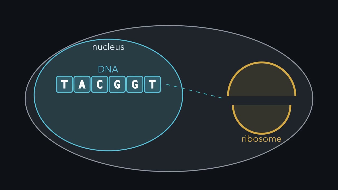 ▶ Watch From Gene Copy to Protein Chain · 2:58
Read the transcript of “From Gene Copy to Protein Chain”
Film transcript: From Gene Copy to Protein Chain
A plant or animal cell keeps its chromosomes inside the nucleus. How can their instructions reach a ribosome outside? Watch the blue DNA row remain behind while a green copy begins to grow. Making a protein from a gene is protein synthesis. The first stage is transcription. An enzyme opens a short stretch of DNA. It uses the template strand to build messenger RNA.
Only the selected gene is copied. Each green RNA block pairs with a blue DNA block. DNA letter A pairs with RNA letter U. T pairs with A. C pairs with G, and G pairs with C. RNA uses U instead of T. Try the next template group. The blue DNA blocks read TAC. Which green messenger RNA blocks will pair with them? Keep the pairing key in view and choose the full group.
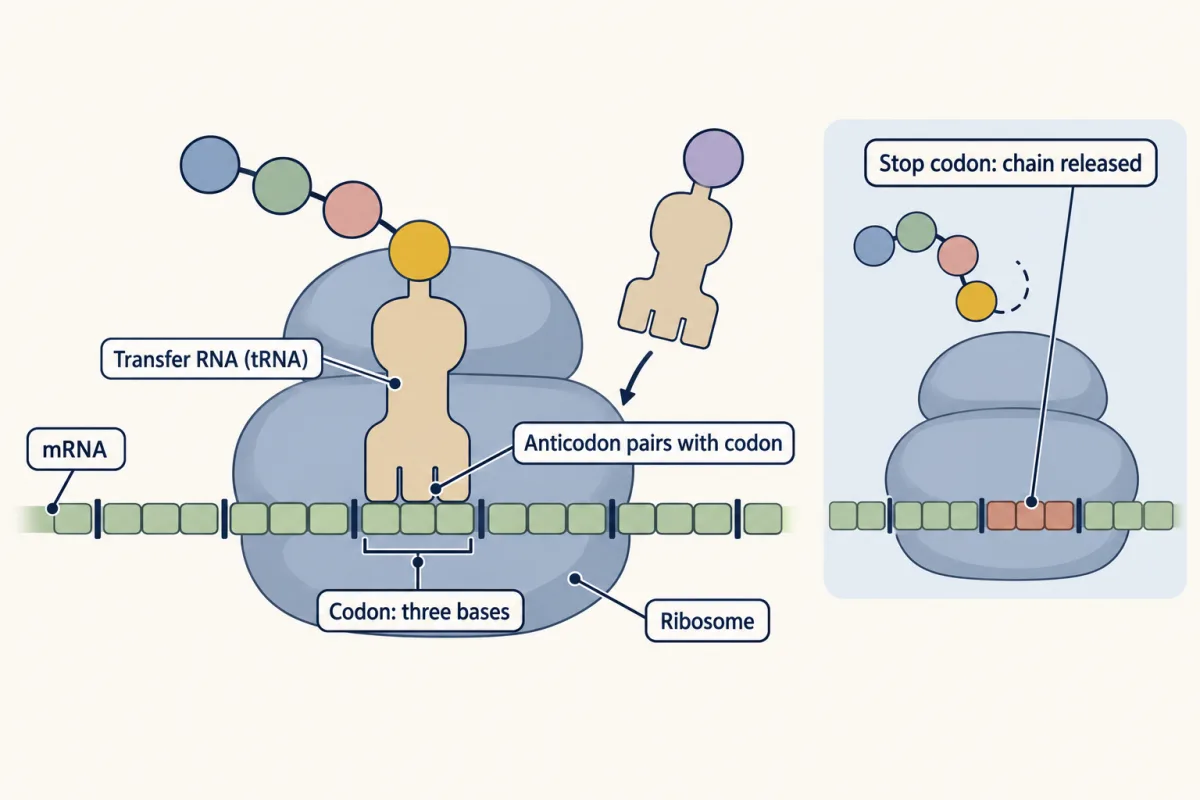
T pairs with A. A pairs with U. C pairs with G. The messenger group is AUG. The enzyme releases the finished message. The green strand peels away, while the blue DNA closes and stays inside. The messenger RNA passes through a pore into the cytoplasm. Translation begins when a gold ribosome grips the message. It reads each codon in order. A codon is a group of three letters that means an amino acid or stop. Purple transfer RNA brings the amino acid matched to each codon. Its anticodon pairs with the codon inside the ribosome. The ribosome joins each red amino acid to the growing chain. At a stop codon, no amino acid is added and the chain is released.
Reading usually begins at the start codon AUG. It adds methionine and fixes the grouping that follows. The ribosome then moves codon by codon. At UGA, the stop sign appears, the reading window opens, and the finished chain floats free. The released chain can fold into a working protein. The message changed form, but its order guided every handoff. In protein-coding genes, DNA order sets mRNA codons and amino acid order. That is how instructions inside the nucleus reach a protein outside.
From the film
- In protein-coding genes, DNA order sets mRNA codons and amino acid order.
- In plant and animal cells, transcription makes an mRNA copy while the chromosomes stay in the nucleus.
- A ribosome reads each mRNA codon, and transfer RNA brings the matching amino acid.
- At a stop codon, no amino acid is added and the chain is released.
A red blood cell is usually a soft, round disc that squeezes through tiny blood vessels. In one gene for hemoglobin, the protein that carries oxygen in red blood cells, a single DNA letter can differ: GTG where most people have GAG. That change can bend red blood cells into stiff sickle shapes. How can one letter out of hundreds reach the shape of a whole cell?
What do you think? Decide before you read.
-
Scientists disagree: Many mutations change nothing about how a protein works. Some even help in one setting. Studies of children in malaria regions found that carrying one copy of the sickle cell allele lowered their chance of serious malaria.
What scientists think
Scientists disagree: Many mutations change nothing about how a protein works. Some even help in one setting. Studies of children in malaria regions found that carrying one copy of the sickle cell allele lowered their chance of serious malaria.
-
Scientists disagree: Comparing DNA with the proteins it codes for shows that many changes leave the protein the same. People with one sickle cell allele and one usual allele (sickle cell trait) usually have no symptoms of sickle cell disease.
What scientists think
Scientists disagree: Comparing DNA with the proteins it codes for shows that many changes leave the protein the same. People with one sickle cell allele and one usual allele (sickle cell trait) usually have no symptoms of sickle cell disease.
-
Scientists disagree: In cells with a nucleus, the DNA stays inside. Cells copy one gene into a short messenger molecule, and that copy travels out to the ribosomes. Classic experiments on amoebas that labelled new RNA found it made in the nucleus and later in the cytoplasm.
What scientists think
Scientists disagree: In cells with a nucleus, the DNA stays inside. Cells copy one gene into a short messenger molecule, and that copy travels out to the ribosomes. Classic experiments on amoebas that labelled new RNA found it made in the nucleus and later in the cytoplasm.
Copy a gene into messenger RNA
- protein synthesis
- transcription
- RNA
- messenger RNA
- translation
- codon
- ribosome
- transfer RNA
- amino acid
- mutation
- substitution
- frameshift
The first film showed the two stages of protein synthesis, making a protein from a gene: transcription copies one gene into messenger RNA (mRNA), and translation reads the mRNA to build a chain of amino acids.
RNA is a nucleic acid, a chain of nucleotides like DNA, with three differences:
| Feature | DNA | RNA |
|---|---|---|
| Strands | two, twisted into a double helix | usually one |
| Sugar | deoxyribose | ribose |
| Bases | A, T, C, G | A, U, C, G (uracil, U, in place of T) |
In transcription, an enzyme reads one DNA strand, the template strand, pairing DNA A with RNA U, T with A, C with G, and G with C. The other DNA strand, the coding strand, matches the mRNA letter for letter, with T where the mRNA has U.
Predict. A template DNA triplet reads CTT. Which mRNA triplet will pair with it?
Check your prediction
C pairs with G, and each T pairs with A, so the mRNA triplet is GAA. Pair with the labelled template; do not copy its letters straight across.
Like DNA, RNA has direction: its two ends differ (biologists call them 5′ and 3′), and it is built and read one way only.
Bacteria have no nucleus, so both stages happen in their cytoplasm.
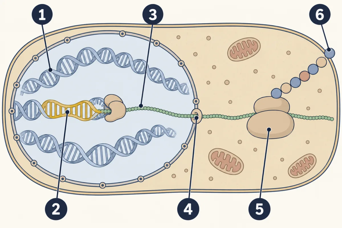
- DNA
- gene
- mRNA
- pores in the nuclear membrane
- ribosome
- chain of amino acids
- DNA
- blank
- blank
- blank
- ribosome
- blank
Word bank: chain of amino acids, gene, mRNA, pores in the nuclear membrane
- blank
- blank
- blank
- blank
- blank
- blank
Check your labels
- DNA
- gene
- mRNA
- pores in the nuclear membrane
- ribosome
- chain of amino acids
Read the message into an amino acid chain
Each codon, a group of three mRNA bases, means one amino acid or “stop.” Amino acids are the small building blocks of proteins; living things use about twenty common kinds. Here is part of the codon chart.
| mRNA codon | Meaning |
|---|---|
| AUG | methionine (Met), start |
| GAG, GAA | glutamic acid (Glu) |
| GUG | valine (Val) |
| UUU, UUC | phenylalanine (Phe) |
| GGC | glycine (Gly) |
| AAA | lysine (Lys) |
| UAA, UAG, UGA | stop |
Predict. A ribosome already reading an mRNA reaches UUU GGC, lined up as two codons. Which two amino acids will it add next?
Check your prediction
The chart gives phenylalanine for UUU and glycine for GGC, in that order.
Now a complete example. A DNA template strand, written from its 3′ end on the left to its 5′ end on the right, reads TAC CTC CTT AAA ACT.
- Pair each base: the mRNA is AUG GAG GAA UUU UGA.
- Translate: Met, Glu, Glu, Phe, then UGA means stop.
- The chain is Met–Glu–Glu–Phe. The stop codon adds nothing.
AUG, the usual start codon, stands for methionine. With four bases, there are 4 × 4 × 4 = 64 possible codons. In the standard code, 61 stand for amino acids, and three (UAA, UAG and UGA) are stops.
In the first film, each transfer RNA (tRNA) paired with a codon through its three-base anticodon. After the stop, the released chain can fold into a working protein.
Explore: transcribe, translate and mutate a gene
The simulation shows a short DNA template as lettered blocks. Build its mRNA, translate it with the codon chart, then make a mutation, a change in DNA base order: swap, add or remove a letter. In cells, mutations come from rare copying errors or damage.
In this sketch: DNA A pairs with RNA U, T with A, C with G, G with C. Reading begins at the first AUG (the usual start codon) and ends at a stop codon. Only the codons in the chart are named; any other shows a ?.
Rule to keep: ribosomes read mRNA three bases at a time, so one added or removed letter shifts every codon after it, while a swapped letter changes at most one codon, unless it removes or creates the AUG where reading begins.
Try this.
- Build the first mRNA codon and explain each pairing.
- Find the start and stop, and check that the stop adds nothing to the chain.
- Predict whether swapping one letter will change zero, one or many amino acids. Swap three different letters and record what happens, including any early stop.
- Add a letter near the start, then remove one. Compare how later codons are grouped.
Specialized cells carry the same genes but use different ones: young red blood cells fill with hemoglobin, while some pancreas cells make insulin.
Predict. Could a bacterium read a suitably prepared gene for a human protein? What fact about codons supports your answer?
Check your prediction
Yes. The genetic code, the set of codon meanings, is almost the same in every living thing, with a few exceptions, including in some bacteria and mitochondria. In work published in 1979, a team put man-made genes for the two human insulin chains into E. coli. The bacteria made the chains. Once joined outside the cells, tests recognized them as insulin.
The second film follows a changed letter further, from one codon to a whole red blood cell.
Read the transcript of “When the Reading Changes”
Film transcript: When the Reading Changes
The first film followed blue DNA into green messenger RNA and a red amino acid chain. If a DNA letter changes, how much of that chain changes? Keep watching the same blocks and reading window. A mutation is a change in DNA base order. A substitution swaps a base. Because several codons can mean the same amino acid, a swap can leave the chain unchanged. Another swap can change an amino acid or create an early stop. An insertion adds bases, and a deletion removes them. The ribosome does not see spaces between codons. It starts at its reading point and groups the message from there. Adding or removing one base inside the coding part shifts that grouping.
This is called a frameshift. Predict what happens when the waiting block enters near the start. Will only its codon change, or will later codons change too? Follow the dividers, not the colours. The inserted block shifts every divider after it. The gold ribosome now reads new codons from that point. Many later amino acids can change, and an early stop may appear. Removing a base can shift the frame in the same way. Now compare a quieter substitution.
Messenger codon GAG means glutamic acid. Change its last letter, making GAA, and it still means glutamic acid. The DNA changed, but this amino acid chain did not. This outcome is called a silent mutation. Now follow the sickle substitution.
The usual coding DNA reads GAG. The sickle allele reads GTG. Messenger RNA therefore reads GUG instead of GAG. The ribosome places valine instead of glutamic acid. That changed spot can alter hemoglobin's behaviour. When oxygen is low, sickle hemoglobin can join into stiff rods. The rods can bend a soft red blood cell. A letter change has reached a whole cell. A changed letter does not guarantee a changed trait. Some substitutions keep the same amino acid. Protein effects also depend on which copies a person has and the environment. Here is the reading rule to keep. In coding DNA, swaps change one codon; frameshifts change later codons.
From the film
- In coding DNA, swaps change one codon; frameshifts change later codons.
- A substitution can leave the chain unchanged, change an amino acid or create a stop.
- In the sickle allele, valine replaces glutamic acid, and low-oxygen hemoglobin can form stiff rods.
- A changed letter does not guarantee a changed trait.
When one letter reaches a whole cell
The second film skipped two things: why valine makes hemoglobin stick, and why one sickle copy usually causes no symptoms.
Glutamic acid carries an electric charge and mixes well with water. Valine has no charge and avoids water. When hemoglobin is low on oxygen, the valine can fit into a water-avoiding patch on another hemoglobin, so the molecules can join into the stiff rods the second film showed.
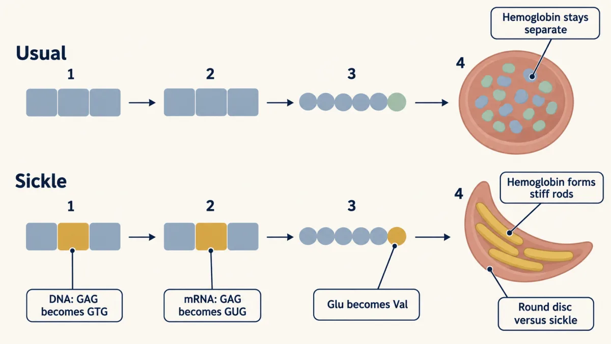
A person has two copies of this gene.
Predict. One person has one sickle allele and one usual allele. Another has a disease-causing change on both copies, including the sickle change. Who usually has sickle cell disease symptoms?
Check your prediction
The second person. One sickle allele with one usual allele is sickle cell trait, which usually causes no symptoms. Sickle cell disease needs a disease-causing change on both copies, at least one of them the sickle change.
Use evidence: when is a mutation harmful?
If the sickle allele can cause disease, why is it common in some places? Compare newborn screening with malaria outcomes.
| Measure | Group or place | Year | Value | Units |
|---|---|---|---|---|
| Sickle cell trait positive screens | 44 US states combined | 2010 | 15.5 | per 1,000 newborns screened |
| Sickle cell trait positive screens | Montana | 2010 | 0.8 | per 1,000 newborns screened |
| Sickle cell trait positive screens | Mississippi | 2010 | 34.1 | per 1,000 newborns screened |
| Sickle cell trait positive screens | Black or African American newborns, 13 states reporting race | 2010 | 73.1 | per 1,000 newborns screened |
| Sickle cell trait positive screens | White newborns, same 13 states | 2010 | 3.0 | per 1,000 newborns screened |
| Protection by sickle cell trait against mild malaria | one group of children, coast of Kenya | published 2005 | 50 | percent |
| Protection by sickle cell trait against hospital admission for malaria | a second group of Kenyan children, same study | published 2005 | 75 | percent |
Sources: rows 1 to 5, a CDC report (Ojodu and colleagues, MMWR, 2014) on newborns screened in 2010 in 44 states; screens were not confirmed diagnoses. Rows 6 and 7, Williams and colleagues (Journal of Infectious Diseases, 2005). Race groups are record labels; the allele follows family ancestry.
Predict. Is the Mississippi screening rate nearer ten times or fifty times the Montana rate? Which malaria outcome shows stronger protection?
Check your prediction
Divide 34.1 by 0.8: the Mississippi rate is about 43 times the Montana rate, nearer fifty times. Protection was stronger against hospital admission (75 percent) than against mild malaria (50 percent).
Can the newborn rows alone tell you why the trait is more common in some groups? No. They show a pattern, not its cause.
Claim, evidence, reasoning
Make a claim that answers the essential question: how can changing one letter change a trait, and is that change always harmful? Use this data.
Start with: Changing one letter in the hemoglobin gene blank, and the effect is blank depending on blank.
Start with: In the Kenyan children, sickle cell trait was blank percent protective against blank. In US newborns, trait screens ranged from blank to blank per 1,000.
Start with: The change GAG to GTG puts blank in place of blank, so hemoglobin blank. With one copy, a person blank, which matters where blank.
Compare with a model answer
One changed letter, GAG to GTG, swaps glutamic acid for valine in beta-globin, so the hemoglobin can form stiff rods and red blood cells can sickle. The effect is not always harmful. In two groups of Kenyan children, one copy of the allele lowered the risk of mild malaria by 50 percent and the risk of hospital admission by 75 percent, while people with one copy usually have no symptoms. In US newborn screening, trait positives ranged from 0.8 per 1,000 in Montana to 34.1 in Mississippi. But the screening data come from one year and are not confirmed diagnoses, and the malaria data come from one region, so neither shows the effect everywhere.
Saved in this browser only.
Design problem
Use the simulation as a model to test this question: which kind of mutation most often changes the amino acid chain, a substitution, an insertion or a deletion? Plan a fair test.
Saved in this browser only.
One way to do it
- Hypothesis: If ribosomes read in threes, then one-base insertions and deletions will change the chain more often than substitutions.
- Independent variable: the kind of mutation (substitution, insertion or deletion of one base).
- Dependent variable: whether the chain differs from the control chain, and how many amino acids changed, compared position by position from the start.
- Constants: the same starting DNA, one mutation per trial, the same codon chart, and only spots inside the coding part, from the start codon up to the stop.
- Control: translate the starting DNA with no mutation, so you know the normal chain.
- Trials: 20 of each kind at randomly chosen spots, with the new base also chosen at random; report the percent that changed the chain.
If insertions and deletions change the chain in almost every trial while some substitutions change nothing, the hypothesis is supported. The limit: a model of one short gene shows the chain, not whether a real folded protein still works.
Going deeper
Counting amino acids in beta-globin
Beta-globin is the hemoglobin chain this gene codes for. In its usual numbering, glutamic acid is the sixth amino acid. Some databases count the starting methionine and call it the seventh.
Silent does not always mean identical
The second film’s silent swap, GAG to GAA, kept the chain the same. The story “Silent, but not always identical” adds a refinement: two messages that make the same amino acid chain can still differ in how long they last, which can change how much protein a cell makes.
Evaluate: check your thinking
The one rule, DNA order sets mRNA codons and amino acid order, connects the scales. One coding letter, GAG to GTG, puts valine in place of glutamic acid, and in low oxygen that hemoglobin can form rods that bend the cell. Other DNA changes can leave the chain unchanged.
Science practices you used: testing predictions with a model, translating a code with a chart, comparing rates in a data table, making a claim from evidence, and planning a fair test with a control and repeated trials.
Your notes
Select any text in the lesson to highlight it or add a note to it. Your highlights and notes are saved in this browser and collected in your notebook.
This browser is not letting the site save anything (a private window or strict privacy settings), so highlights and notes will be lost when you leave the page.
How we measure it
Newborn screening
A few drops of blood from a newborn's heel are tested for unusual forms of hemoglobin and other conditions. Counting the positive screens across many births shows how common a trait such as sickle cell trait is among the newborns in a state or a group.
Cell-free protein synthesis
Cells are broken open and the juice, which still holds ribosomes and tRNA, is mixed with an RNA and amino acids that carry a radioactive label. Measuring how much label ends up in protein shows which RNA directs which amino acids into a chain.
Where this lesson fits
Before thisReview first
- DNA: Structure and CopyingThis course · Lesson 16
You need DNA's base pairing and strand order from there to pair mRNA bases with the template.
- What Am I Made Of?This course · Lesson 2
You need to know proteins are chains of amino acids from there to follow what translation builds.
After thisStudy next
- Engineering GenesThis course · Lesson 22
Bacteria make human insulin because the nearly universal code lets them read a human gene.
- Variation and SurvivalThis course · Lesson 27
Mutations that change proteins, like the sickle change, are one source of the variation selection acts on.
- Meiosis and VariationThis course · Lesson 19
It shows how mutated alleles like the sickle allele are passed on and shuffled into children.
Suggested by an AI model from the lessons themselves, then checked. How we use AI
Finish line
Test yourself
8 questions
Practice and MCAT-style, with explained answers.
Next · Lesson 19
Meiosis and Variation
If two parents give every child half of their chromosomes, why do their children come out different from each other?
Continue →