Genetics and Molecular Biology · Unit 3: DNA: copying, changing, repairing
New to this? Review it at high school level: From DNA to Protein
Mutation: Where Variation Comes From
Rewritten on : new text, flashcards and film. Your notes and highlights stay, but the text around them may have moved.
Ten bacterial cultures meet the same virus: why does one leave 52 resistant colonies while five leave none?
Pause when the reading reaches a film, and offer to play it. Saved on this device.
Watch the film now, or keep listening
Builds on:DNA Replication: Semiconservative Copying and Its MachineryProbability, Crosses and the Chi-Square TestDNA Is the Genetic MaterialWhy these?
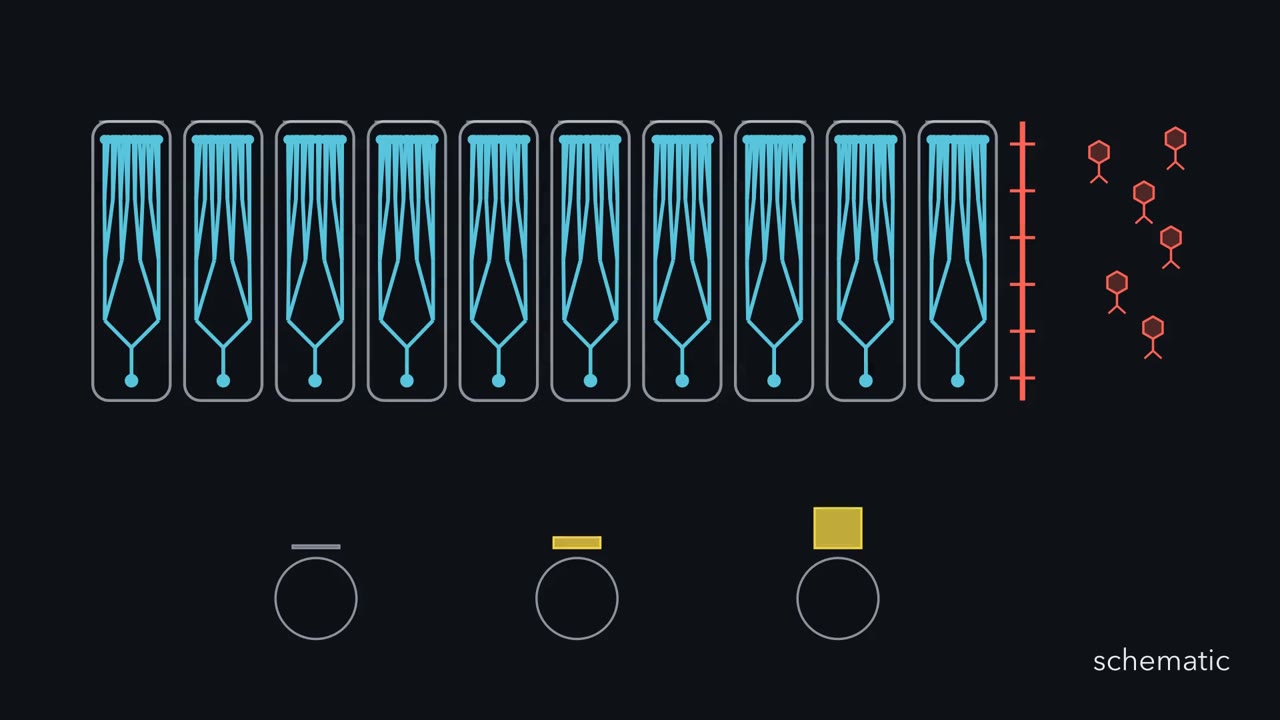 ▶ Watch Before the Phage Arrives · 3:37
Read the transcript of “Before the Phage Arrives”
Film transcript: Before the Phage Arrives
Identical bacterial cultures grow, then meet the same bacterial virus, called a phage. Why do some plates show no survivors while another shows a jackpot? Each tube began alike, and every plate receives the same phage. Every culture starts from the same bacterial strain. They receive the same food and grow to similar sizes. The selecting phage has not arrived yet. The growing branches record who descended from whom. During one division, a heritable DNA sequence change appears in one cell. That cell and its descendants carry the variant as the tree keeps growing. The changed cell cannot know whether that variant will later help. The change merely enters the population.
Timing changes the family size. An early event leaves a large yellow branch. A late event leaves only a few yellow tips. Some cultures get no event at all. Outcomes depend on whether a resistance mutation occurred and, if so, when. Another explanation was possible.
Perhaps the phage itself makes resistance after contact. Then each exposed cell gets a similar small chance, independent of its family history. The competing models therefore predict different shapes across cultures. If variants arise during growth, what pattern should appear after exposure? Similar survivor counts, or uneven counts with jackpots and empty plates? Use the family trees, not the final plates, to decide.
Now open the gate. Phage removes susceptible blue cells. Resistant yellow cells remain and form colonies. Exposure changes which variants are visible, not their earlier family histories. The phage acts as a filter applied after growth. The growth model predicts this shape: several empty cultures, some small counts, and an occasional jackpot. Contact-induced resistance predicts much tighter scatter. Averages alone can hide this crucial difference in shape. Luria and Delbrück compared many independent cultures with repeated samples from one bulk culture. Independent cultures scattered far beyond ordinary sampling and plating variation. Repeated bulk samples reveal how much scatter handling alone can produce.
The contrast tests timing. This result has a boundary. It shows that phage resistance arose before phage exposure in this system. Mutation rates can still change with cellular conditions. Changing a rate is not the same as directing a useful mutation. Mutation creates heritable DNA variants; selection changes how common they become. The sequence change starts a branch. The environment changes the branch's representation afterward. Keep origin and sorting separate whenever you interpret adaptation.
From the film
- Mutation creates heritable DNA variants; selection changes how common they become.
- In the fluctuation-test system, resistant variants arose while cultures grew, before phage exposure.
- An early mutation can leave many resistant descendants, while a late mutation leaves few.
- Selection can make a rare pre-existing variant easy to see.
Classify the sequence change, then its consequence
A mutation is a heritable change in the DNA sequence. Here, point mutation means a change to one base pair or the addition or removal of a few bases. Classify it twice: by the DNA change, then, in a coding sequence, by the protein effect.
By the DNA change. A transition swaps a purine for a purine (A and G) or a pyrimidine for a pyrimidine (C and T). A transversion swaps a purine for a pyrimidine or the reverse. Each base has one transition partner and two transversion partners, so there are 4 possible transitions and 8 possible transversions. Even so, in many organisms studied, transitions are the more common kind. An insertion or deletion (together, an indel) adds or removes bases.
Predict. A G:C base pair becomes T:A. Is that a transition or a transversion? Can this alone tell you its protein effect?
Check your prediction
A transversion: G, a purine, is replaced by T, a pyrimidine. Its protein effect depends on whether it lies in a coding sequence and which codon it changes.
By the protein effect. Inside a coding sequence:
- A synonymous (silent) change gives a codon for the same amino acid.
- A missense change gives a different amino acid. Some missense changes leave the protein working; the label does not tell you the severity.
- A nonsense change turns an amino-acid codon into a stop codon.
- A frameshift is an indel whose length is not a multiple of three, so every codon downstream is read in different groups.
- An in-frame indel adds or removes a multiple of three bases. The downstream frame is kept, and the protein gains or loses amino acids.
Think of a sentence written in three-letter words: THE CAT ATE THE RAT. Change one letter and one word changes. Delete one letter and every group after it changes. Delete one whole word and the later groups stay aligned.
Predict. A deletion removes exactly three bases from the middle of a coding sequence. Must it shift the reading frame?
Check your prediction
No. Three bases make one codon, so the frame is kept and later codons stay grouped as before. The coding sequence loses one codon’s worth of bases. A deletion aligned with a codon usually removes one amino acid; one that straddles two codons can also change the junction amino acid, and if the junction becomes a stop codon, the protein is cut much shorter.
“Silent” describes the codon, not the consequence. Exons also carry signals for splicing, mRNA stability and translation speed, so a synonymous change can still lower the amount of protein. How the code is read is the subject of the genetic code lesson.
Does selection cause the mutation? Luria and Delbrück
The first film showed both hypotheses and the test. In numbers: repeated samples of one bulk culture measure sampling and plating error alone; induction predicts a variance close to the mean; mutation first predicts one far larger. Luria (experiments) and Delbrück (theory) used E. coli B and phage T1, virus α in their 1943 Genetics paper. Independent cultures scattered far beyond sampling error, so they concluded that, in their system, resistance arose independently of the virus.
From scatter to a mutation rate
Ten independent 0.2 mL cultures and one bulk culture of E. coli each grow to about 2 x 10⁸ cells per 0.2 mL. Each independent culture and ten 0.2 mL bulk samples are plated on T1. Assume every mutant survives plating and is counted, mutants grow like the parent, no mutations arise after plating, and divisions roughly equal the final cell number.
Independent cultures: 0, 0, 1, 0, 3, 0, 0, 52, 2, 2. Bulk samples: 4, 8, 6, 3, 9, 5, 7, 6, 8, 4.
- Means. Both sets sum to 60, so both means are 6.0.
- Variances. Sample variances are about 262 for the independent cultures and 4.0 for the bulk samples (arithmetic under Going deeper).
- Interpretation. The bulk variance is close to its mean, as Poisson sampling predicts. The independent variance is about 44 times its mean. Mutation before exposure fits; induction does not.
- Rate. Five of ten cultures have no mutants, so p0 = 0.5. If mutation events per culture follow a Poisson distribution with mean m, then p0 = e−m.
m = -ln(p0) = -ln(0.5) = 0.69 mutation events per culture
rate = m / divisions = 0.69 / (2 x 10⁸) = 3.5 x 10⁻⁹ per cell per division
Mutant frequency is mutant cells divided by all cells; mutation rate counts mutation events per division. In the jackpot culture, frequency is 52 / (2 x 10⁸) = 2.6 x 10⁻⁷, about 75 times the rate, from what was probably one early event. Confusing them makes one lucky culture look like a mutator, a strain with an elevated mutation rate.
Predict. Suppose the jackpot clone gains 100 more resistant descendants, but no new mutation event occurs. Does p0, and therefore this zero-class rate estimate, change?
Check your prediction
No. That culture was already nonempty, so p0 is unchanged. Mutant frequency rises; this rate estimate does not.
Rates do change with a cell’s condition, as in pulses of mutagenesis after alkylation damage in single E. coli cells, and debate over stress and mutation continues; none of this reverses the 1943 result for phage resistance.
| Hypothesis | Mean | Variance | Variance ÷ mean | Cultures with none |
|---|
Rule to keep: if the virus induced resistance, counts from separate cultures scatter only as much as sampling allows (variance close to the mean); if mutation comes first, jackpots and many empty plates make the variance far larger than the mean. This is a sketch of the logic using the lesson’s worked-problem numbers (about 2 x 10^8 cells per culture, 0.69 mutation events per culture, induced mean 6), not a model of a real strain. In this sketch a mutation after a fraction f of the growth leaves about 1/f resistant cells (f random, capped at 2 x 10^8), so clone sizes here are not measured values; the lesson’s jackpot was 52. The two arms’ means are not matched.
Replica plating: seeing the mutants before the test
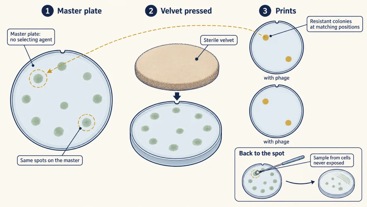
The fluctuation test is an argument from statistics. In 1952 Joshua and Esther Lederberg gave a direct one. They pressed sterile velvet onto a master plate grown without a selecting agent, then printed its colonies in the same positions onto fresh plates with phage or streptomycin.
Predict. If resistant cells were already present on the master, what spatial pattern should appear on separate selective prints?
Check your prediction
Resistant colonies should appear at matching positions. Each match points back to the same colony on the unexposed master plate.
That is what the E. coli K-12 prints showed. The Lederbergs sampled those master-plate spots, whose cells had never touched the phage or drug, and purified resistant cultures through repeated growth and re-plating. Replica plating also became a workhorse for finding mutants that fail to grow on one medium, the start of many screens like those in complementation and pathways.
Both experiments fit the rule: mutation creates heritable DNA variants; selection changes how common they become. The second film follows how a variant begins.
Read the transcript of “When Damage Becomes Mutation”
Film transcript: When Damage Becomes Mutation
The first film separated a variant's origin from selection. Now a DNA strip is damaged; is the cell mutated yet? Follow how that variant begins. The red mark appears before any daughter sequence has formed. A mutation is a heritable change in DNA sequence.
Heritable matters here. A temporary chemical lesion is damage, but it is not automatically a changed sequence. The sequence must change and persist when the cell divides. Changes can begin in two broad ways. Copying can place the wrong block. Or chemistry can damage a block before the next copy. Either route can end without a mutation. Damage enters a fork.
Repair may restore the original block. If repair misses it, copying may preserve the original sequence or produce a different one. The lesion's presence does not choose the outcome. Before the strip reaches that fork, predict: must a red damage mark become a mutation? Trace every possible path before choosing.
No. On one path, repair removes the lesion and restores the blue block. After copying, both daughter strips keep the original sequence. The cell had damage, yet no mutation remains. On another path, copying places a wrong block opposite the lesion. At the next copy, that wrong block templates a full base-pair change. The damaged strand goes to a separate daughter, where repair or bypass can follow. In the changed lineage the lesion is gone, but its consequence persists. Internal chemistry and ordinary copying create spontaneous mutations without an applied mutagen. Outside agents are mutagens when they raise mutation. Both routes end the same way.
A change becomes a mutation only if repair or copying leaves a heritable sequence change. The effect on a protein may range from none to loss of function, and occasionally a changed or gained function. Those consequences come after the inherited sequence change. Only after a heritable variant exists can an environment change its representation. The first film followed that later sorting step with phage resistance.
Selection cannot reach backward and request that earlier change. Damage becomes mutation when repair or copying leaves a heritable sequence change. Repair can erase the damage first. The inherited sequence, not the lesion, is the mutation. A red mark alone never satisfies the definition. The lesson now names the specific sources of these changes. Then the Ames test asks whether a chemical raises mutations in bacteria. Those details belong in the text that follows.
From the film
- Damage becomes mutation when repair or copying leaves a heritable sequence change.
- Repair can restore the original sequence before a change is inherited.
- Spontaneous mutations arise from internal chemistry and ordinary copying; mutagens are outside agents that raise mutation.
- A mutation is the inherited sequence change, not the first chemical lesion.
Where mutations come from: spontaneous and induced
Spontaneous mutations arise from the cell’s own chemistry and copying, with no applied mutagen.
- Replication errors. DNA polymerase sometimes inserts the wrong base (see DNA replication).
- Deamination. Cytosine loses its amino group and becomes uracil, which repair enzymes recognize as foreign. 5-Methylcytosine deaminates to thymine, a normal base, so the G:T mismatch is harder to resolve correctly. CpG sites, where cytosines are often methylated in vertebrates, are hot spots for C:G to T:A transitions. The chemistry is in nucleotide metabolism.
- Depurination. The bond between a purine and its sugar breaks, leaving a site with no base to copy.
- Slippage. In repeated bases, the new strand can loop out or slip back during copying, adding or removing repeat units.
Induced mutations come from outside agents, called mutagens.
- Base analogs such as 5-bromouracil are built into DNA in place of a normal base and mispair, giving transitions.
- Alkylating agents such as ethyl methanesulfonate (EMS) add groups to bases. EMS chiefly gives G:C to A:T transitions.
- Intercalators such as proflavine slip between base pairs and favor single-base insertions and deletions, so frameshifts.
- Ultraviolet light joins adjacent pyrimidines into dimers. Each dimer is damage until repair or copying past it leaves a heritable sequence change.
- Ionizing radiation breaks DNA strands and damages bases.
Hermann Muller reported in Science in 1927 that X-rays greatly raise the mutation rate in Drosophila, in experiments that counted new lethal mutations on the X chromosome. He received the Nobel Prize in 1946. How cells fix damage before it becomes mutation is the subject of DNA repair and recombination.
The Ames test and what it can and cannot say
Bruce Ames’s test uses Salmonella strains that cannot make histidine because of a mutation in a histidine gene. On a plate with only a trace of histidine, they divide a few times and stop unless a new mutation restores histidine synthesis. Each revertant colony marks such an event, sometimes at the original site and sometimes at a second, compensating site. TA100 reverts mainly by base-pair substitution; TA98 reverts mainly by frameshift. Plates run with and without S9, a rat-liver enzyme fraction, because many carcinogens become reactive only after liver metabolism.
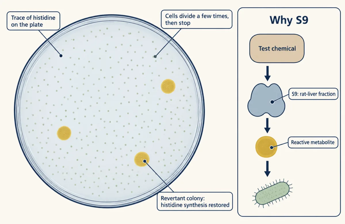
Read an Ames result in a fixed order: compare with spontaneous background, look for a dose response, compare strains, then compare with and without S9.
A positive result shows that a chemical, or a metabolite, mutates bacteria; it does not measure cancer risk in people. A negative result does not prove safety, because some carcinogens act without mutating DNA or through processes bacteria lack.
Design problem
Predict. TA100 gives about 120 spontaneous revertants per plate and TA98 about 30. Compound X is tested at three rising doses. TA100 without S9: 125, 118, 130. TA100 with S9: 240, 480, 950. TA98 with S9: 30, 28, 35. What does X do, and what does the result not show?
Saved in this browser only.
One way to do it
TA100 without S9 stays at about 120 at every dose, so X itself shows no detectable mutagenic activity in TA100 at these doses. TA100 with S9 rises to about 2, 4 and 8 times background: a dose-dependent rise in base-pair substitutions after liver metabolism. TA98 with S9 stays at about 30, so there is no detectable frameshift activity in TA98.
Conclusion: a metabolite of X is a base-substitution mutagen in Salmonella. This does not show that X causes cancer in people, since the test measures bacterial mutation with rat-liver enzymes.
Going deeper
The arithmetic behind the variances
For the independent cultures, the squared deviations from 6 are 36, 36, 25, 36, 9, 36, 36, 2,116, 16 and 16, summing to 2,362. Divided by 9, the sample variance is about 262. For the bulk samples, the squared deviations sum to 36, so the variance is 36/9 = 4.0.
Tautomers and mismatch shapes
Rare alternative forms of bases, called tautomers, were proposed long ago as one cause of mispairing. Nuclear magnetic resonance (NMR) work has since detected short-lived tautomeric and ionized forms that let a wrong pair take the shape of a correct one, though how much they add to mutation in living cells is still being worked out.
Ames validation counts
In 1973 Ames and colleagues reported that liver homogenate converted 18 tested carcinogens into frameshift mutagens. In McCann and colleagues’ 1975 set, 156 of 174 tested carcinogens were positive, a figure for that set of chemicals, not a general probability that a carcinogen will test positive.
Your notes
Select any text in the lesson to highlight it or add a note to it. Your highlights and notes are saved in this browser and collected in your notebook.
This browser is not letting the site save anything (a private window or strict privacy settings), so highlights and notes will be lost when you leave the page.
How we measure it
Fluctuation analysis
Grow many small independent cultures from few cells, plate each one entirely on a selective medium, and count mutant colonies per culture. The scatter across cultures tells you whether mutants arose during growth, and the distribution gives an estimate of the average number of mutation events per culture and so the mutation rate.
Replica plating
Press sterile velvet onto a master plate and print its colonies, in their positions, onto plates with different media or agents. A colony that fails to grow on one print, or grows only there, marks a likely mutant at that spot on the master, which you can pick without ever exposing it and then confirm by retesting.
Bacterial reverse-mutation (Ames) test
Expose amino-acid-requiring tester strains to a chemical, with and without a liver enzyme fraction, on plates with only a trace of the amino acid. Count revertant colonies against the spontaneous background; a dose-related rise flags the chemical, or a metabolite, as a mutagen in bacteria.
Mutation accumulation with whole-genome sequencing
Pass separate lines through repeated single-cell bottlenecks (each round restarted from one cell) so that selection barely acts, then sequence the genomes and count the changes. It measures mutation across the whole genome instead of at one selectable gene.
Practice questions on this lesson (3) → MCAT-style, original, each with an explanation
Where this lesson fits
Before thisReview first
- DNA Replication: Semiconservative Copying and Its MachineryThis course · Lesson 13
You need to know how polymerase copies DNA to see how copying errors become mutations.
- Probability, Crosses and the Chi-Square TestThis course · Lesson 3
The zero-class rate estimate and variance-to-mean reasoning here rely on probability and counts scattering around expectations.
- DNA Is the Genetic MaterialThis course · Lesson 12
You need to know DNA carries heritable information before treating changes in its sequence as heritable variants.
After thisStudy next
- DNA Repair and RecombinationThis course · Lesson 15
It shows how cells fix damage before it becomes a mutation, and which repair failures leave one.
- The Genetic Code and TranslationThis course · Lesson 18
Silent, missense, nonsense and frameshift effects are read from the codon table and reading frame taught there.
- Population Genetics: Hardy-Weinberg and Evolutionary ForcesThis course · Lesson 25
It follows how new mutations rise or fall in frequency through selection and drift.
Elsewhere on the siteFilms, simulations and method films in other courses
- Where Differences BeginFilm · Life Science
It shows how chance mutations add new genetic differences that selection can later act on.
- How Populations ChangeFilm · Biology
It shows mutation making new alleles and how inherited variation changes populations across generations.
- From Gene Copy to Protein ChainFilm · Biology
It shows how one changed DNA letter alters codons and the amino acid chain.
Suggested by an AI model from the lessons themselves, then checked. How we use AI
Finish line
Test yourself
8 questions
Practice and MCAT-style, with explained answers.
Next · Lesson 15
DNA Repair and Recombination
If ultraviolet light joins two neighboring bases in your skin-cell DNA, how can the cell restore the original sequence?
Continue →← Lesson 13 · DNA Replication: Semiconservative Copying and Its Machinery