Genetics and Molecular Biology · Unit 3: DNA: copying, changing, repairing
DNA Repair and Recombination
Rewritten on : new text, flashcards and film. Your notes and highlights stay, but the text around them may have moved.
If ultraviolet light joins two neighboring bases in your skin-cell DNA, how can the cell restore the original sequence?
Pause when the reading reaches a film, and offer to play it. Saved on this device.
Watch the film now, or keep listening
Builds on:DNA Replication: Semiconservative Copying and Its MachineryMutation: Where Variation Comes FromComplementation and Gene Function: From Beadle and Tatum to BenzerWhy these?
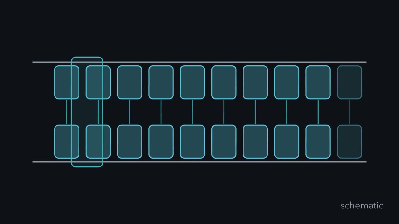 ▶ Watch How DNA Chooses a Repair Route · 3:38
Read the transcript of “How DNA Chooses a Repair Route”
Film transcript: How DNA Chooses a Repair Route
One DNA letter is wrong among billions. How can a cell repair the right spot without rewriting good DNA? Watch these two blue rows. They carry paired information. A red mark changes one block on the upper row, while the lower row stays intact. The problem's shape determines the first move.
Repair outcomes depend on what is damaged and what intact information remains. Sometimes the original base is still recognizable beneath an added chemical group. Direct reversal removes that change in place. The backbone stays uncut, and no partner strand serves as a template. This route fits only damage with chemistry an enzyme can directly undo. A small damaged base needs a different move. Base excision repair removes one block from the upper row. The lower row guides a replacement into that position, and the strand is sealed. One damaged block becomes one restored green block. Most surrounding DNA remains untouched.
Repair machinery works locally, preserving the surrounding sequence. Ultraviolet damage can join neighboring bases and bend the row. Nucleotide excision repair responds to the distorted stretch. Two cuts lift out a short patch instead of one base. The intact row guides several replacements across the gap. The same partner supplies information.
Lesion chemistry and distortion choose the pathway, which sets how much is removed. Nucleotide excision repair responds to the distorted stretch. Now both letters are ordinary, yet they pair incorrectly. Neither looks damaged. If the strands carry no history, which base should repair replace? Choose the upper block or the lower block.
The mismatch alone cannot answer. Repair needs another clue identifying the newly made strand. During replication, the template existed before the error. Signals that tell new DNA from old direct removal past the mismatch. The template then guides a corrected patch. Here, trustworthy information includes strand identity, not just the partner's letters. That extra label solves the ambiguity. Carry the same picture through the choices. A reversibly damaged base is restored in place. A small lesion loses one base. A bulky distortion loses a patch. A mismatch loses part of the strand identified as new. Each route limits its work to the problem it recognizes.
The excision routes copy the intact partner to restore the strand. If repair misses a lesion, replication may turn damage into a stable sequence change. Repair can also change sequence when information is missing. That is why finding damage is only half the problem. The cell must also know what information remains trustworthy. Follow that question whenever you choose a repair route.
From the film
- Repair outcomes depend on what is damaged and what intact information remains.
- Direct reversal restores some damaged bases without cutting the DNA backbone.
- Base excision repair removes one damaged base; nucleotide excision repair removes a damaged stretch.
- Mismatch repair also needs a signal that identifies the newly made strand.
This page adds the lesions, proteins and strand signals behind the first film’s sorting of one-strand problems.
The one idea. Repair outcomes depend on what is damaged and what intact information remains.
Scope: sequence-level repair here. Direct reversal needs no template; excision copies a partner as in DNA replication; mismatch repair needs strand identity; a double-strand break may have only its ends unless a homologous template exists. Repair need not restore the starting sequence; end joining, for example, can lose a few bases at a break.
Damage is constant: the lesions
A lesion is a change to DNA’s chemical structure, not yet a mutation, a heritable sequence change. A lesion usually becomes one when replication converts it into a normal-looking base pair on both strands.
Water and body temperature alone make some cytosines lose an amino group and become uracil, which pairs with adenine. If it remains, one round of replication puts A opposite U; the next can leave A·T where G·C had been.
The main kinds:
- Spontaneous chemistry. Besides deamination, purine bases fall off their sugars, leaving an abasic (AP) site, a sugar with no base.
- Oxidation and alkylation. Reactive oxygen produces 8-oxoguanine, which can pair with adenine. Alkylating agents add methyl groups; O6-methylguanine pairs with thymine.
- Ultraviolet light. UV joins two neighboring pyrimidines on the same strand into a cyclobutane pyrimidine dimer or a 6-4 photoproduct. Both distort the helix, the 6-4 photoproduct much more than the dimer, and both stall polymerases.
- Strand breaks. Ionizing radiation and collapsed replication forks can break both strands at once.
- Replication errors. The polymerase pairs normal bases wrongly or slips on repeats (see mutation).
Reversing and cutting out: direct reversal, BER and NER
Direct reversal undoes the chemistry in place, without cutting the backbone. Photolyases use blue and near-UV light to split pyrimidine dimers back into separate bases. They work in bacteria, yeast and plants, but humans lack the photolyases that reverse UV dimers, so your skin cannot use this route. O6-methylguanine methyltransferase (MGMT) moves the methyl group from the guanine to a cysteine in its own active site, which restores the base and inactivates the protein for good. One molecule repairs one lesion, so it is called a suicide enzyme.
Base excision repair (BER) handles most small, non-bulky base lesions that direct reversal does not: uracil, 8-oxoguanine, some alkylated bases and AP sites. Each DNA glycosylase recognizes one damaged base or a few related ones, flips it out and cuts its bond to the sugar, leaving an AP site. AP endonuclease cuts the backbone there. Polymerase beta (in human cells) inserts the correct nucleotide from the partner strand and removes the leftover sugar fragment; a ligase seals the nick. This short-patch route replaces one nucleotide; a long-patch route replaces several.
Some glycosylases, including the one that removes 8-oxoguanine, also cut the backbone; AP endonuclease then trims the cut end.
Predict. A cell has working uracil-DNA glycosylase but no working AP endonuclease. After cytosine deamination, which intermediate should accumulate: uracil in DNA, AP sites with an intact backbone, or sealed repaired DNA?
Check your prediction
AP sites with an intact backbone should accumulate. Glycosylase removes uracil, but AP endonuclease cannot make the next cut. The glycosylase exception means this result does not fit every BER substrate.
Nucleotide excision repair (NER) handles bulky, helix-distorting lesions: UV dimers, 6-4 photoproducts and adducts from drugs such as cisplatin. Because it recognizes distortion rather than one chemical group, one pathway handles many lesions. TFIIH, a complex with helicase activity, opens the DNA and helps verify the lesion before the dual cut and refill shown in the first film.
Human NER has two entry routes. XPC surveys the genome for distorted DNA, helped by UV-DDB at weakly distorting cyclobutane dimers. In a transcribed gene, RNA polymerase stalled at a lesion flags the damage. XPF-ERCC1 makes the 5′ cut and XPG the 3′ cut.
Predict. A lesion barely distorts the helix but strongly stalls RNA polymerase in an active gene. Which recognition route gives NER its clearest starting signal?
Check your prediction
The transcription-coupled route: the stalled polymerase supplies the signal. Both routes then feed the same opening, cutting and refill steps.
Fixing the copier’s mistakes: mismatch repair
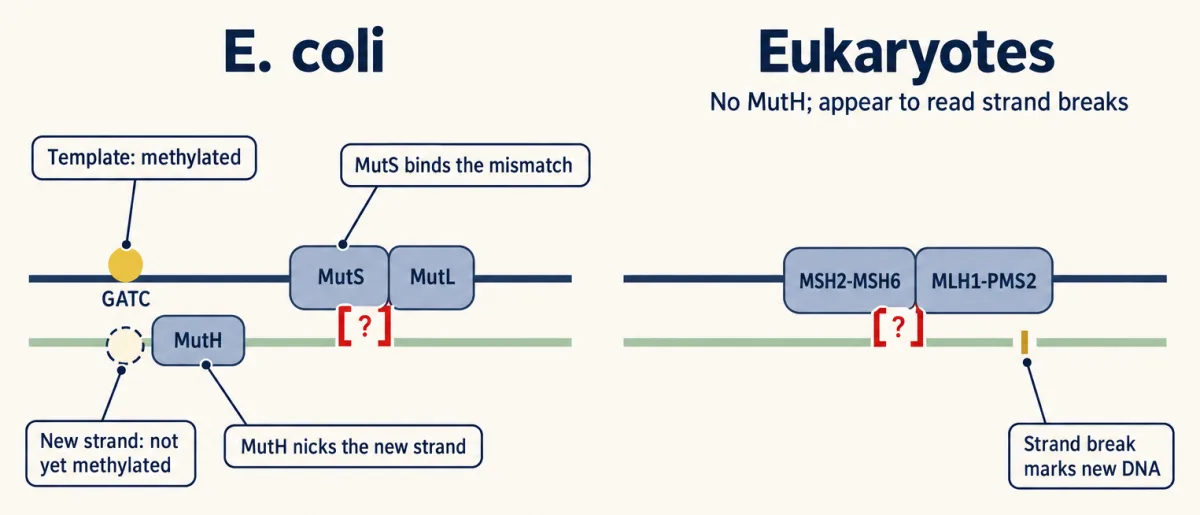
A mismatch is two normal bases paired wrongly, such as G with T. The first film showed why the new strand’s base is the one replaced; cells must mark that strand.
In E. coli the new strand carries a mark of absence: Dam methylase methylates adenine in GATC sequences only after the fork has passed, so for a window DNA is hemimethylated, methylated on the template strand only. MutS binds the mismatch, MutL joins it and they activate MutH, which nicks the unmethylated strand at GATC. From the nick, UvrD helicase and exonucleases remove the strand past the mismatch; polymerase resynthesizes the tract and ligase seals it. In Paul Modrich’s purified system, a GATC site about 1 kilobase away still directed repair.
That GATC signal is limited to E. coli and its relatives. Eukaryotes keep MutS and MutL relatives (MSH2 paired with MSH6, and MLH1 paired with PMS2 in humans) but have no MutH. They appear to read strand breaks instead: new DNA has free ends, at the tip of the growing strand and between Okazaki fragments. Human mismatch repair also corrects small loops left when the polymerase slips on a repeat.
Predict. In a purified human system, must the nick be on one particular side of the mismatch to direct repair?
Check your prediction
No. A nick on either side could direct repair in the purified system. This supports strand breaks as signals but does not settle every in-vivo strand signal.
Worked problem. The numbers here are round and chosen for illustration, not measured for one polymerase. Assume a replicative polymerase inserts a wrong base about once per 105 nucleotides, proofreading removes 99% of those errors, and mismatch repair removes 99.9% of what is left. Count errors per daughter genome: each daughter cell inherits a diploid genome of about 6.4 x 10⁹ base pairs, one strand of each pair newly made. (One S phase makes two daughter genomes, so the whole cell’s totals are double.)
Predict. Before calculating, remove mismatch repair from this model. Should the errors left per daughter genome stay below one, rise to tens, or rise to hundreds?
Check with the calculation
Step 1. Turn each percentage into a fraction that survives. Proofreading lets 1% through, 10−2. Mismatch repair lets 0.1% through, 10−3.
Step 2. Multiply the stages, because each acts on what the last one missed.
final error rate = 10−5 x 10−2 x 10−3 = 10−10 per base pair
Step 3. Scale to one daughter genome.
6.4 x 109 bp x 10−10 = 0.64 errors per daughter genome
Step 4. Remove mismatch repair. The rate is now 10−5 x 10−2 = 10−7 per base pair, and 6.4 x 109 x 10−7 = 640 errors per daughter genome.
So with these numbers, losing mismatch repair raises the error rate a thousandfold, from under one error per daughter genome to hundreds.
Every route so far worked on one strand while its partner stayed whole. The second film asks what a cell can use when a break cuts both strands.
Read the transcript of “Two Ways to Mend a Broken Chromosome”
Film transcript: Two Ways to Mend a Broken Chromosome
The first film followed repair while one strand still held a usable partner. What can a cell do when a break cuts both strands? The blue chromosome now separates into two ends. No continuous partner crosses the gap. The cell can work with those ends, or search for matching DNA elsewhere.
A broken chromosome can join its ends or copy a matching template. Non-homologous end joining chooses the ends already present. Homologous recombination copies a matching template, usually the sister chromatid. Neither name guarantees one outcome. The ends' condition and the available template shape what happens next. Start with end joining. Teal Ku rings grip both blue ends and recruit partners that hold them together. Clean, compatible ends may be joined without changing their sequence. Ends that do not fit need trimming or filling before ligation. That processing can remove or add bases, so accuracy depends on the break. End joining needs no matching template.
It can therefore operate throughout the cell cycle. Before DNA replication, that matters because no sister chromatid exists. After replication, another option becomes available beside it. Here are two cells, one before replication and one after. Which cell can copy from a sister chromatid? Watch where you expect a matching gold chromosome to appear.
After replication, each chromosome has a sister chromatid. That matching copy makes homologous recombination useful during and after replication. The broken blue chromosome aligns beside the gold sister. Before replication, the sister is absent, so this sister-templated route is unavailable. Homologous recombination first reshapes the break into single-stranded tails. A recombinase coats a tail and helps it search the gold sister. At matching DNA, the tail invades. Its end primes new green DNA, copying information from the intact template. The repaired chromosome keeps the sister's sequence across the gap. Copying does not force a crossover. Often the extended strand leaves the donor and pairs with the other broken end.
Another branch can form crossed junctions before separation. Both begin by using matching DNA as information. Reset the same break. End joining preserves clean compatible ends, but processing may change them. Homologous recombination can restore missing sequence from a sister, but only when that template exists. The choice is not a contest between bad and good repair. It responds to available information and cell-cycle context. When both strands break, ask what remains. Are two joinable ends the only local evidence? Is an intact sister nearby to copy? Those questions predict the route and its possible outcome.
From the film
- A broken chromosome can join its ends or copy a matching template.
- End joining needs no template and can work throughout the cell cycle.
- End joining can be exact when compatible ends need no processing.
- Homologous recombination usually copies the sister chromatid in S and G2.
Broken chromosomes: end joining and homologous recombination
A double-strand break cuts both strands and can split a chromosome. The one idea still decides: a cell with only two ends joins them; one with an intact template can copy it.
Non-homologous end joining (NHEJ) works with the ends it has. Ring-shaped Ku binds each end and recruits the kinase DNA-PKcs. If the ends do not fit, nucleases such as Artemis and polymerases trim or fill them, and XRCC4 with DNA ligase IV joins the result. NHEJ uses no template, so if the ends had to be processed, a few bases may be gained or lost at the joint. Calling it simply error-prone overstates the case: clean, compatible ends are often rejoined exactly.
Homologous recombination (HR) copies matching DNA. The second film’s 3′ single-stranded tails come from resection: nucleases trim the 5′ ends. The recombinase (RecA in E. coli, Rad51 in eukaryotes) invades the matching duplex, making a displacement loop (D-loop) where the 3′ end primes synthesis. The best template is the sister chromatid, made during replication, so HR acts mainly in S and G2; in meiosis, breaks are repaired from the homologous chromosome.
The second film’s usual finish, in which the extended strand leaves the D-loop and pairs with the other end, is synthesis-dependent strand annealing (SDSA); it forms no crossover. In another branch, second-end capture links the two duplexes through two Holliday junctions, four-stranded crossings. Before chromosome separation, these junctions are dissolved (no crossover) or cut by resolvases, giving a crossover or not depending on which strands are cut.
This double-junction branch makes most meiotic crossovers. Related machinery, started by programmed breaks and using meiosis-specific proteins, makes the crossovers that chromosomes and meiosis introduces and linkage mapping measures. Repair of a CRISPR cut appears in forward and reverse genetics.
NHEJ and HR are the two main routes. Single-strand annealing and alternative end joining by polymerase theta use short matching sequences on resected ends and usually delete DNA.
Match the repair to the damage: one helix strand is damaged, so cut it out and copy the partner; a break through both strands leaves no partner, so join the ends or splice from the sister. This is a sketch of the logic, not a timeline.
When repair fails: three human conditions
Unrepaired damage also triggers checkpoints, signaled by the kinases ATM and ATR and enforced partly by p53 (see stress signals). Three inherited conditions show what happens when a repair pathway is lost.
Predict. Compare an affected person with two nonworking xeroderma pigmentosum (XP) alleles to a Lynch syndrome carrier with one working MSH2 allele. Whose normal cells should already lack the relevant repair pathway?
Check your prediction
The person with XP: every cell has two nonworking copies of one XP gene (an NER gene in most forms). One working MSH2 allele usually supports repair in the carrier’s normal cells; losing it can remove repair in a tumor lineage.
Xeroderma pigmentosum (XP) is autosomal recessive: affected people have two nonworking alleles of one gene. Most forms disrupt nucleotide excision repair (NER), so UV dimers in skin cells go unrepaired, and the condition is associated with sunlight-induced skin cancers. Cell fusion sorted patients into several complementation groups, each a different NER gene. One form, the XP variant, has normal excision. Its cells cannot replicate efficiently past UV damage, and in 1999 two groups, Masutani and colleagues and Johnson, Prakash and colleagues, found mutations in the gene for DNA polymerase eta, a bypass polymerase, in XP variant cell lines.
Lynch syndrome is a dominantly inherited predisposition to colorectal, endometrial and other cancers, with risks that vary by gene. Carriers are heterozygous for a germline variant in MLH1, MSH2, MSH6 or PMS2, or have a deletion at the 3′ end of EPCAM that silences the neighboring MSH2. One working allele is usually enough for repair, so their normal cells repair mismatches. Typically a tumor arises in a cell lineage that loses or inactivates the second allele and with it mismatch repair, though some cancers in carriers keep mismatch repair. The link was made in 1993, when Fishel and colleagues and Leach and colleagues each reported MSH2 mutations in such families. The two-hit logic belongs to cancer genetics.
BRCA1 and BRCA2 were cloned by Miki and colleagues (1994) and Wooster and colleagues (1995). Both are needed for efficient homologous recombination (HR): Brca1-deficient mouse stem cells repaired chromosome breaks poorly by HR, and BRCA2-mutant human and mouse cell lines showed 6-fold to more than 100-fold less homology-directed repair, depending on the line. Carriers inherit one nonworking allele in an autosomal dominant pattern of risk for breast and ovarian cancers, and tumors typically lose the remaining allele. HR-deficient cells are unusually sensitive to PARP inhibitors, an example of synthetic lethality.
Design problem
Fibroblasts from four unrelated XP patients, P1 to P4, each show low unscheduled DNA synthesis after UV. You fuse cells in pairs and score the fused cells after UV: P1+P2 normal, P1+P3 low, P1+P4 normal, P2+P3 normal, P2+P4 low, P3+P4 normal. How many genes are involved? What would you do next to identify them?
Saved in this browser only.
One way to do it
Use the logic of complementation and pathways: XP is recessive, so a fused cell repairs normally only if each partner supplies the gene the other lacks.
- P1+P3 stays low, so P1 and P3 are defective in the same gene. P2+P4 stays low, so they share a gene.
- Every pair across those groups (P1+P2, P1+P4, P2+P3, P3+P4) is normal, so the two groups are different genes.
- Answer: two complementation groups, {P1, P3} and {P2, P4}, and so at least two genes.
To identify them, transfect cells of each group with cloned candidate NER genes, one at a time, and look for the gene that restores UV-induced repair synthesis in one group but not the other. Then sequence that gene in the group’s patients, expecting two nonworking alleles in each. A patient sensitive to UV but with normal unscheduled DNA synthesis would point instead to the XP variant, a defect in bypass by polymerase eta.
Going deeper
How excision repair was rebuilt
Tomas Lindahl’s 1974 E. coli uracil-releasing activity left the backbone uncut; his group’s 1996 human system of four purified proteins filled a one-nucleotide patch.
Aziz Sancar and Dean Rupp showed NER’s dual incision in 1983 with purified E. coli UvrA, UvrB and UvrC. The removed E. coli fragment is about 12 to 13 nucleotides long; in a human cell-free system it was 27 to 29 nucleotides.
Your notes
Select any text in the lesson to highlight it or add a note to it. Your highlights and notes are saved in this browser and collected in your notebook.
This browser is not letting the site save anything (a private window or strict privacy settings), so highlights and notes will be lost when you leave the page.
How we measure it
Unscheduled DNA synthesis
Cells outside S phase are irradiated with ultraviolet light and given labeled thymidine. Any label they take up comes from filling the gaps left by excision repair, so the amount measures how well the cells cut out and replace damage.
Cell-fusion complementation
Cells from two patients with the same recessive repair defect are fused into one cell with both nuclei. If repair returns, each patient supplies the gene product the other lacks, so their mutations are in different genes; if not, they are in the same gene.
Reconstitution from purified proteins
A repair reaction is rebuilt in a test tube from separately purified proteins and a DNA substrate with a defined lesion. It shows which proteins are sufficient for the reaction and lets each step be read separately, though it does not show how the reaction runs on chromatin in a living cell.
Site-specific break reporter
The yeast endonuclease I-SceI cuts a recognition site placed once in a chromosome, inside an inactive copy of a reporter gene such as GFP; a nearby fragment of the same gene serves as the donor. Repair of the break by homologous recombination restores a working reporter, so the fraction of reporter-positive cells measures homology-directed repair at that one site.
Practice questions on this lesson (5) → MCAT-style, original, each with an explanation
Where this lesson fits
Before thisReview first
- DNA Replication: Semiconservative Copying and Its MachineryThis course · Lesson 13
You need semiconservative copying and proofreading from there to see where replication errors and the template strand come from.
- Mutation: Where Variation Comes FromThis course · Lesson 14
It separates a lesion from a heritable mutation and shows how errors and damage become new alleles.
- Complementation and Gene Function: From Beadle and Tatum to BenzerThis course · Lesson 10
The XP fusion design problem uses complementation groups to count genes, as taught there.
After thisStudy next
- Cancer as a Genetic DiseaseThis course · Lesson 21
It takes up Lynch syndrome and BRCA carriers and the loss of the second allele that lets repair-defective tumors arise.
- Forward and Reverse Genetics: Screens, RNAi and CRISPRThis course · Lesson 24
A Cas9 cut is a double-strand break, and its outcome depends on end joining versus homology-directed repair.
Elsewhere on the siteFilms, simulations and method films in other courses
- One Strand Is EnoughFilm · Biology
It shows how each strand serves as a template, the same intact information that excision repair copies.
- Mitosis stepper and phase counterSimulation · Biology
It shows S and G2 phases, when sister chromatids exist for homologous recombination to copy.
Suggested by an AI model from the lessons themselves, then checked. How we use AI
Finish line
Test yourself
10 questions
Practice and MCAT-style, with explained answers.
Next · Lesson 16
Transcription: From DNA to RNA
A gene has two DNA strands. What chooses which one the polymerase reads?
Continue →