Genetics and Molecular Biology · Unit 5: Tools and genomes
New to this? Review it at high school level: How the DNA Model Was Built
Sequencing and Genomes
Rewritten on : new text, flashcards and film. Your notes and highlights stay, but the text around them may have moved.
A deeply sequenced genome can have almost no random gaps and still break at one repeat. How can both be true?
Pause when the reading reaches a film, and offer to play it. Saved on this device.
Watch the film now, or keep listening
Builds on:DNA Replication: Semiconservative Copying and Its MachineryRecombinant DNA, Cloning and PCRMutation: Where Variation Comes FromWhy these?
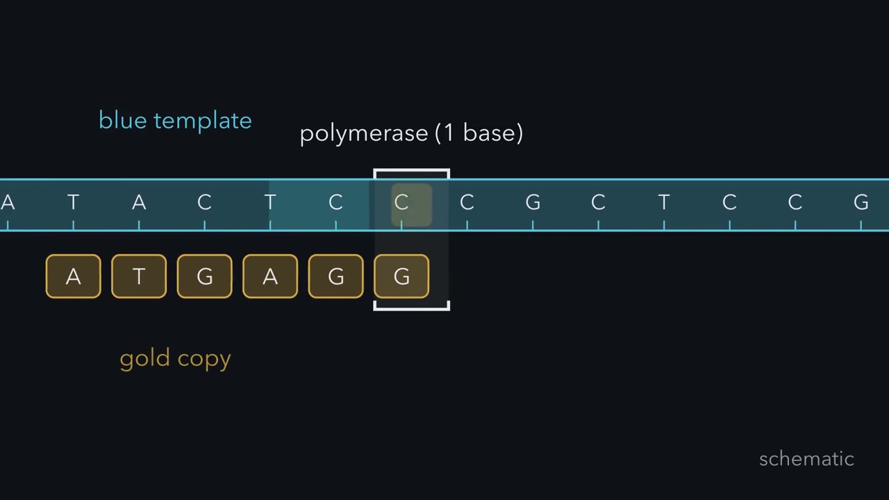 ▶ Watch How Chemistry Becomes a Read · 3:32
Read the transcript of “How Chemistry Becomes a Read”
Film transcript: How Chemistry Becomes a Read
How can a machine read DNA when a polymerase handles only the next base? Watch one blue template cross the frame. A gold copy grows beside it. The machine never sees an entire chromosome. It must turn local copying events into an ordered read.
Sanger sequencing makes many copies from the same starting primer. Most copies take ordinary nucleotides and keep growing. Occasionally, a red dideoxynucleotide joins the copy. It lacks the usable end needed for another addition. That copy stops permanently, while other copies stop at other positions. The stopped copies now sort from shortest to longest. Each red endpoint carries the identity of its last base. Reading those endpoints in length order spells the newly made strand. The template runs in the opposite direction, so finding its sequence also requires reversing the complement.
Now imagine replacing the permanent red stop with a removable teal pause. What must happen before polymerase can copy the next base? The machine images the colour while every copy in a cluster is paused. Then it removes both the dye and the block. The free end can accept another nucleotide. Repeating this add, image, and reset cycle reports one new base from every cluster each time. This is not Sanger sequencing performed in parallel.
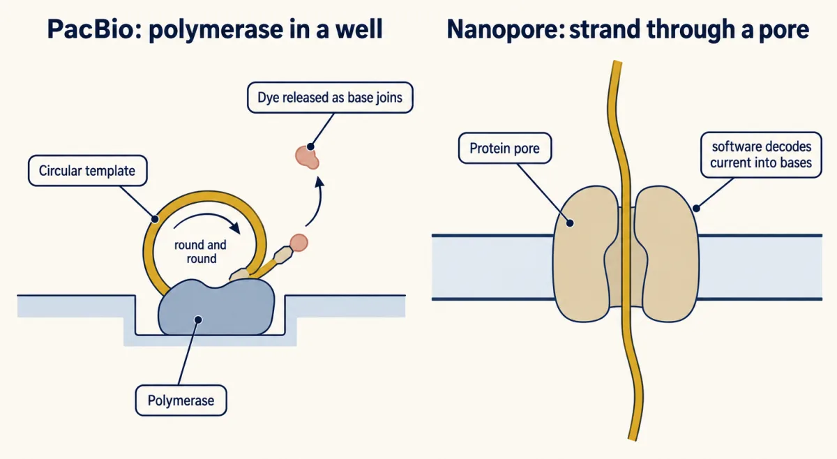
Sanger reads a permanent ladder of fragment lengths. Short-read sequencing follows temporary pauses through repeated cycles. Millions of clusters can work together, and both ends of each fragment can be read. Yet each reported stretch remains short. Long-read methods follow single molecules instead. In one method, a polymerase circles the same template repeatedly, letting repeated passes support a consensus.
In the other, a strand moves through a pore while changing ionic current. Software converts that moving signal into bases. Both approaches preserve longer-range context. These methods do not merely differ in speed. The chemistry, the DNA fragments loaded, and base-calling software together shape read length, error pattern, and context. A read is still evidence from one fragment, not a chromosome.
Later analysis must decide where it belongs. Genome results depend on what each read records and where it fits. Sequencing turns molecular events into ordered base calls. A polymerase stop, a cycle of coloured pauses, or a changing current becomes an ordered string. The chemistry shapes what evidence reaches the computer. That distinction will guide every calculation and genome comparison that follows. The next question is whether those strings fit one genomic path or several.
From the film
- Sequencing turns molecular events into ordered base calls.
- Sanger sequencing reads permanent stops sorted by fragment length.
- Short-read sequencing records one reversible pause per cycle across many clusters.
- Long-read methods follow single molecules and preserve longer-range context.
- Genome results depend on what each read records and where it fits.
Two drafts of the human genome were published in February 2001. The first assembly in which every chromosome but Y ran end to end without a gap came in 2022. As the first film showed, faster machines alone could not close the gaps: a sequencer reports reads, the base sequences of single DNA fragments, and reporting a base is not locating it in a genome.
The one idea. Genome results depend on what each read records and where it fits.
Scope: sequencing, alignment, assembly and variant calling. Read evidence alone does not say what a variant means for a person.
Reading DNA one base at a time: Sanger’s stopped copies
For the first film’s stopped copies, you supply a single-stranded template, a short primer (DNA that pairs with the template and gives the polymerase a 3’ end to extend), the four ordinary deoxynucleotides, and a small amount of dideoxynucleotides (ddNTPs). A ddNTP lacks the 3’ hydroxyl that the next nucleotide must bond to, the chemistry you met in nucleotide metabolism.
Frederick Sanger, Steve Nicklen and Alan Coulson published the method in 1977, using DNA from the phage phiX174. In the classical version, four tubes each held one kind of ddNTP, and the radioactive fragments ran in four lanes of a gel that separates pieces differing by a single nucleotide. Later versions give each terminator its own fluorescent dye, so one reaction holds all four, and a capillary sorts fragments by size while a detector reads each colour. Shortest to longest spells the new strand 5’ to 3’; the template is its reverse complement.
Predict. A detector reports the shortest fragments in order as A, C and G. What are the new strand and its template, each written 5’ to 3’?
Check your prediction
The new strand is 5’-ACG-3’. Its antiparallel template is 3’-TGC-5’, or 5’-CGT-3’. Complementing without reversing gives the template backwards.
Sanger reads are accurate, but each reaction reads one region, commonly up to about 800 to 1,000 bases, and the first few dozen bases are often poor. The sequence in the 2001 drafts came mostly from this chemistry, run on many instruments at once.
Reading millions at once, short and long
Short-read sequencing runs the first film’s add, image and reset cycle on hundreds of millions of fragments at once. Genomic DNA is broken into fragments a few hundred bases long and fitted with adapters. In the Illumina method, which David Bentley and colleagues used to sequence a human genome in 2008, each fragment is copied in place on a glass flow cell into a cluster of identical copies. The pause comes from a reversible terminator, a removable block on the 3’ end of each dye-carrying nucleotide; cleaving off the dye and the block regenerates the 3’ hydroxyl for the next cycle.
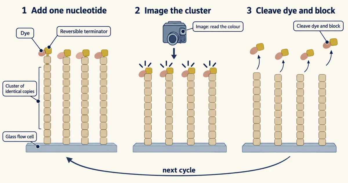
Reading each fragment from both ends, a paired-end read, gives two short sequences and roughly how far apart they lie. Reads are short (35 bases from each end in the 2008 human genome; commonly 100 to 150 now), and the errors in the study of one such instrument were mostly substitutions.
In PacBio instruments the polymerase sits at the bottom of a tiny well, with the dye on the nucleotide’s terminal phosphate so it is released as the base joins; going round the circular template many times gives HiFi reads, in a 2019 study of one human genome averaging 13.5 kilobases at 99.8% accuracy, with leftover errors mostly small insertions or deletions in runs of one base. In Oxford Nanopore instruments the pore is a protein; a 2018 study of human DNA reported ultra-long libraries in which about half the sequence lay in reads of roughly 100 kilobases or longer. The trade-off: short reads give many accurate bases per run but cannot span long repeats; long reads span most of them, though the largest repeat arrays still need ultra-long reads and special assembly methods.
That trade-off sets up the second film: once the reads exist, can their combined evidence determine one genome?
Read the transcript of “When Reads Become a Genome”
Film transcript: When Reads Become a Genome
The first film turned molecular events into reads. How do millions of reads become one genome? Some overlap unique places. Others seem to fit several. The computer must separate missing evidence from evidence that cannot be placed.
Start with sampling. Coverage is the mean number of reads lying over each base. As more gold reads land at random, blue gaps shrink. The mean does not promise equal stacks everywhere. A position can still receive many reads, few reads, or none. Under ideal random sampling, the unread fraction falls exponentially as mean coverage rises. Adding reads therefore attacks a sampling problem. It makes an unseen position less likely, although real libraries are less even. Now the same red repeat block appears at two places along the blue ribbon. A short gold read from inside that block matches both places equally. Its bases may be accurate, yet its address is ambiguous.
More copies of that same short evidence do not create a unique address. Another pile of short reads is ready to land on the repeats. Will a deeper pile tell which flanking regions belong together? The pile grows, but both placements remain possible. More reads fill sampling gaps; reads that span a repeat place it. Now one long gold read crosses the red repeat and reaches blue unique sequence on both sides. That span links the repeat copy to its flanks. De novo assembly joins reads without a reference, first producing contigs or an assembly graph. Reference alignment asks where reads fit on a reference path or graph already chosen. Now the reference branches.
A read missing from one path can fit another. What counts as a reported difference changes with the reference. Long accurate reads and ultra-long spans closed difficult gaps in one human assembly. A pangenome then added alternative paths from many chromosome copies. Completeness asks whether sequence is missing. Representation asks whether one path captures human variation. Reference choice changes two stages. First, it changes whether a read has a close place to align. Then it changes which base or segment is labelled different. A variant call is therefore a comparison, not a claim about rarity, selection, or health. Genome results depend on what each read records and where it fits.
Was the region sampled, and was the evidence placed uniquely? Extra reads help the first. Reads that span the repeat help the second. A richer reference gives reads from missing sequence a place to land. Those are different limits, and they need different fixes.
From the film
- More reads fill sampling gaps; reads that span a repeat place it.
- Coverage is a mean, so random sampling can leave some bases unread.
- An internal read from an exact repeat may fit more than one place.
- A reference helps place reads but also shapes which differences are reported.
- Genome results depend on what each read records and where it fits.
Coverage and assembly: two different limits
Reads aligned to a reference are placed on it; reads assembled de novo, without one, are joined by their overlaps, first into an assembly graph or contigs, continuous consensus sequences. Scaffolds place contigs in order and orientation, with estimated gaps between them.
Coverage (depth), the second film’s mean stack of reads over each base, is all the bases read, spread over the genome:
In words Coverage is the number of reads times the read length, divided by the genome length.
- C“C, coverage”
- the mean number of reads over each base, written as a fold (30-fold, 30x)
- N“N”
- the number of reads
- L“L”
- the read lengthbases
- G“G”
- the haploid genome lengthbases
Sanity check Bases on top and bases below cancel, so coverage is a plain number. Double the reads or the read length and coverage doubles; sequence a genome twice as big with the same reads and it halves.
Try one number
Round numbers to keep the arithmetic easy (not from a real project): 1,000 reads of 100 bases on a 10,000-base piece of DNA.
Total bases read: 1,000 × 100 = 100,000. Spread over 10,000 bases: 100,000 / 10,000 = 10.
Each base is covered 10 times on average, 10-fold coverage.
Need a refresher? powers and roots · units and prefixes
Worked problem. Assume reads start at random positions, all reads are usable, and the haploid human genome is G = 3.1 x 10⁹ bases (an estimate rounded for the calculation).
Predict, then calculate. How many 150-base reads give 30-fold mean coverage? First decide: should it rise or fall if each read were longer? Then multiply both sides by G (C x G = N x L) and divide by L.
N = C x G / L = 30 x 3.1 x 109 / 150 = 9.3 x 10¹⁰ / 150 = 6.2 x 10⁸ reads
That is 620 million reads, or 310 million read pairs of 2 x 150 bases, and 93 billion bases. Check: 6.2 x 108 x 150 = 9.3 x 10¹⁰, and dividing by 3.1 x 10⁹ gives 30. Longer reads mean fewer of them: doubling L to 300 bases halves N to 3.1 x 10⁸. (Going deeper works the powers of ten step by step; see also powers.)
Predict, then calculate. At C = 5, what fraction of bases should be unread, and how many bases is that in this genome? The second film’s falling curve comes from the Poisson model: with random starts, the number of reads over a base follows a Poisson distribution with mean C. The fraction with zero reads is then:
In words The fraction of bases with no read is e to the minus C.
- e−C“e to the minus C”
- the Poisson chance of zero reads at a base. e is about 2.718, and e−C means 1 divided by eC; for C = 3 that is 1 divided by e × e × e.
- C“C”
- the mean coverage (fold)
Sanity check At C = 0, e0 = 1: with no reads, every base is unread. Each extra fold of coverage multiplies the unread fraction by e−1, about 0.37, so it falls fast but never reaches exactly zero.
Try one number
At C = 3: e−3 is about 0.37 × 0.37 × 0.37, about 0.05 (0.0498 on a calculator).
So at 3-fold coverage about 5% of the genome would get no read at all.
Need a refresher? e and ln · powers and roots
At three depths:
C = 1: e−1 = 0.37, so 37% unread
C = 5: e−5 = 0.0067, so 0.0067 x 3.1 x 109 = about 21 million bases unread
C = 30: e−30 is about 9 x 10⁻¹⁴, far less than one base
So 30-fold is a mean, not a guarantee; Going deeper adds real libraries and the model’s source.
Put numbers on the second film’s repeat: two identical 6-kilobase copies. No 150-base read, and no pair from a 400-base fragment, reaches from unique sequence on one side across the repeat to its far edge. The assembler cannot determine which flanking sequences belong together, so it may collapse the copies or break the contig. More reads fill sampling gaps but add no new span; reads that span a repeat place it. A 15-kilobase read can hold one whole copy plus several kilobases of unique sequence on each side, pinning that copy in place.
That is the rule in short: ask what each read records, then where it fits. The exact-repeat model is deliberately simple: differences between copies, linked pairs and other long-range evidence can sometimes place repeats no single read spans. Some human-specific gene duplications described in the human brain expansion lesson sit in long near-identical repeats, one reason they were hard to study.
| In this draw | Observed | Expected |
|---|
Depth decides how many bases go unread (e−C), but only a read that reaches unique sequence on both sides of a repeat can place it; this is a sketch of the logic on a toy 60-kilobase ring (so the ends behave like the middle), not a real assembler, a real assembler needs more unique flank than one base, and real libraries cover the genome less evenly.
The human reference: gaps, a finished genome and a pangenome
The 2001 papers, from the public International Human Genome Sequencing Consortium and from Celera Genomics, were drafts. The consortium’s “finished” sequence of 2004 covered about 99% of the euchromatin (the gene-rich, less tightly packed part of the genome) and still had 341 gaps, many associated with segmental duplications; centromeres and other heterochromatin were not part of its target. The same paper estimated 20,000 to 25,000 protein-coding genes, far fewer than the 2001 estimate. The current standard reference, GRCh38, descends from it: a mosaic of DNA clones from several people, not any one person’s genome.
Many remaining gaps were the worked example’s repeat problem at chromosome scale, in large centromeric arrays and near-identical segmental duplications. The Telomere-to-Telomere Consortium closed them using the CHM13 cell line, whose two copies of each chromosome are nearly identical, and a combination of PacBio HiFi and Oxford Nanopore ultra-long reads. Their 2022 assembly, T2T-CHM13, is 3.055 billion bases and gapless for every chromosome except Y, which CHM13 lacks. A complete Y from a different person, HG002, followed in 2023.
One complete genome still represents one person. In 2023 the Human Pangenome Reference Consortium released a draft pangenome, the second film’s branching reference: 47 people’s genomes, each with its two chromosome copies assembled separately, joined into a graph in which each person’s copy is a path. It added 119 million bases of variable euchromatic sequence missing from GRCh38.
What varies between two genomes, and why the reference matters
Differences come in sizes. A single-nucleotide variant (SNV) changes one base. A small indel inserts or deletes a few bases. A structural variant is larger, by a common convention 50 bases or more, and includes deletions, duplications, inversions, insertions and translocations; when a deletion or duplication changes the number of copies of a segment, it is a copy-number variant. The chromosome-scale versions are in chromosome changes, and how such changes arise is in mutation.
In the 1000 Genomes Project’s 2015 analysis of 2,504 people from 26 populations, a typical genome differed from the reference at 4.1 to 5.0 million sites, and structural variants affected more bases than SNVs and small indels together. Each is a comparison with a reference. Where the reference carries a rare allele, most people look “variant” there; where a sequence is missing from the reference, reads from it have no correct place to align, so they go unmapped or land on similar sequence elsewhere. In the Human Pangenome Reference Consortium’s benchmarks, short-read workflows using the pangenome made 34% fewer small-variant errors and found 104% more structural variants per haplotype (2.04 times as many) than workflows using GRCh38; those figures describe the workflows tested, not every data set.
Short reads detect large variants indirectly, from depth (copy number changes the number of reads), discordant pairs (two ends of a fragment mapping farther apart than the fragment could be, or in an unexpected orientation) and split reads (one read aligning in two pieces across a junction).
Predict and design
Predict. Short paired-end reads from a patient sample are aligned to the reference. The fragments are normally 400 ± 50 bases long. Depth is 30-fold across a chromosome segment but 15-fold across one 10-kilobase stretch within it. Forty read pairs map with their two ends 10.4 kilobases apart across that stretch, and several reads split, aligning partly to each edge. What does this pattern show, and how would you confirm it?
One way to do it
Half depth means one copy instead of two: one chromosome has lost the stretch and the other has not, a heterozygous deletion. The discordant pairs give its size: ends that should lie about 400 bases apart map 10.4 kilobases apart, so about 10.4 - 0.4 = 10 kilobases are missing from the chromosome they came from. The split reads mark the two breakpoints to the base.
Confirm it two ways. Design PCR primers just outside the two breakpoints. On the intact chromosome they lie about 10.4 kilobases apart, too far for a standard short PCR, but on the deleted chromosome they lie a few hundred bases apart, so a short product appears only from the deleted allele; sequence it to read the junction. Long reads across the region would show both alleles directly.
Then ask whether the deletion is unusual at all. Check population variant collections and the pangenome haplotypes: if many people lack this stretch, the reference may carry a less common insertion, and the “deletion” is common. Finding a variant says nothing on its own about any effect on health.
Going deeper
The powers of ten in the coverage problem
The full arithmetic behind the read count is 30 x 3.1 = 93, so 30 x 3.1 x 109 = 93 x 109 = 9.3 x 10¹⁰. Then 9.3 / 150 = 0.062, and 0.062 x 1010 = 6.2 x 10⁸ reads.
Where the ideal coverage model breaks
Real libraries cover some regions, such as very GC-rich ones, less evenly than the Poisson model assumes. The treatment here follows Eric Lander and Michael Waterman’s 1988 analysis of random clones; the same reasoning applies to ideal random shotgun sampling.
Your notes
Select any text in the lesson to highlight it or add a note to it. Your highlights and notes are saved in this browser and collected in your notebook.
This browser is not letting the site save anything (a private window or strict privacy settings), so highlights and notes will be lost when you leave the page.
How we measure it
Sanger chain-termination sequencing
A polymerase copies one template from a primer with ordinary nucleotides plus a small amount of dideoxynucleotides, each type carrying its own fluorescent dye. Capillary electrophoresis sorts the stopped copies by length, and the dye at each length names the base. It reads one region at a time, commonly up to about 800 to 1,000 bases.
Short-read sequencing by synthesis
Fragments with adapters are amplified into clusters on a flow cell, then copied one base per cycle with fluorescent reversible terminators, imaging every cluster after each cycle. Hundreds of millions of short reads, often from both ends of each fragment, come from one run.
Long-read single-molecule sequencing
PacBio instruments watch a single polymerase copy a circular molecule many times; Oxford Nanopore instruments read the change in ionic current as one DNA strand passes through a protein pore. Reads run to tens of kilobases or longer, long enough to span most repeats; the largest repeat arrays need ultra-long reads and special assembly methods.
Read alignment, depth and de novo assembly
Reads are either aligned to a reference genome, where depth and read-pair geometry reveal variants, or assembled de novo by their overlaps into contigs and scaffolds. Coverage statistics say how evenly and how completely the genome was sampled.
Method films · the methods library
Practice questions on this lesson (3) → MCAT-style, original, each with an explanation
Where this lesson fits
Before thisReview first
- DNA Replication: Semiconservative Copying and Its MachineryThis course · Lesson 13
Sanger and short-read sequencing both rely on polymerase extending a primer, so you need how new strands are built here.
- Recombinant DNA, Cloning and PCRThis course · Lesson 22
Library preparation and amplification of DNA fragments use the cloning and PCR ideas taught there.
- Mutation: Where Variation Comes FromThis course · Lesson 14
Variants called against a reference are the mutations whose origins and types are described there.
After thisStudy next
- Forward and Reverse Genetics: Screens, RNAi and CRISPRThis course · Lesson 24
Genome sequence and reads are used there to find which gene to target and to read what an edit leaves.
- Population Genetics: Hardy-Weinberg and Evolutionary ForcesThis course · Lesson 25
Variants found by sequencing many genomes are the allele frequencies this lesson analyses.
- Complex Traits, Heritability and GWASThis course · Lesson 26
GWAS builds on genotypes and variant calls from sequencing, and on the reference used.
Elsewhere on the siteFilms, simulations and method films in other courses
- Molecular clock datingMethod film
Sequence differences between genomes accumulate over time, and this method dates splits from them.
- Radioactive and stable isotope tracersMethod film
Radioactive labels were used to read the classical four-lane Sanger gel fragments.
Suggested by an AI model from the lessons themselves, then checked. How we use AI
Finish line
Test yourself
8 questions
Practice and MCAT-style, with explained answers.
Next · Lesson 24
Forward and Reverse Genetics: Screens, RNAi and CRISPR
How do you find what a gene does: start from a trait and hunt for the gene, or start from a gene you name and see whether the cell cares?
Continue →