Physiology · Unit 4: Breathing
Lung Mechanics and Surfactant
Rewritten on : new text, figures and flashcards. Your notes and highlights stay, but the text around them may have moved.
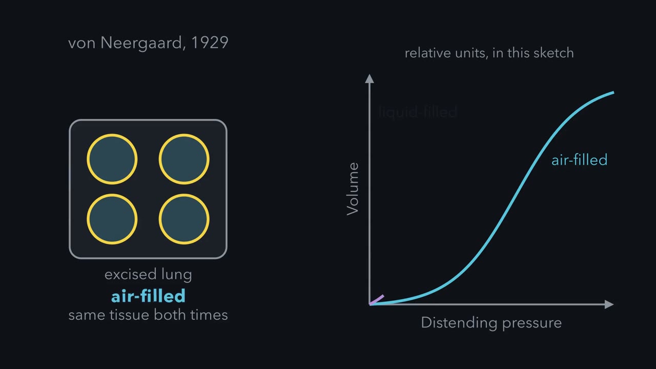
Fill an excised lung with saline instead of air and it takes far less pressure to inflate. What was the air adding?
Pause when the reading reaches a film, and offer to play it. Saved on this device.
Watch the film now, or keep listening
 ▶ Watch Why Small Alveoli Need Surfactant · 2:37
Read the transcript of “Why Small Alveoli Need Surfactant”
Film transcript: Why Small Alveoli Need Surfactant
Fill an excised lung with saline instead of air, and it takes far less pressure to inflate it to the same volume. The tissue is the same both times. In 1929, Kurt von Neergaard compared an isolated pig lung filled with air and with a gum solution. The liquid removes the air-liquid surface that lines every alveolus. He concluded that surface tension, not tissue elasticity, accounts for most of the lung's recoil. So how hard is it to keep one alveolus open? Laplace's relation answers it for a single spherical air-liquid surface. The pressure equals 2T divided by r. T is the surface tension, and r is the radius. Take a radius of 100 micrometres and a tension of 70 mN/m, with no surfactant.
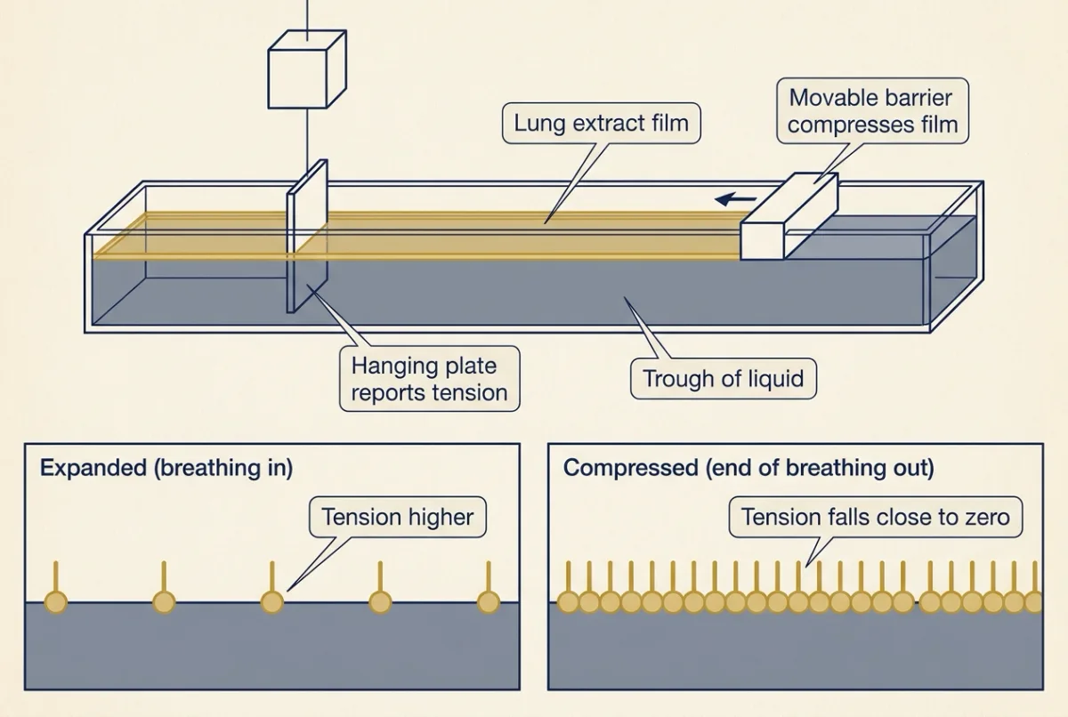
These are illustrative values. The pressure is 14.3 cmH2O. Now halve the radius to 50 micrometres, at the same tension. The pressure doubles, to 28.6 cmH2O. Join the two alveoli to one airway. At equal tension, the small alveolus would empty into the large one. Surface tension makes the smaller space harder to hold open. Surfactant changes that. In the 1950s, John Clements put films from rat, cat and dog lung extracts on a surface balance. A movable barrier compresses the film, and a thin plate hanging in the surface reports the tension. The tension depended on area. It fell from about 46 to about 10 dynes per cm as the film was compressed. The name surfactant came later.
Give the alveoli a lining whose tension falls as the area falls. These values are illustrative, and the tension in the small alveolus is made up. At 100 micrometres, with surfactant, tension is 20 mN/m. The pressure is 4.1 cmH2O. At 50 micrometres, tension is 5 mN/m. The pressure is 2.0 cmH2O, below the large alveolus. The small one no longer empties. This emptying is a result of theory and of films studied in vitro. Whether a film prevents it depends on how steeply tension changes with area. Real alveoli are also tethered. Neighbours share walls, so one alveolus cannot shrink alone like a free bubble. Keep this rule. Surface tension makes small alveoli harder to hold open, and surfactant lowers tension most when the alveolus is small.
Fill an excised lung with saline instead of air and it takes far less pressure to inflate it to the same volume. The tissue is the same both times.
Predict. What does the air add that saline does not?
Check your prediction
An air-liquid surface. Saline removes the wet surface that lines every alveolus but leaves the tissue in place; with air, that surface is present, and its tension pulls each alveolus smaller. The gap between the two inflations is the part of the lung’s recoil that comes from surface tension, and it is a large part.
The lung makes surfactant, a film that lowers the tension of its wet lining. A baby born before its lungs make enough has a stiff lung whose alveoli tend to collapse at the end of each breath: respiratory distress syndrome (RDS). Gas exchange and breathing gave the first look at surfactant and compliance; here they get numbers.
The one idea to carry through this lesson. A breath has two pressure drops. Across the lung’s wall, transpulmonary pressure (alveolar minus pleural) equals the lung’s recoil at its current volume: exactly when no air flows, and very nearly during quiet breathing. That recoil is tissue plus the surface tension of the wet alveolar lining. Along the airways, the gap between atmospheric and alveolar pressure is what moves air, and it grows with flow and with airway resistance. Pleural pressure, set by the breathing muscles and the chest wall, has to pay for both. Surfactant shrinks the surface part of the recoil; disease changes the recoil or the resistance.
The rule is for the lung alone; the chest wall is a second spring (limits in Going deeper, “Limits of the one idea”).
One breath, two pressure drops
The visceral pleura covers each lung and the parietal pleura lines the chest wall, with a thin film of fluid between. The stretched lung recoils inward; the chest wall springs outward. At functional residual capacity (FRC), the volume left after a quiet expiration, the two recoils balance. The lung pulls the inner layer in and the chest wall pulls the outer layer out; the sealed fluid film between them cannot expand, so its pressure falls below atmospheric. Picture two springs hooked together, one stretched and one compressed, resting where their forces cancel.
Three pressures describe a breath. Each is measured against atmospheric (0), in centimetres of water (cmH2O), the unit lung physiologists use because breathing pressures are small and a water column reads them easily.
- Alveolar pressure is the pressure of the gas inside the alveoli.
- Pleural pressure is the pressure in the thin fluid film between lung and chest wall, just outside the lung.
- Transpulmonary pressure is alveolar minus pleural pressure: inside minus outside, the pressure across the lung’s wall.
Why inside minus outside? Think of a balloon. Its rubber stays stretched only while the air inside pushes harder than the air outside, by just the pull of the rubber; let the difference fall and the balloon shrinks until its rubber pulls only that hard. The lung works the same way, which is why transpulmonary pressure tracks recoil: a bigger lung recoils harder, so it needs a bigger transpulmonary pressure.
The second pressure drop runs along the airways: air flows in only while alveolar pressure is below atmospheric, and out only while it is above, and the faster the flow or the narrower the airways, the bigger that drop.
Now follow one quiet breath in a healthy adult at rest. Recoil, the lung’s elastic recoil at that moment’s volume, equals transpulmonary pressure on every row of this simplified table (to within a small tissue-friction term during flow; row 2: −1 − (−7.5) = 6.5).
| Moment | Air flow | Alveolar | Pleural | Transpulmonary (= recoil) |
|---|---|---|---|---|
| End of a quiet expiration (FRC) | none | 0 | about −5 | about 5 |
| Middle of inspiration | in | about −1 | about −7.5 | about 6.5 |
| End of inspiration | none | 0 | about −8 | about 8 |
| Middle of expiration | out | about +1 | about −5.5 | about 6.5 |
With no flow (FRC, end-inspiration), alveolar pressure is 0 and pleural pressure is simply minus the recoil: −5, then −8 once the lung is more stretched. In between, the diaphragm contracts, pleural pressure falls, the lung expands with the chest and its gas spreads into more room, so alveolar pressure dips below atmospheric and air flows in. Expiration at rest needs no muscle: the diaphragm relaxes and recoil squeezes the alveolar gas above atmospheric. In quiet breathing pleural pressure stays below atmospheric throughout; it turns positive only when the expiratory muscles contract, as in a forced expiration or a cough.
So the fall in pleural pressure pays for two things. At mid-breath it has fallen 2.5 cmH2O: 1.5 pays for stretching the lung (recoil 5 to 6.5) and about 1 pays for moving air through the airways. By end-inspiration flow has stopped, and the whole 3 cmH2O fall is elastic. A sicker lung shifts that split: a stiff lung needs more for the first, and narrowed airways need more for the second. The inspiratory fall in pleural pressure also helps blood return to the heart (cardiac output and venous return).
Predict. A stab wound opens the pleural space to the outside air. What happens to pleural pressure, to transpulmonary pressure, and to the lung?
Check your prediction
Pleural pressure rises to 0, atmospheric. Alveolar pressure is also 0 once flow stops, so transpulmonary pressure falls to 0, less than the lung’s recoil at any normal volume. The lung shrinks until its recoil is zero, which is close to collapse. The chest wall, no longer pulled inward by the lung, springs outward. This is a pneumothorax.
Compliance: the slope of the recoil curve
Plot lung volume against transpulmonary pressure, with no air flowing, and you trace the lung’s recoil curve. Its slope, how stretchy the lung is, is compliance.
In words Compliance equals the change in volume divided by the change in distending pressure.
- C“C, compliance”
- the stretchiness. It is measured at zero flow, so that only elastic forces act. For the lung alone it is written CL.L per cmH2O
- ΔV“delta V”
- the change in volume (Δ, “delta”, means “change in”)L
- ΔP“delta P”
- the change in distending pressure; for the lung, the change in transpulmonary pressurecmH2O
Sanity check A stiff lung needs a big pressure change for a small volume change, so its compliance is small; a floppy lung is the reverse. Elastance, the stiffness, is the inverse: 1 / C.
Try one number
Worked problem part 1 below finds that a 0.50 L breath raises transpulmonary pressure by 3 cmH2O: compliance = 0.50 / 3 = 0.17 L per cmH2O.
The elastance of the same lung is the flip, 3 / 0.50 = 6 cmH2O per L: it takes 6 cmH2O to add each litre.
Need a refresher? rates and “per” · units and prefixes
Worked problem, part 1: compliance and elastic work. The quiet breath above, tidal volume 0.50 L, zero flow at functional residual capacity (FRC) and at end-inspiration, a straight pressure-volume line, lung only.
- Alveolar pressure is 0 at both points, so transpulmonary pressure is 0 − (−5) = 5 cmH2O at FRC and 0 − (−8) = 8 cmH2O at end-inspiration.
- Lung compliance = 0.50 / 3 = 0.17 L per cmH2O (the card’s example), near the textbook adult figure of about 0.2.
- The 5 cmH2O present at FRC is held by the chest wall’s outward spring, so count only what the breath adds: the extra pressure grows from 0 to 3 cmH2O, and the extra elastic work on the lung alone, the area under the pressure-volume line, is a triangle: 0.5 x 3 cmH2O x 0.50 L = 0.75 cmH2O x L.
- Halve compliance to 0.083 L per cmH2O: the same breath needs 6.0 cmH2O and 0.5 x 6.0 x 0.50 = 1.5 cmH2O x L, double.
A stiffer lung costs more work for every breath.
What the air adds: surface tension
A static pressure-volume curve is made by inflating an excised lung in steps, holding each until flow stops, then deflating it the same way. The saline curve lies far to the left: at any volume it needs much less pressure. And it shows little hysteresis, while the air curve shows a lot: at the same pressure, the air-filled lung holds more volume on deflation than on inflation.
Saline removes the surface and leaves the tissue, so the pressure gap between the curves at one volume is the surface part of recoil. In 1929 Kurt von Neergaard compared an isolated pig lung filled with air and with a gum solution, and concluded that surface tension, not tissue elasticity, accounts for most of the lung’s recoil. Mead’s laboratory later found the same in excised lungs: surface tension supplies much of the recoil, and the air-saline gap widens as volume rises. The hysteresis also comes mainly from the air-liquid interface, through the surfactant film and through collapsed airspaces and small airways reopening, with a smaller share from tissue; it is larger for breaths starting near residual volume.
An air-water surface acts like a stretched sheet; its surface tension, T, is force per length, in mN per m (equal to dynes per cm). Pulling along the curved surface, it pulls the alveolus smaller, adding to the recoil that transpulmonary pressure must match. How much pressure? Laplace’s relation:
In words The pressure equals two times the surface tension, divided by the radius.
- P“P”
- the pressure across one spherical air-liquid surface that holds it openPa (1 cmH2O = 98.07 Pa)
- T“T, surface tension”
- force per length along the surfaceN per m (1 mN per m = 1 dyne per cm)
- r“r”
- the radius of the spherem
Sanity check No tension, no pressure needed. Double the tension and the pressure doubles. Halve the radius and the pressure doubles: a small bubble is harder to hold open than a big one at the same tension.
Try one number
Units first: N per m divided by m is N per m², which is a pascal (Pa).
Sizes: 100 micrometres = 0.1 mm = 1.0 x 10⁻⁴ m, that is 0.0001 m. Dividing by such a small number gives a large result, which is why tiny alveoli need sizeable pressures. Worked problem part 2 below puts in the numbers.
Need a refresher? rates and “per” · units and prefixes · powers and roots
This is for one surface. A soap bubble has two surfaces and needs 4T/r; an alveolus has one. The sphere simplifies polyhedral alveoli, so treat every number below as a crude one-sphere model.
Predict. Line an alveolus of radius 100 micrometres with plain water (T = 70 mN per m, no surfactant). A healthy lung has a transpulmonary pressure of about 5 cmH2O at functional residual capacity (FRC). Is that enough to hold it open?
Check your prediction
No. P = 2 x 0.070 / (1.0 x 10⁻⁴ m) = 1,400 Pa, which is 14.3 cmH2O, almost three times the 5 cmH2O available. In this crude model a water-lined sphere would have to grow to a radius of about 286 micrometres before 5 cmH2O could hold it. Something must be lowering the tension.
Worked problem, part 2: how hard is it to keep an alveolus open? Illustrative values, static, 1 cmH2O = 98.07 Pa.
The prediction above is step 1: r = 100 micrometres, plain water, 14.3 cmH2O.
- With surfactant, take a mid-range T = 0.020 N per m: P = 400 Pa = 4.1 cmH2O, 3.5 times smaller. That is the size of the surface term in this model, not a measured share; it is below the 5 cmH2O available at FRC, which also has to cover the tissue recoil, so real alveolar tension at end-expiration must be lower still.
- Halve r to 50 micrometres at 0.070 N per m: P = 2,800 Pa = 28.6 cmH2O. At equal tension the small alveolus would empty into the large one.
- Let tension fall with area (made-up): at 50 micrometres, T = 0.005 N per m, so P = 2 x 0.005 / 5.0 x 10−5 = 200 Pa = 2.0 cmH2O, below the 400 Pa of the 100-micrometre alveolus in step 2. The small one no longer empties.
Steps 3 and 4 are a model of two free bubbles joined to one airway. Real alveoli share walls with their neighbours, so one cannot shrink alone like a free bubble (tethering), and whether a real film prevents the emptying depends on how steeply its tension falls (Going deeper, “Small into large”).
In this sketch, the status appears here.
In this sketch: each alveolus is one free spherical air-liquid surface, static, so P = 2T/r (the lesson’s relation for one surface), with 1 cmH₂O = 98.07 Pa. The values 70 and 20 mN per m are the lesson’s illustrative tensions. The rule T = 20 mN per m × (r / 100 µm)², is made up for the sketch; it gives the lesson’s made-up 5 mN per m at 50 µm. The 5 cmH₂O is the lesson’s transpulmonary pressure for a whole healthy lung at FRC; comparing it with one sphere’s surface term is only a crude sense of scale. The radius ranges are chosen for the sketch, and the small alveolus is never set larger than the large one (the other slider moves along).
Real alveoli are tethered polyhedra: neighbours share walls, so one alveolus cannot shrink alone like a free bubble. The spheres are a simplification. Drawing: circle size is to scale with radius, and a thicker outline means higher tension.
Keep this: at equal tension the smaller alveolus has the higher pressure and would empty into the larger one; whether a film prevents that depends on how steeply tension changes with area.
About the film at the top of this page. “Why Small Alveoli Need Surfactant” works through Laplace’s relation for one and then two alveoli, the free-bubble model above. Surfactant’s tension is not one number (next section); the film’s 20 and 5 mN per m are illustrative points on its range. Respiratory distress syndrome (RDS) and its treatment are not in the film.
Surfactant lowers tension most when the alveolus is small
What it is. Surfactant is about 90 percent lipid, mainly phospholipid, and 10 percent protein, secreted by alveolar type 2 cells, which store it in lamellar bodies. Its main lipid is dipalmitoylphosphatidylcholine (DPPC; see lipids). Phospholipid alone adsorbs too slowly for a breathing lung; surfactant protein B (SP-B) speeds adsorption.
What it does. Its tension changes with area. Expanded in inspiration, the film’s tension rises. Compressed at end-expiration, it falls close to 0 mN per m on modern surface balances, and in situ estimates at low lung volume are also very low. So tension is lowest when alveoli are smallest, and the smallest airspaces, which a constant tension would close first, stay open. And with a smaller surface part of the recoil, the same breath needs a smaller pleural swing and less work.
How we know. In 1957 John Clements, in Maryland, put films from extracts of rat, cat and dog lungs on a modified Wilhelmy balance, where a movable barrier compresses the film and a thin plate hanging in the surface reports the tension. Tension fell from about 46 to about 10 dynes per cm as the film was compressed. He called it a surface-active material; it later became known as pulmonary surfactant. Richard Pattle in England had inferred a low-tension lining two years earlier, from lung foam that stayed stable for hours.
Why it matters: the classic experiment. Hyaline membrane disease (HMD) is the lung pathology of respiratory distress syndrome in newborns, most often preterm ones. Mary Ellen Avery saw Clements’s balance in December 1957; in their 1959 paper she and Jere Mead put extracts of lungs from infants who had died, with or without HMD, on a surface balance and recorded the lowest tension as each film was compressed. If HMD lungs lacked the lining, their extracts should not reach low tension. They did not: a mean of about 30 dynes per cm in extracts from nine infants with HMD, against about 8 in extracts from infants over about 1,200 g who died of other causes.
Predict. Extracts from smaller infants who died of other causes, without HMD, also stayed high, about 26 dynes per cm. Does that weaken or strengthen the claim that a lack of surfactant causes HMD?
Check your prediction
It weakens it as proof of cause, though not the link. Immature lungs lacked the low-tension lining with or without the disease, so immaturity and disease were entangled. And post-mortem, injured lungs left open whether the deficiency followed from the disease or from death. Avery and Mead concluded only that HMD is associated with absence or late appearance of a substance that lets the lung surface reach low tension at low volume.
The case for cause came later, from animal studies and from treatment. Randomized controlled trials in the 1980s and 1990s, and their meta-analyses, established that surfactant replacement lowers mortality and pneumothorax in preterm infants. Liggins and Howie’s 1972 trial showed that antenatal glucocorticoid treatment can speed fetal lung maturation (pregnancy, birth and lactation), and antenatal glucocorticoids lower RDS and neonatal death. Antenatal steroids for women at risk of early preterm birth, and replacement surfactant for preterm infants with RDS, are now standard care.
The pressure balance explains what RDS looks like. A large surface term means a stiff lung, so each breath needs a large negative swing in pleural pressure. That pulls the infant’s soft chest wall inward with each breath (retractions), and at low volume the alveoli close.
The modern experiment. If surfactant needs correct packaging as well as its proteins, a packaging defect should cause deficiency even at term. After an SP-B mutation in infants (1994) and SP-B knockout mice (1995) tied the protein to survival, Shulenin, Nogee, Whitsett, Dean and colleagues in 2004 sequenced ABCA3, an ATP-binding cassette transporter gene, in infants born at or near term with severe surfactant deficiency of unknown cause, and found mutations in most: the packaging, not only the proteins, is needed in humans (limits in Going deeper, “The genetics”).
Reading the balance in the clinic
Spirometry measures what moves, not what stays
A spirometer records volume against time. Four volumes do not overlap: tidal volume, inspiratory reserve volume (the extra that can be inhaled after a tidal breath), expiratory reserve volume (the extra that can be exhaled) and residual volume (RV, what stays after a maximal expiration). Capacities are sums: inspiratory capacity (tidal plus inspiratory reserve), functional residual capacity (FRC) (expiratory reserve plus RV), vital capacity (all but RV) and total lung capacity (TLC, all four).
Gas that never leaves the lung is invisible to a spirometer, so RV, FRC and TLC need other methods. These are helium dilution (a known amount of helium spreads into the lung and is diluted), nitrogen washout (collect the nitrogen washed out by pure oxygen) and body plethysmography (Boyle’s law in a sealed box). In obstructive disease the first two reach only gas that communicates with the airways, so they can underestimate RV, FRC and TLC.
Volumes vary with height, age and sex, so one person’s value is judged against reference equations from healthy populations, which give a lower limit of normal.
In the forced test, from TLC, blow out as hard and fast as possible: the total is the forced vital capacity (FVC), and the volume in the first second is FEV1.
Forced expiration: the same balance, run hard
Pleural pressure still pays for recoil and airflow together. In a forced expiration the expiratory muscles push pleural pressure above atmospheric. Alveolar pressure is then recoil plus pleural pressure, so blowing harder raises the pressure that drives air out.
But the airways sit in the same pleural space. As air flows out, friction uses up pressure along the airways, so the pressure inside them falls until it equals pleural pressure, and downstream of that point the positive pleural pressure squeezes the deformable airways: dynamic compression.
Predict. At mid-volume, a healthy person who is already blowing out moderately hard blows harder still. Does expiratory flow rise?
Check your prediction
Past a modest effort, no. Extra pleural pressure raises alveolar pressure, but squeezes the airways downstream of the squeeze point by just as much. The airway upstream of that point is driven by the lung’s recoil alone. So at middle and low volumes, maximal expiratory flow is set by recoil and the resistance of the airways upstream of the squeeze point, not effort. At high volumes flow still rises with effort.
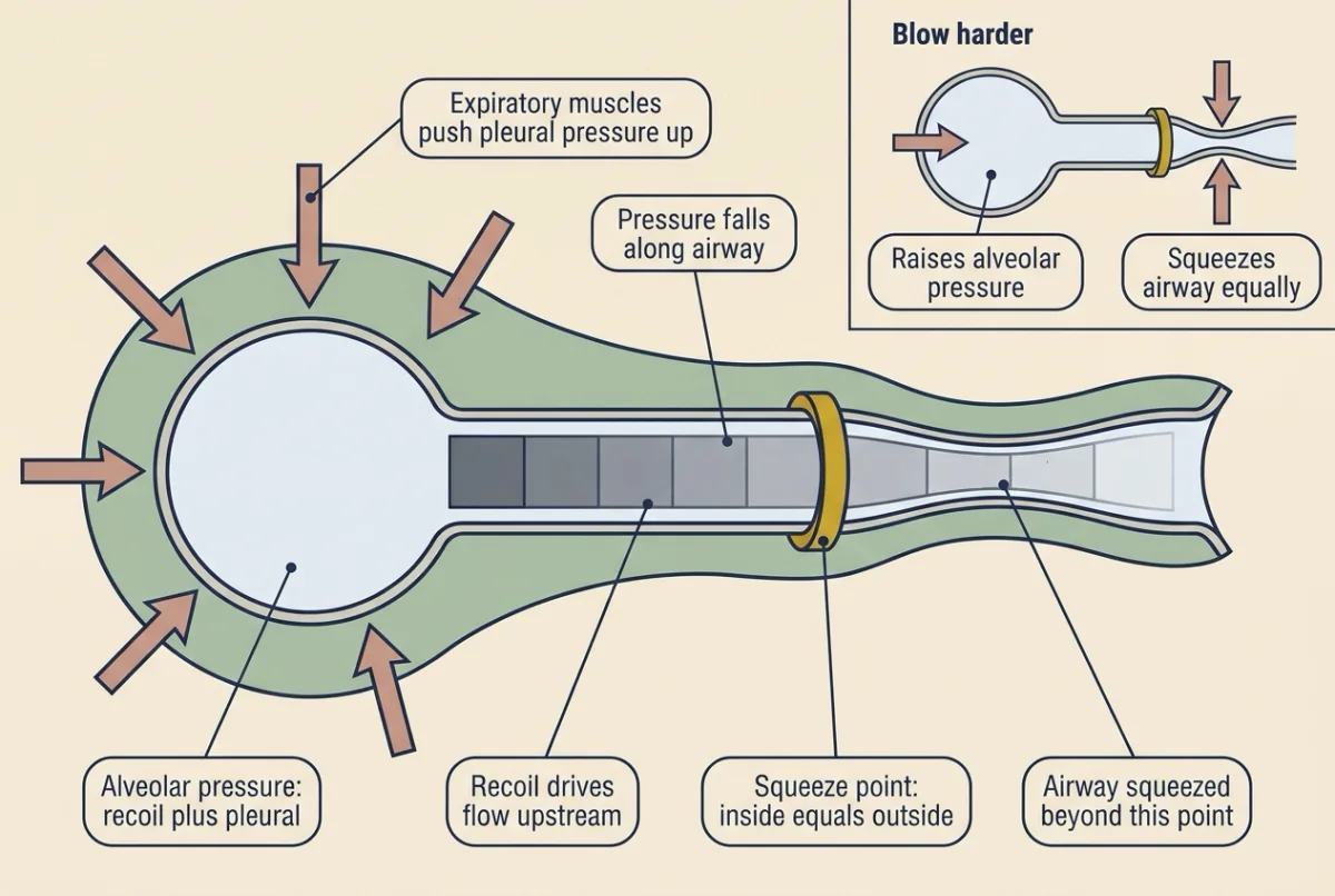
So a weak recoil (emphysema) or narrowed airways upstream of the squeeze point (asthma, chronic bronchitis) means a low maximal flow: the obstructive pattern. In health, most airway resistance sits in the larger airways, not the small ones.
Obstructive and restrictive patterns
An obstructive pattern lowers FEV1 more than FVC, so FEV1/FVC falls. A restrictive pattern lowers TLC; FEV1 and FVC fall together and the ratio is normal or high. Both are judged against reference limits, and a low FVC alone does not prove restriction: it goes with a low TLC less than half the time.
Worked problem, part 3: two made-up spirograms with made-up lower limits. Volunteer 1: FEV1 1.5 L, FVC 3.0 L, ratio 0.50, below a limit of 0.68: obstructive. Volunteer 2: FEV1 2.2 L, FVC 2.6 L, ratio 0.85, within limits, but plethysmographic TLC 3.9 L, below a limit of 4.6 L: restrictive, which the low FVC alone would not have shown.
Three diseases, read through the one idea as changes to the recoil curve:
- Emphysema: too little recoil. Destroyed alveolar walls lower elastic recoil, so compliance rises. The resting balance with the chest wall moves to a larger volume, so FRC rises; airways close earlier in expiration, so RV rises; and with less recoil opposing the inspiratory muscles, TLC usually rises. Low recoil means low maximal flow, and without radial traction small airways compress earlier, so FEV1 falls more than FVC and the ratio falls.
- Fibrosis: too much tissue recoil. Compliance and TLC fall. High recoil drives high flow for the volume, and stiff tissue holds airways open, so the ratio is normal or high. The elastic work of a breath of a given size rises, as in part 1, so breathing shifts to small, fast breaths.
- Respiratory distress syndrome (RDS): too much surface recoil. The main problem is the surface term, not the tissue, until surfactant is supplied.
Breathing costs elastic work (part 1) and resistive work, and the central drive picks a tidal volume and rate that tend to minimise the total (control of breathing; for respiratory muscles competing with the legs for blood, exercise physiology).
Design problem
You have excised lungs from adult rabbits, a syringe pump and a pressure transducer. Show how much of the lung’s recoil comes from surface tension, and test what depleting surfactant does.
Saved in this browser only.
One way to do it
Record a static air curve to a fixed volume and back, as described above, then refill the degassed lung with saline and repeat. Predict a saline curve far to the left with little hysteresis; the gap at one volume estimates the surface-tension share.
Controls: the same volume history before each curve, randomized air-saline order in a second set, constant temperature, lung weight before and after (edema).
Lavage a third set to deplete surfactant: predict an air curve shifted right, with higher opening pressure and more hysteresis, and an unchanged saline curve. Add back natural surfactant: partial recovery. Conditional SP-B-deficient mouse lungs would give a genetic version.
Limits: no chest wall, saline may alter tissue, one species and age. This is von Neergaard’s logic, and that of Mead’s laboratory.
In one line: the pressure across the lung matches its recoil, the wet lining supplies much of it, and surfactant makes each breath cheap and keeps a newborn’s lungs open.
Going deeper
The main line above does not depend on these details.
Limits of the one idea
During flow, lung tissue adds a little friction, so transpulmonary pressure slightly exceeds pure elastic recoil during inspiration and falls slightly short of it during expiration. “Recoil at its current volume” means the curve for the direction of the breath (hysteresis). Pleural pressure also varies by region. If flow rises and falls smoothly through inspiration, the most negative pleural pressure, about −8.3 cmH2O, comes slightly before end-inspiration, where flow and recoil terms overlap; −8 is the end-inspiration value. Some texts, and most ventilator papers, define transpulmonary pressure with airway-opening pressure in place of alveolar pressure; the two are equal only at zero flow.
Pressure units and where pleural pressure is read
1 mmHg = 1.36 cmH2O, so −5 cmH2O is about −3.7 mmHg, and the −3 to −4 mmHg often quoted for pleural pressure is the same thing. Pleural pressure is less negative at the base of the upright lung than at the apex. An esophageal balloon in the lower esophagus is the usual way to estimate it, and it reports the region near the esophagus, not the whole pleural space.
Lung and chest wall together
Lung and chest wall are in series: one breath must stretch both, so their stiffnesses (elastances) add. In compliances, 1/Crs = 1/CL + 1/Ccw, where Crs is the whole respiratory system’s compliance, CL the lung’s and Ccw the chest wall’s. The whole system is less compliant than either part.
Worked problem part 1 counted only the extra elastic work on the lung. Since 1 cmH2O x L = 98.07 Pa x 0.001 m3 = 0.098 J, that is about 0.074 J per breath, and the stiffer lung’s about 0.15 J. For a linear curve from the relaxed functional residual capacity (FRC), elastic work per breath on the whole respiratory system is 0.5 x VT² / Crs; with a chest-wall compliance of 0.2 L per cmH2O, the same breath costs about 1.4 cmH2O x L. Halving the lung’s compliance doubles the lung’s part (0.75 to 1.5 cmH2O x L) and raises the total by about half, to about 2.1.
Helium dilution and typical volumes
Rebreathe helium in a closed circuit. The helium is not absorbed, so the amount before, C1 x V1 in the spirometer, equals the amount after, spread through spirometer and lung, C2 x (V1 + V lung). Divide both sides by C2 and subtract V1: V lung = C1 x V1 / C2 − V1. For scale, one open textbook gives a tidal volume of about 500 mL and a TLC of about 6,000 mL in adult men and 4,200 mL in adult women: rough averages, which is why individuals are judged against reference equations instead.
Small into large
The small-into-large emptying, or “Laplace instability”, is a result of theory and of films in vitro. Whether a small alveolus empties into a larger one depends on how steeply tension changes with area, the film’s surface dilatational modulus, E*. In analyses of DPPC and clinical surfactant monolayers in vitro, uniform inflation is predicted to be stable when 2E* minus T is positive; a tension that falls with area, but not steeply enough, still lets the small one empty. Real alveoli are also tethered: in Wilson and Bachofen’s structural model, alveolar walls are platforms for surface tension, and neighbours share walls, so one alveolus cannot shrink alone like a free bubble. How much film instability matters once tethering is counted is an open question.
Pattle and Clements
Pattle, the physicist who inferred the low-tension lining in 1955, worked at Porton Down. Clements cited him, but Clements’s own estimates from lung bubbles put the lowest tension at 10 to 15 dynes per cm, not the under-1 value Pattle had inferred.
From association to treatment
Nebulized DPPC alone showed no clear benefit in 1960s trials. In the early 1970s Enhorning and Robertson found that natural surfactant improved lung expansion in preterm rabbits. In 1980 Fujiwara and colleagues reported improved oxygenation in ten preterm infants given a modified bovine surfactant, with no untreated controls. The randomized trials that followed supplied the controls.
The genetics
The 1994 surfactant protein B (SP-B) mutation was a frameshift, found in full-term infants who died of respiratory failure; the 1995 knockout mice, with both copies of the SP-B gene disrupted, died at birth. The 2004 ABCA3 study sequenced 21 infants, found mutations in 16 (76 percent), and reported abnormal lamellar bodies in lung tissue from four. The study does not show what ABCA3 carries, how each mutation acts, or that preterm respiratory distress is genetic. Later, in adult mice whose SP-B could be switched off, high surface tension injured the alveolar epithelium before edema formed.
Where airway resistance sits
Airway resistance is the pressure difference per unit flow. Poiseuille’s fourth-power rule assumes steady laminar flow in a rigid tube (blood and vessels), and airways are deformable. Small airways are so numerous, in parallel, that classic measurements put all airways under 2 mm in diameter at roughly 10 to 20 percent of airway resistance in health. The squeeze-point account of forced expiration used above is the classic teaching model; wave-speed theory is the fuller account.
How the airways condition and clean the air
Inspired air reaches the alveoli at 37 degrees C, saturated with water vapour (47 mmHg), which ventilation and perfusion subtracts from barometric pressure in the alveolar gas equation. A separate rule, Henry’s law, says dissolved gas is proportional to partial pressure (also in ventilation and perfusion). Nasal hairs trap large particles; mucus from goblet cells and glands catches smaller ones, and cilia sweep it toward the pharynx on a watery layer set by epithelial ion transport, including cystic fibrosis transmembrane conductance regulator (CFTR) (membrane transport). Alveolar macrophages take up most particles that reach the alveoli. A panting dog uses its airway surfaces for evaporative heat loss (thermoregulation).
Your notes
Select any text in the lesson to highlight it or add a note to it. Your highlights and notes are saved in this browser and collected in your notebook.
This browser is not letting the site save anything (a private window or strict privacy settings), so highlights and notes will be lost when you leave the page.
How we measure it
Static pressure-volume curves of excised lungs
A degassed lung is inflated in steps with a syringe, each step held until flow stops, while a transducer records the distending pressure; deflation is recorded the same way. Repeating the curve with the lung filled with saline removes the air-liquid surface, so the gap between the two curves estimates how much recoil surface tension supplies.
Surface film balance
Lung extract fills a shallow trough and its surface-active material forms a film at the surface, a movable barrier compresses and expands the film, and a thin plate hanging in the surface (a Wilhelmy plate) reports the tension. Plotting tension against area shows whether the film reaches low tension when compressed.
Spirometry, gas dilution and body plethysmography
A spirometer records volume breathed against time, giving tidal volume, vital capacity, FVC and FEV1. Helium dilution, nitrogen washout or a sealed body box (Boyle's law) add the gas that never leaves the lung, giving residual volume, FRC and TLC; in obstruction, dilution and washout can underestimate them.
Esophageal balloon manometry
A thin balloon catheter in the lower esophagus records a pressure used as a surrogate for pleural pressure. With mouth or alveolar pressure, it gives transpulmonary pressure and lets lung compliance be separated from chest-wall compliance; it reports the region around the esophagus, not the whole pleural space.
Practice questions on this lesson (3) → MCAT-style, original, each with an explanation
Where this lesson fits
After thisStudy next
- Ventilation, Perfusion and Gas ExchangeThis course · Lesson 13
It builds on this lesson's airflow and alveolar pressure ideas to explain how gas exchange is matched to flow.
- Control of Breathing, Altitude and DivingThis course · Lesson 14
It uses the elastic and resistive work of breathing here to explain how drive picks tidal volume and rate.
- Exercise: The Whole Body at WorkThis course · Lesson 30
It applies breathing work and ventilation limits from this lesson to the whole body under maximal effort.
Elsewhere on the siteFilms, simulations and method films in other courses
- Area, thickness and rateSimulation · Organismal Biology
Air-versus-water exchange surfaces show how area and thickness set gas flow across the lung wall.
- Why a Mouse Needs PlumbingFilm · Organismal Biology
Surface-to-volume scaling explains why a lung needs a large exchange surface made of tiny alveoli.
Suggested by an AI model from the lessons themselves, then checked. How we use AI
Finish line
Test yourself
9 questions
Practice and MCAT-style, with explained answers.
Next · Lesson 13
Ventilation, Perfusion and Gas Exchange
A resting adult moves about 6 litres of air and 5 litres of blood through the lungs every minute, yet neither number tells you what the blood leaves with. What does is whether air and blood arrive at the same places in the same proportion, and when they do not, gas exchange suffers.
Continue →