Cell Biology · Skim version
Vesicle Budding, Targeting and Fusion
Read the full lesson → Skim the whole course →
The guiding idea
A yeast mutant keeps making secretory enzymes at 37 °C but cannot release them. Which step of delivery broke, and how could you tell?
A vesicle delivers cargo in three handoffs that can each fail on its own: a coat buds it, a Rab and a tether catch it, and SNARE proteins, which NSF later pulls apart, fuse it.
From the film: Three Handoffs for a Vesicle
- The sec1 mutant keeps making enzymes at 37 °C, but they pile up inside vesicles that fail to fuse with the surface.
- A vesicle's trip has three handoffs: a coat buds it, a Rab-GTP with a tether catches it, and SNAREs guided by an SM protein fuse it.
- Zipping the four SNARE strands pulls the membranes together, and the zip itself uses no ATP.
- A pile of vesicles alone cannot name the failed step, but Sec1's known job as the fusion SM protein does.
- A fast inhibitor such as GDI stops working once its target state is past, which helps time the steps.
From the film: Reading Where Cargo Stops
- The fraction secreted is the enzyme outside the cell divided by all of it, so a block can be measured.
- If trapped enzyme leaves once the cells return to 25 °C with synthesis blocked, it was waiting to be delivered, not dead.
- Double mutants put the blocks in order, but a pile shows where transport stops, not when or where else a protein acts.
- Because NSF acts at several steps, a sec18 mutant reveals only its earliest blocked step.
- HOPS forms lacking the Vps11 or Vps18 beta propeller still caught Ypt7-bearing lipid spheres but failed to promote fusion.
-
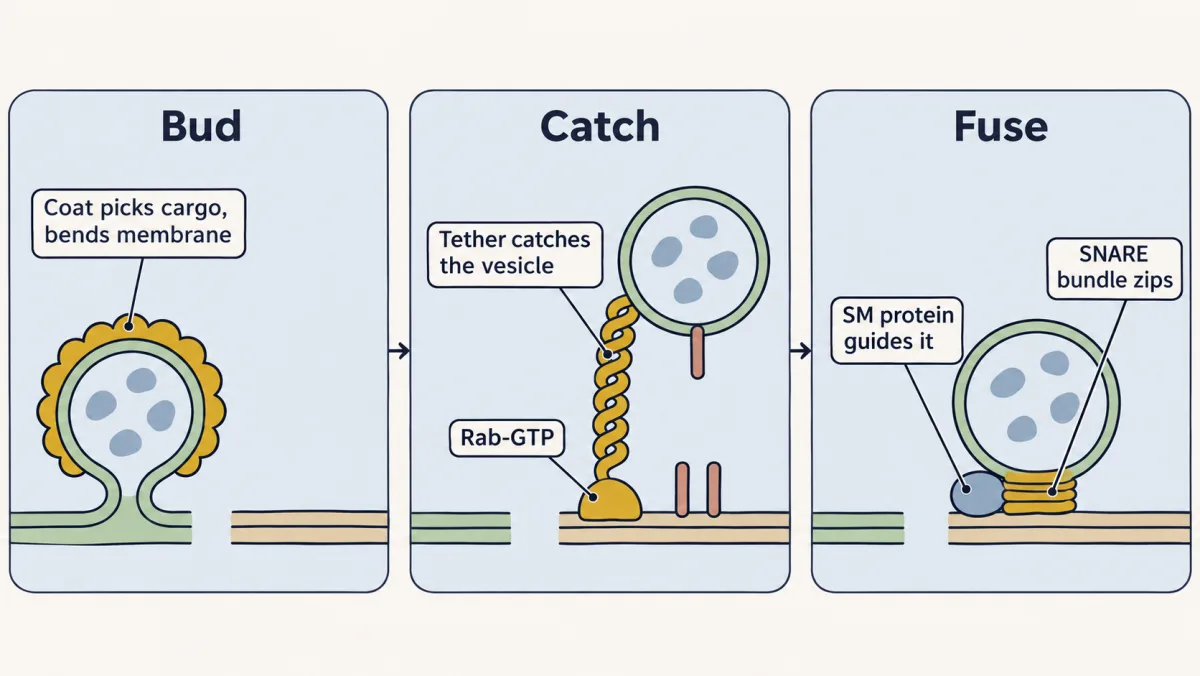
A vesicle's trip is three separate handoffs: a coat buds it and picks its cargo, a Rab-GTP and a tether catch it at the right membrane, and a SNARE bundle guided by an SM protein fuses it. Each can fail on its own, which is why mutants and inhibitors can tell them apart. AI-generated illustration -
Step 1, budding: a coat picks the cargo and bends the membrane
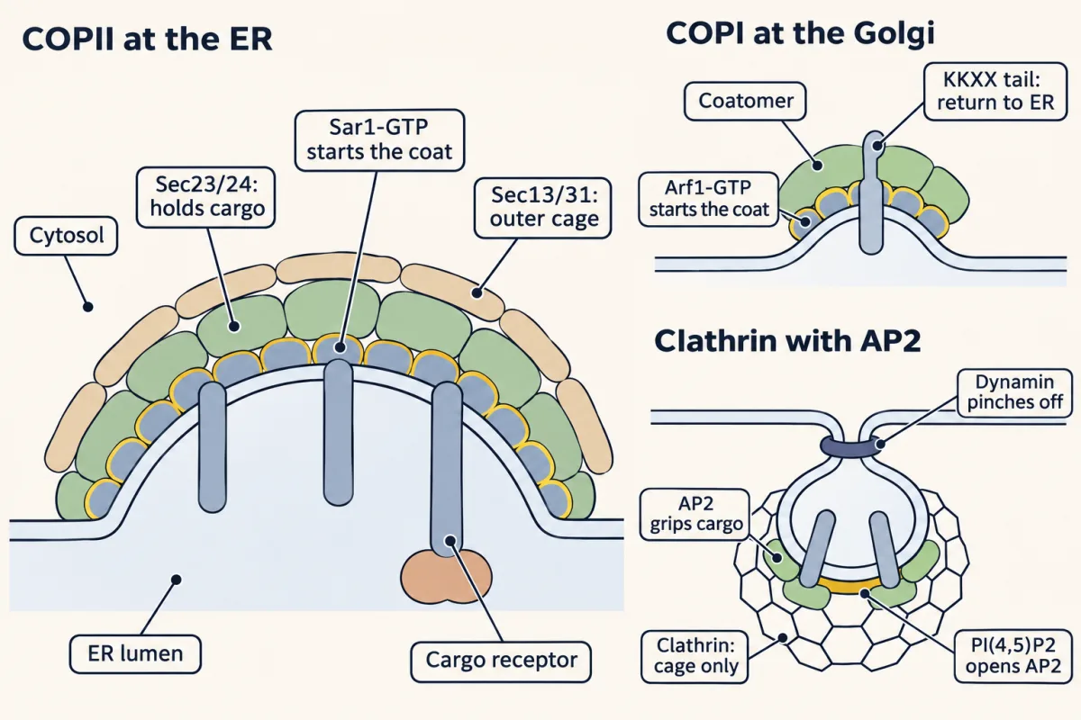
Each coat is started by a switch or a lipid (Sar1-GTP, Arf1-GTP, PI(4,5)P2) and does two jobs, choosing cargo and bending the membrane. In COPII, Sec23/24 holds cargo under a Sec13/31 cage; coatomer grips KKXX return tails; AP2 grips cargo while clathrin only builds the cage, and dynamin pinches the bud off. AI-generated illustration -
Step 2, targeting: a Rab and a tether catch the vesicle
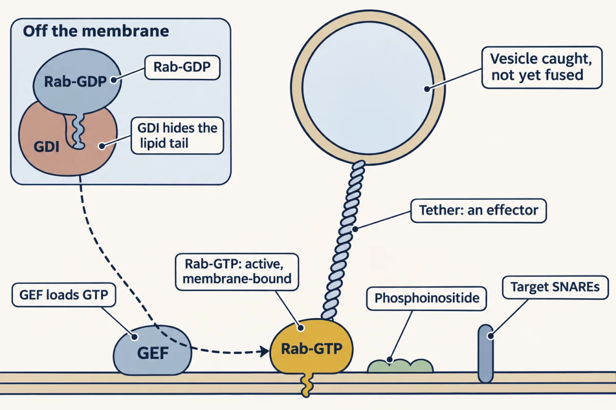
GDI carries Rab-GDP in the cytosol; at the right membrane a GEF loads GTP, and Rab-GTP recruits effectors such as a tether that catches the vesicle. No single molecule is the address: the Rab, the membrane's lipid and the SNAREs each narrow the choice. AI-generated illustration -
Step 3, fusion: SNAREs zip, an SM protein guides them, NSF resets them
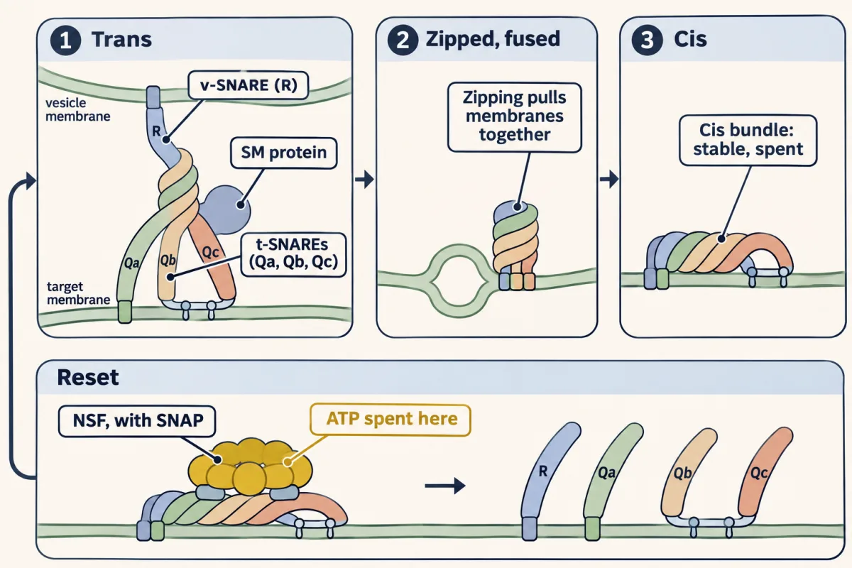
SNARE strands from two membranes zip into one four-helix bundle, and the zip itself pulls the bilayers together without ATP. The spent cis bundle sits in one membrane until NSF, through SNAP, spends ATP to pull it apart for the next round. AI-generated illustration -
Putting the handoffs in order: mutants
-
Splitting catch from fuse: test tubes
-
Going deeper
Where this lesson fits
Before thisReview first
- Protein Sorting into CompartmentsThis course · Lesson 7
You need to know how signals send proteins into compartments, since the vesicles here carry that sorted cargo.
- Membrane Organization and TopologyThis course · Lesson 4
Vesicle lumen versus cytosol and the cytosolic face of each protein are used throughout this lesson.
- Experiments That Distinguish MechanismsThis course · Lesson 3
The sec-mutant and cell-free reasoning here applies the fractionation and ordering logic taught there.
After thisStudy next
- The Secretory PathwayThis course · Lesson 12
It follows cargo through the ER and Golgi using the budding, tethering and fusion steps learned here.
- Endocytosis, Recycling and DegradationThis course · Lesson 13
Clathrin, AP2 and dynamin budding from the plasma membrane are used again to take cargo into the cell.
- Molecular Motors and CargoThis course · Lesson 15
Motors move vesicles along tracks to the target that the Rabs and tethers then catch.
Elsewhere on the siteFilms, simulations and method films in other courses
- Counting failuresSimulation · Neuroscience
Vesicle fusion at a nerve terminal is a regulated SNARE-driven release step.
- Pulse-chase labellingMethod film
Following a labelled batch of secreted enzyme through compartments is the same approach used to time delivery steps.
Suggested by an AI model from the lessons themselves, then checked. How we use AI