Sensory Systems · Unit 3: Temperature, pain and itch
Hot, Warm, Cool and Cold
Rewritten on : new text, flashcards and film. Your notes and highlights stay, but the text around them may have moved.
Mice lacking the cold-and-menthol receptor TRPM8 failed completely to learn to report forepaw warming in a 10-day task. Why would a cold receptor matter for warmth?
Pause when the reading reaches a film, and offer to play it. Saved on this device.
Watch the film now, or keep listening
Builds on:Five Ways to Turn a Stimulus into a CurrentSensory Neurons by Their Genes: the Atlas EraWhy these?
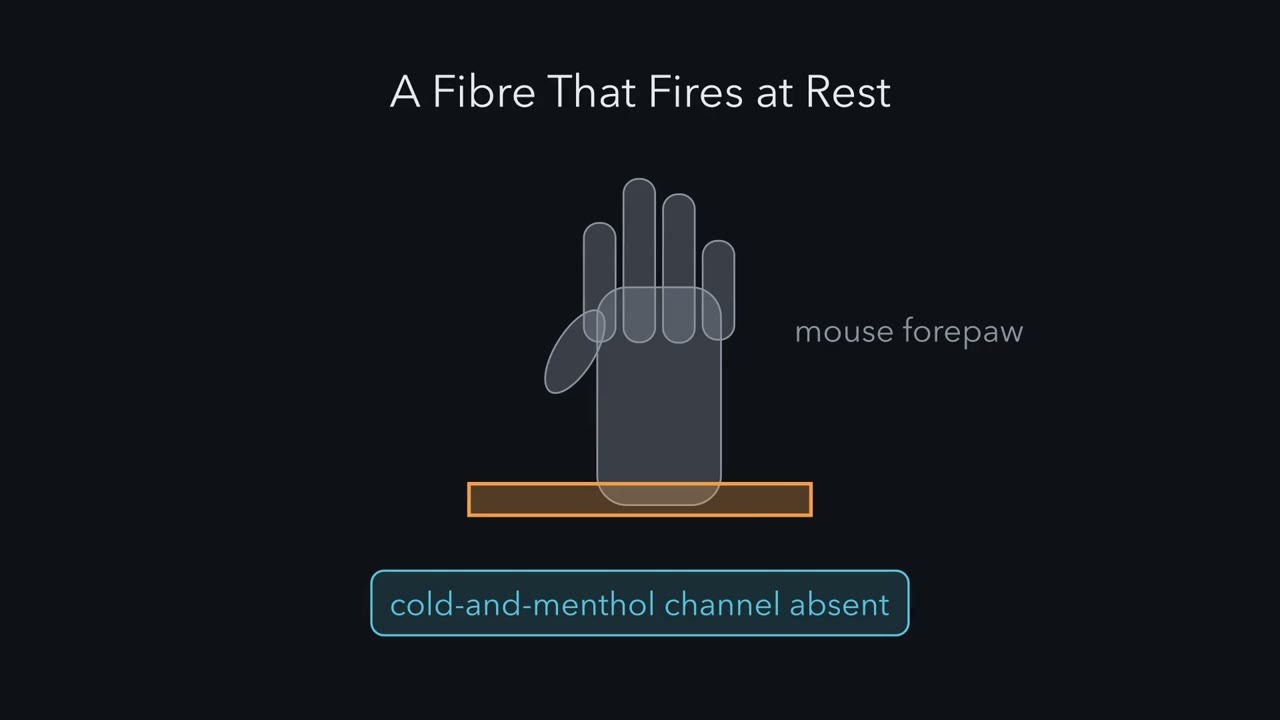 ▶ Watch A Fibre That Fires at Rest · 2:45
Read the transcript of “A Fibre That Fires at Rest”
Film transcript: A Fibre That Fires at Rest
Mice that lack a cold-and-menthol channel were trained to report gentle warming of a forepaw. They failed completely. Why would a cold channel matter for warmth? A transducer turns a stimulus into an electrical signal. For temperature, the candidates are ion channels. A line is one kind of sensory fibre and what it reports. A channel sits in a line. Most fibres in this story are C fibres. They are thin, unmyelinated and slow. For gentle cooling, the main established transducer is TRPM8. A fibre that is silent at rest can report a change only by starting to fire. In 2020, one study recorded single fibres from mouse forepaw skin kept alive with its nerve.
Awake forepaw skin measured 26 to 28 degrees. So they also used a bath at 27. Suppose a fibre already fires slowly at rest. What can it report that a silent fibre cannot? It can report change in both directions. Faster for one, slower for the other. In the 27 degree bath, one population did exactly that. These were cool-sensitive C fibres. With no stimulus, they fired slowly and steadily. Cooling makes such fibres fire faster. Warming made them fire slower.
A second kind, warm-excited C fibres, had little ongoing firing. Warming made them fire faster. In the usual 32 degree bath, no such fibres had been seen. Semicircular canal fibres in the inner ear use a similar design. They fire at rest, so rotation can speed them up or slow them down. The one idea: skin temperature is read from a few lines at once, not one channel per feeling. A line that fires at rest can signal both ways. The cool line speeds up on cooling and slows on warming. So a channel that supplies a line's firing matters for everything that reads that line. That is how losing a cool channel can cost warmth. This is a mouse forepaw result.
The circuit that reads the lines has not been identified. The picture to keep: a fibre that fires at rest can say warmer by going quiet.
From the film
- Skin temperature is read from a few afferent lines at once, not one channel per feeling.
- A cool line that fires at rest can report warming by slowing down.
- In the mouse forepaw, losing a channel that supplies a line's firing can cost warmth, though the circuit that reads the lines has not been identified.
Mice lacking transient receptor potential melastatin 8 (TRPM8), the cold-and-menthol receptor, were trained for 10 days to report a gentle warming of the forepaw. They failed completely. Why would a cold receptor matter for warmth?
If TRPM8 is the cold receptor, a mouse without it should be bad at cold, not at warm. The answer this lesson builds is that temperature is not read off one channel at a time. It is read off the activity of several groups of sensory fibres at once, including a group that reports warming by going quiet.
A few terms first. A transducer is the protein that turns a stimulus into an electrical signal; for temperature, the transducers are temperature-gated ion channels, which open or close as temperature changes, and no single channel reads out every temperature. A line here means one kind of afferent fibre (a sensory fibre carrying signals in from the skin) together with what it reports. A channel sits in a line; it is not itself a feeling. Innocuous temperatures, warm or cool, do not threaten the tissue; noxious ones damage or threaten to damage it, and usually hurt.
Touch and pain covers transient receptor potential vanilloid 1 (TRPV1), TRPM8 and transient receptor potential ankyrin 1 (TRPA1) at the level of necessity and sensitization, and ion channels as receptors covers how gates open and leaves heat gating open as a question. This lesson starts where those stop: how warmth, cool and noxious heat are encoded by populations of afferents and read by cortex, and what the gating evidence can and cannot say.
The map: ranges, fibres and channels
Two kinds of afferent fibre carry the signals in this lesson. C fibres are thin, unmyelinated and slowly conducting. Aδ fibres (“A-delta”) are thinly myelinated and conduct faster. Five channels come up. Transient receptor potential vanilloid 1 (TRPV1) opens with noxious heat and with capsaicin, the hot molecule in chilli; transient receptor potential melastatin 8 (TRPM8) opens with cooling and with menthol; TRPM3 and transient receptor potential ankyrin 1 (TRPA1) are two more channels tested here for noxious heat; TRPM2 is a candidate for warmth. Four temperature ranges come up, each with its own fibres and candidate channels.
| Range | What a mouse in this lesson does | Fibres that carry it | Channels tested here |
|---|---|---|---|
| Cold | still avoids surfaces below 10 °C without TRPM8 | not taken up here | not TRPM8 alone; not identified here |
| Cool | reports a 0.5 °C cooling step | cool-sensitive C fibres, including a population that fires at rest | TRPM8 |
| Warm | reports a 1 °C warming step | warm-excited polymodal C fibres speed up; the cool fibres slow down | TRPM8 needed in this task; TRPV1 and TRPM2 knockouts blunt detection |
| Noxious heat | withdraws the paw | C and Aδ afferents | TRPV1, TRPM3 and TRPA1, in parallel |
Where did the channels come from? From cloning screens. Michael Caterina and colleagues in David Julius’s laboratory expressed pools of sensory-neuron cDNA in cells that do not normally respond to capsaicin and screened for capsaicin-evoked calcium influx (Nature, 1997). The clone they recovered, TRPV1, encoded a nonselective cation channel that was also activated by noxious heating, which led them to suggest that it transduces painful thermal stimuli in vivo. Five years later, David McKemy, Neuhausser and Julius cloned a cold- and menthol-responsive receptor, TRPM8, from a trigeminal-neuron library made from newborn Sprague Dawley rats and characterised it in HEK293 cells and Xenopus oocytes (Nature, 2002). In the same year, Andrea Peier and colleagues in Ardem Patapoutian’s laboratory reported cold and menthol activation of heterologously expressed TRPM8 (Cell, 2002). Julius and Patapoutian shared the 2021 Nobel Prize in Physiology or Medicine for discoveries of receptors for temperature and touch.
A heterologous expression screen puts a gene into a foreign cell that does not normally carry it. It tests molecular sufficiency: this protein, in this foreign cell, responds to this stimulus. It does not test whether an intact animal needs the protein, whether other proteins do the same job, or where in the protein the temperature is sensed; the field spent the next twenty years on those three questions. Need is tested by removing the gene. Mice lacking TRPM8 have profoundly reduced cold responses in neurons and fibres, yet they still avoid surfaces below 10 °C (Bautista and colleagues, Nature, 2007). That is why TRPM8 is called the main established transducer of innocuous cooling, not of all cold.
Lines that fire at rest
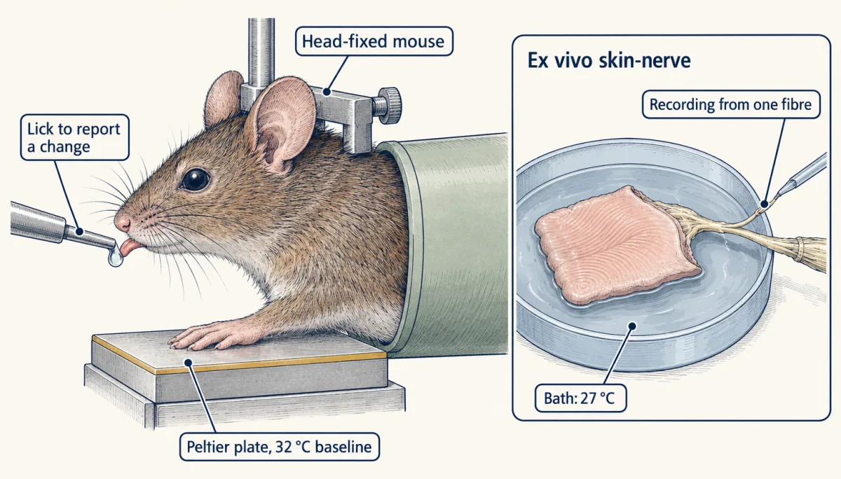
The answer to the puzzle came from one 2020 study that ran two preparations side by side. Paricio-Montesinos and colleagues, working with Gary Lewin and James Poulet among others (Neuron, 2020), trained mice to report temperature steps on the forepaw (the next section) and recorded single afferent fibres in a forepaw glabrous (hairless) skin-nerve preparation: a patch of skin kept alive ex vivo, outside the animal, with its nerve attached, so spikes from single fibres can be recorded while the bath temperature is controlled. Temperature ramps ran at 1 °C per second.
Such recordings are usually run with the bath near body-surface temperature, 32 °C. Thermal imaging showed awake forepaw skin at 26 to 28 °C, so the investigators also used a 27 °C bath, while the patch of skin under the stimulator was held at the same 32 °C baseline used in the behaviour. That choice changed what they saw.
Predict. A fibre that is silent at rest can report a change only by starting to fire. What can a fibre that already fires slowly and steadily at resting skin temperature report that a silent fibre cannot?
Check your prediction
A change in either direction: faster for one, slower for the other. In the 27 °C bath, about 19% of the temperature-sensitive fibres recorded were cool-sensitive C fibres with ongoing activity, firing with no stimulus at 0.2 to 6 spikes per second (Hz). At the usual 32 °C bath, no such fibres had been seen.
The signature response
Two kinds of C fibre answered warming, in opposite directions.
| Cool-sensitive C fibre | Warm-excited polymodal C fibre | |
|---|---|---|
| Skin at rest | fires slowly and steadily (0.2 to 6 Hz in the 27 °C bath) | little ongoing firing |
| Skin cools | fires faster | not part of this story |
| Skin warms | fires slower | fires faster |
Polymodal means the fibre also responds to other kinds of stimulus, such as pressure. The table is a constructed summary of the two kinds of response, not one recording.
The left column is the signature of this system: a fibre that fires at rest can signal by slowing down. Semicircular-canal afferents in the inner ear use a similar design: they fire at rest, so one fibre can signal head rotation about its axis one way by speeding up and the other way by slowing down (the vestibular system). How much a slowdown can tell depends on how much firing there was to lose. In a simple counting model, if warming removes the same fraction of spikes, a fibre that fired faster at rest says more; a fixed absolute drop does not follow that rule.
The one idea to carry through this lesson. Skin temperature is read from a few afferent lines at once, not from one channel per feeling. A line that is already firing at resting skin temperature can signal in both directions: the TRPM8-dependent cool line speeds up when the skin cools and slows down when it warms. So a channel that supplies a line’s activity matters for every sensation that reads that line, which is how losing a cool channel can cost warmth. And more than one channel can carry the same signal, which is how acute noxious heat survives the loss of any one of transient receptor potential vanilloid 1 (TRPV1), TRPM3 or transient receptor potential ankyrin 1 (TRPA1).
Its scope: gentle warm and cool detection in the mouse forepaw and, in its second half, acute withdrawal from noxious heat in mice. It is not a human result, it names no central circuit (none was identified), and another preparation points the other way on warmth. Where inside a channel temperature is sensed is a different question, taken up at the end.
Measuring detection: hits, false alarms and d′
The behaviour came first in that study. Adult mice of both sexes were trained, head-fixed (the head held still so the stimulus stays in place), to report a temperature step on the forepaw from a 32 °C baseline, delivered by an 8 by 8 mm Peltier surface, a small plate whose temperature can be stepped electrically. The mouse licked to report a change. Trained mice reported a warming step of 1 °C; a separately trained cooling group reported a cooling step of 0.5 °C.
How sensitive is a mouse in such a task, when it also licks sometimes for nothing? A lick on a warming trial is a hit. A mouse that licked on every trial would score hits without detecting anything, so the task includes catch trials, with no temperature change; a lick on a catch trial is a false alarm. The study’s analysis uses the signal-detection index d′ (“d-prime”), which compares the two rates after z, the inverse standard-normal cumulative distribution, converts each into a z score. In the standard equal-variance model with a stable criterion, a change in readiness to lick shifts both z scores equally, so their difference measures sensitivity apart from that readiness. In real sessions d′ remains useful, but it does not automatically remove learning, motor or lapse effects.
d′ = z(hit rate) − z(false-alarm rate)
Worked problem. Here is a constructed session, not published data: 100 warming trials and 100 catch trials with no temperature change, with licks on 80 warming trials and 20 catch trials.
- Hit rate H = 80/100 = 0.80. False-alarm rate F = 20/100 = 0.20.
- z(0.80) = 0.8416. z(0.20) = −0.8416. (The two are mirror images because 0.80 and 0.20 are symmetric about 0.5.)
- d′ = 0.8416 − (−0.8416) = 1.6832, so d′ ≈ 1.68, dimensionless.
It is a sensitivity index, not a temperature: 1.68 is not 1.68 °C, and nothing about the stimulus size enters the formula.
Predict. A second mouse licks on 80 warming trials and on 80 catch trials. Its hit rate is the same 0.80. Is it detecting warmth?
Check your prediction
No. Its d′ ≈ 0, because it licks as often when nothing happens as when the plate warms. Separating sensitivity from criterion, how freely the animal licks, is why the index is used and why catch trials are built into the design.
| Step | Expected (from the sliders) | Drawn (simulated session) |
|---|---|---|
| Licks, warming / catch | ||
| H, hit rate | ||
| F, false-alarm rate | ||
| z(H) | ||
| z(F) | ||
| d′ = z(H) − z(F), dimensionless |
In this sketch: independent trials at fixed lick rates, an equal-variance normal model, a head-fixed mouse reporting with licks from a 32 °C baseline; the session is constructed, not published data. A rate of 0 or 100 licks is moved to 0.5 or 99.5 licks in 100 so that z is finite; the formula itself is undefined there, and that convention is this sketch’s, not the lesson’s. Rule to keep: d′ is a sensitivity index, not a temperature and not the hit rate.
Take away the cool line
Read the transcript of “Take Away the Cool Line”
Film transcript: Take Away the Cool Line
If warming quiets the cool line, what happens to warmth when that line is gone? First, the task. A head-fixed mouse rests a forepaw on a small plate at 32 degrees. It licks to report a change. Trained mice reported a warming step of 1 degree. A mouse that licked on every trial would score hits without detecting anything. So some trials are catch trials, with no temperature change. A lick then is a false alarm. An index called d′ compares the two. It separates detection from how freely the mouse licks. Licking as often on catch trials as on warming trials gives a d′ prime near zero.
Now the knockouts. TRPM8 is the main channel behind the cool fibres' response to cooling. Remove it. What happens to the cool fibres' resting firing? And can the mouse still report warming? Without TRPM8, the cool fibres lose their ongoing firing. Warming has nothing left to silence. And the mice failed completely to report warming, even after 10 days of training.
Not because heat opens TRPM8. Removing it removes the firing whose fall is part of the warmth signal. Now remove heat channels instead. Mice without TRPV1, or without TRPM2, still detected warming. Even mice lacking three heat channels still detected warming, with reduced sensitivity. Heat-channel loss blunts. TRPM8 loss abolished the cold report in this task. And TRPM8 knockout mice still had warm-excited fibres, in normal proportions. If each line alone were enough to drive a report, the remaining warm responses should have supported one. In these mice, they did not.
The findings support a role for reduced cool input. Warm-excited input may also contribute, but its separate effect on sensitivity was not isolated. How the brain combines the two lines is not known. One gap remains. The resting cool firing was recorded in isolated skin, not in a behaving mouse. The skin under the stimulator sat at the same 32 degrees. Joining the two is still an inference, not a recording. Another preparation points the other way. In mouth-warming experiments, blocking TRPV1 removed the imaged warmth responses. So does gentle warmth rest mainly on warm fibres, or on quieter cool fibres? That is not settled. Keep the bath in mind.
A resting condition is part of the stimulus, not a neutral background.
From the film
- Without TRPM8, mice failed to report forepaw warming in this task, even after 10 days of training.
- TRPM8 loss removes the cool fibres' resting firing in isolated skin, so warming has no ongoing firing left to silence.
- Loss of heat channels blunted warmth detection, whereas TRPM8 loss abolished it in this task.
- Whether gentle warmth rests mainly on warm fibres or on quieter cool fibres is not settled.
The same study tested knockout mice in the forepaw task. Transient receptor potential melastatin 8 (TRPM8) is the main channel behind the cool fibres’ response to cooling.
Predict. Take TRPM8 away. What should happen to the cool fibres’ ongoing firing, and to the mouse’s ability to report warming? Then predict the same for mice that lack transient receptor potential vanilloid 1 (TRPV1), the heat- and capsaicin-activated channel.
Check your prediction
| Mouse | Cool fibres’ ongoing firing | Reports warming in this task? |
|---|---|---|
| Wild type | present, and warming reduces it | yes, down to a 1 °C step |
| No TRPM8 | lost | no: failed completely after 10 days of training |
| No TRPV1 | not reported here | yes, blunted; still detected a 1 °C step |
| No TRPM2 | not reported here | yes, blunted; threshold 2 °C rather than 1 °C |
| No TRPV1, TRPA1 or TRPM3 (all three) | not reported here | yes, with reduced sensitivity |
Without TRPM8 the cool fibres lose their ongoing firing, so warming has nothing left to reduce. The warmth deficit arises not because TRPM8 is heat-gated, but because removing it removes the cool-afferent firing whose reduction is one of the two parts of the warmth signal. (TRPM8-null mice also could not report cooling with the larger plate.) Channel loss on the warm side, single or triple, blunted detection without abolishing it.
What does failing mean here? The TRPM8-null mice were trained for 10 days with warming stimuli from 32 to 42 °C, and their hit and false-alarm rates stayed similar throughout. That alone does not identify the cause, since a mouse can fail to learn for other reasons. Two observations support a sensory explanation: the same mutants readily learned to report mechanical stimuli to the forepaw, and in wild-type mice a TRPM8 antagonist applied to the forepaw skin reduced warm detection, which recovered by the next day.
Notice how lopsided that is. TRPM8-null mice still had warm-excited fibres, in proportions similar to wild type. On the simplest picture, each line an independent alarm sufficient to drive a report, those fibres should have let the mouse detect warming less well, not fail. In this task the retained warm-excited responses were not enough, and the results support an important role for reduced cool input. Warm-excited input may also improve sensitivity, but its separate contribution was not isolated. The result does not say how the brain combines the two lines: a cool-required, warm-augmenting readout is one possible model, and a weighted sum with a threshold or a nonlinear interaction could also fit.
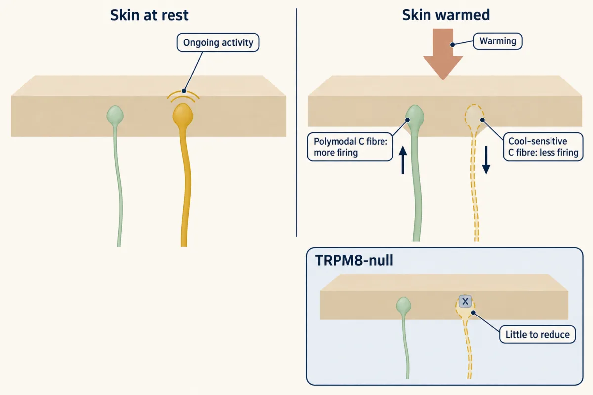
Whatever the computation, something downstream must treat a fall in the cool line as evidence of warming. These recordings did not identify that circuit, so write the finding as reduced cool input. A named disinhibitory circuit, a central synapse that inverts the sign of the cool signal, is an extension of the data, not a finding in them.
Two cautions keep the question open.
Isolated skin. The ongoing cool-fibre firing was recorded in isolated skin in a 27 °C bath, not in a behaving mouse. So the step from “warming slows cool fibres in the dish” to “the mouse uses that slowing” is an inference that joins two preparations, not a recording made during the task.
Another preparation. Yarmolinsky and colleagues imaged trigeminal ganglion cell bodies (the sensory neurons of the face and mouth) in anaesthetized mice expressing GCaMP5 during oral thermal stimulation (Neuron, 2016). Warmth sensors were approximately 10% of the heat-responsive cells they sampled, and acute antagonism of TRPV1 eliminated the imaged warmth responses over 35 to 43 °C in that preparation. This is a statement about sampled trigeminal ganglia during oral stimulation, not about mouse skin elsewhere or about people. It sits awkwardly beside the forepaw study, in which TRPV1-null mice still detected a 1 °C warming step. The two also differ in method: one blocked TRPV1 acutely with a drug in an anaesthetized animal, the other removed the gene from the start, and a germline knockout leaves time for development to compensate. Whether innocuous warmth rests on TRPV1-positive warm fibres or on reduced TRPM8-dependent cool input is not settled, and the answer may differ between body sites.
Three channels for one burn: redundancy, not a chain
Read the transcript of “Three Channels, One Burn”
Film transcript: Three Channels, One Burn
Mice without TRPV1 keep much of their response to painful heat. So what else senses the burn? In 2011, TRPM3 was found to contribute heat sensitivity to some small mouse sensory neurons. Mice lacking it avoided noxious heat less well. TRPA1 was a third candidate. Suppose the three worked in series, each passing the signal to the next. What would each single knockout do?
In series, every single knockout would abolish the withdrawal. They do not. Now suppose they work in parallel. What would it take to remove the reflex? All three at once. In 2018, mice lacking all three lacked acute withdrawal from noxious heat, in the reported assays.
The channels are redundant for this reflex. Remove one, and the others carry the signal. Remove all three, and the acute reflex has nothing left. But the same mice still preferred moderate temperatures. Withdrawal and preference are different behaviours. Whatever supports preference is not exhausted by these three channels. Set this beside the cool line. The cool fibres' resting firing depends on TRPM8. So losing TRPM8 cost warmth. Here, three channels can each carry the heat signal. So losing TRPV1 alone did not abolish the burn. In mice, acute heat withdrawal survives the loss of any one of three channels.
From the film
- In mice, acute withdrawal from noxious heat survives the loss of any one of TRPV1, TRPM3 or TRPA1.
- The three channels act in parallel: only mice lacking all three lacked acute withdrawal in the reported assays.
- The same mice still preferred moderate temperatures, so withdrawal and preference are different behaviours.
Mice lacking transient receptor potential vanilloid 1 (TRPV1) keep much of their response to painful heat, so other heat sensors had to exist. Joris Vriens, Thomas Voets and colleagues reported that TRPM3 contributes heat sensitivity to a subset of small mouse sensory neurons, and that TRPM3-deficient mice had impaired avoidance of noxious heat (Neuron, 2011). Transient receptor potential ankyrin 1 (TRPA1) was a third candidate.
Predict. Suppose the three channels worked in series, each needed to pass the signal to the next. What would each single knockout do to acute withdrawal from noxious heat? Now suppose they work in parallel, each able to carry the signal on its own. What would single knockouts do, and what would it take to remove the reflex?
Check your prediction
In series, each single knockout would abolish withdrawal, and single knockouts do not. In parallel, single knockouts would leave the reflex and only the loss of all three would remove it. Vandewauw and colleagues made that combinatorial test (Nature, 2018), pairing recordings from isolated mouse sensory neurons and from cutaneous C and Aδ afferents ex vivo with withdrawal behaviour in vivo. Mice lacking TRPV1, TRPM3 and TRPA1 together lacked acute withdrawal from noxious heat in the reported assays. The channels are functionally redundant for this assay: remove one and the others carry the signal, remove all three and the acute reflex has nothing left.
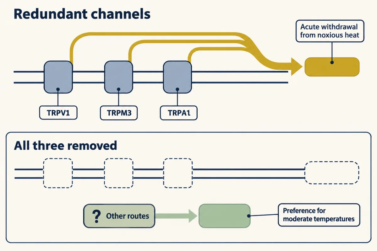
The same animals retained a preference for moderate temperatures, and students regularly mangle that result. It means that whatever supports temperature preference is not exhausted by these three channels. Acute nociceptive withdrawal and graded thermal preference are different behaviours with different requirements, and a mouse that cannot do the first can still do the second.
Set this beside the cool line: losing transient receptor potential melastatin 8 (TRPM8) cost warmth because the cool line’s resting firing depends on it, whereas no single heat channel is needed for the burn.
Reading the lines: spinal cord, ganglia and insular cortex
Between the ganglion and the cortex sit the spinal dorsal horn and the thalamus, and the codes already differ there: in mouse spinal cord, heat-responding neurons encode absolute skin temperature without adapting and receive TRPV1-positive input, while cold-responding neurons adapt quickly and encode temperature change (Ran, Hoon and Chen, Nature Neuroscience, 2016).
Wang and colleagues imaged dorsal root ganglion (DRG) somata, the sensory neurons for the body below the head, with two-photon microscopy during hindpaw stimulation in anaesthetized mice (Cell Reports, 2018). Raising the temperature recruited more heat-responsive neurons, a graded population signal. Cooling responses were largely ungraded, which the authors read as suggesting combinatorial cold coding: which cells respond, rather than how hard they respond. This study and the trigeminal imaging above used different ganglia, different stimulus sites and different anaesthesia, so differences in tuning between them are not yet differences between body regions.
Centrally, Vestergaard, Carta, Güney and Poulet imaged cortex in awake mice during forepaw Peltier stimulation (Nature, 2023). Posterior insular cortex responded to both cooling and warming and was somatotopically arranged (neighbouring parts of the body mapped to neighbouring cortex), while primary somatosensory cortex (S1) showed a widefield response only to cooling, with only sparse warm-responding neurons under two-photon imaging.
Predict. They then silenced posterior insula optogenetically, by driving inhibitory neurons with light in VGAT-ChR2 mice (whose inhibitory neurons carry a light-gated channel, so light silences the surrounding cortex), during the detection task. If the insula takes part in reporting temperature, should silencing it hurt warming and cooling detection alike? Should a small cooling step and a large one suffer alike?
Check your prediction
Neither. Inhibiting posterior insula reduced warming-detection hit rates at every warming amplitude tested. It reduced cooling hit rates for 0.5 to 2 °C steps, but spared the tested 10 °C step.
Design problem
What can that pattern establish, and what does it not?
Saved in this browser only.
One way to do it
It establishes a causal contribution to the behaviour tested, in this preparation, at those amplitudes; because light-driven inhibition is acute and its spread incomplete, it is not proof of strict necessity. It shows less about exclusivity: the authors also inhibited primary somatosensory cortex alone, with a smaller effect, and S1 together with the insula, but other subcortical routes may carry thermal information and were not silenced.
The spared 10 °C cooling step is the informative asymmetry, and it does not show that the insula contributes nothing there. Two readings fit it. A parallel route may be sufficient when the stimulus is large, so the insula is dispensable there. Or performance at 10 °C may be at a ceiling, where removing part of the signal leaves enough to cross threshold. To separate them, vary the amplitude finely around the spared step, and add a second task where the mouse must tell two large steps apart rather than simply detect one, so the ceiling is pushed away.
The control to ask for is motor, and here the authors supply it. Driving inhibitory neurons in cortex while an animal must lick can reduce licking for reasons that are not sensory. Before reading reduced hit rates as a sensory deficit, show that false-alarm rates and lick kinematics on unaffected trial types are unchanged, or that the same light leaves an equally demanding non-thermal detection task intact. Here, inhibiting posterior insula left licking for free water and licking in an acoustic detection task unchanged, and it increased, rather than reduced, licking on no-go trials. The weaker effect of S1 inhibition is a regional comparison, not the motor control.
And the scope: mouse cortex, awake, forepaw, with a licking report. No human thermal localisation follows.
A different question: where is the sensor inside the channel?
Everything so far has been about lines. A separate question is what, inside a channel such as transient receptor potential vanilloid 1 (TRPV1), makes it open with heat. It is a different level of explanation, and still open.
Q10 is the ratio of a process’s rates measured 10 °C apart, and a steep Q10 is often treated as the signature of a temperature sensor. It is not a channel constant. Apparent Q10 is read from a plot of the logarithm of current against inverse temperature. For TRPV1 that plot has a shallow limb at lower temperatures and a steep limb at higher temperatures, each with its own apparent Q10. For rat TRPV1 expressed in HEK293T cells, recorded at −60 mV, increasing activation by resiniferatoxin, a TRPV1 agonist, reduced the apparent high-temperature Q10 from approximately 38 to approximately 3, while the low-temperature value stayed near 1.7 (Kwon and colleagues, Nature Communications, 2022). The same protein gives different numbers depending on how much agonist has already opened it, so Q10 characterises a state and an interval.
One possible reason is simple: a channel that is already mostly open cannot open much more. In an illustrative two-state model whose thermal gating is held fixed, adding an agonist drops the Q10 of the open probability steeply while the Q10 of the opening equilibrium itself does not change (Going deeper). So saturation alone can produce a drop of this size, and the drop by itself does not show a change in the sensing mechanism.
Structures sharpen the question without closing it. The first near-atomic cryo-EM structure of TRPV1, in 2013, was of a minimal construct. Full-length channels in lipid nanodiscs, imaged across temperatures in 2021, support a global conformational change followed by outer-pore rearrangement during heat-dependent opening with capsaicin present. But those activated states needed an agonist, in constructs and lipids the experimenters chose, and the authors of the 2022 ensemble work cautioned that multiple cryo-EM classes may include off-pathway states rather than only sequential intermediates.
There is also a theoretical reason not to expect a tidy sensor module. Opening is an equilibrium between closed and open states. If the heat absorbed on opening were fixed, a plot of the logarithm of that equilibrium constant against inverse temperature would be a straight line. Clapham and Miller proposed that a large gating-associated change in heat capacity can account for steep TRP-channel temperature sensitivity. A heat-capacity change makes the heat absorbed on opening itself change with temperature, so the line curves. Because the argument is independent of mechanistic detail, it does not require the temperature dependence to sit in one domain; the steepness could be spread across the protein. On that view, looking for the sensor the way one looks for a capsaicin pocket may be the wrong search.
Nociceptors and the sodium channels of pain takes up what happens after transduction, and when pain persists takes up what happens when these systems are altered by injury.
Going deeper
How much can a fall in firing say?
This is an illustrative model, not data from the study. Count one fibre’s spikes over T seconds and treat the counts as Poisson (variance equal to the mean). Define a count-separation index D = |μrest − μwarm| / √[(σrest² + σwarm²)/2], the separation in units of the root-mean-square standard deviation. It is computed like d′, but not from hits and false alarms, and it is not the most an ideal observer could extract. Complete silencing from a rate r gives D = rT / √(rT/2) = √(2rT) for r > 0; when both conditions are silent, D is set to 0 by continuity.
| Resting rate | D, one fibre falling silent (T = 1 s) |
|---|---|
| 0 Hz | 0.00 |
| 0.2 Hz | 0.63 |
| 2 Hz | 2.00 |
| 6 Hz | 3.46 |
The table assumes complete silencing at each selected resting rate. The study observed warming-induced suppression, including silencing for some stimuli (a 22 to 32 °C warming step, for example), but these rows are not measured responses at each rate, nor evidence that every small warming step silences the fibre. Partial falls follow the same logic: halving a 2 Hz rate gives D = (2 − 1) / √1.5 ≈ 0.8165 for one fibre, and 0.8165 × √20 ≈ 3.65 for 20 independent fibres responding the same way. If warming removes the same fraction f of spikes, D = f√(rT) / √(1 − f/2); a fixed absolute drop instead gives D = 0.82 for 2 to 1 Hz but 0.43 for 6 to 5 Hz in 1 s. In this model, a cool fibre anywhere in the 0.2 to 6 Hz range recorded in the 27 °C bath could carry information about warming if warming reduced its firing enough. A silent fibre that starts firing at rate r gives the same D as one falling from r to silence, so in that matched comparison ongoing firing buys signalling in both directions, not extra sensitivity.
Why two independent alarms do not fit
Again illustrative, not measured. Take an optimal observer combining two cues that are Gaussian and independent given the stimulus, each with the same variance on warm and reference trials, and assume the retained cue’s statistics do not change when the other is removed. Then d′ = √(d′cool² + d′warm²). The component values are invented, not measured sensitivities of the two populations: try cool and warm values of 1.2 and 1.2, 1.5 and 0.8, or 1.0 and 1.5. Wild type then sits at d′ 1.70, 1.70 or 1.80, and a TRPM8-null mouse that kept the warm line would keep d′ 1.2, 0.8 or 1.5: blunted, not abolished. The reported result is complete failure. That excludes only this example’s assumptions, not every additive readout.
A culture comparison
Preparation matters even in a dish. In dissociated dorsal root ganglion (DRG) cultures from male mice aged 6 to 25 weeks, warmth-sensitive neurons were about 26 ± 9% of neurons in overnight cultures and about 6 ± 3% after three days (Abd El Hay and colleagues, eLife, 2025). That roughly fourfold difference between culture protocols, measured in different groups of animals, is enough to put two laboratories into apparent disagreement about the same biology.
Insular imaging details
Vestergaard, Carta, Güney and Poulet imaged cortex in awake Thy1-GCaMP6s mice older than two months. The grand-average insular calcium response began at about 80 ms for cooling and about 320 ms for warming, a difference the authors compare with the short-latency Aδ-fibre cool responses and longer-latency C-fibre responses recorded in earlier peripheral work. The forepaw study did not establish that Aδ fibres mediate its small innocuous-cooling reports.
Q10, saturation and apparent enthalpy
Q10 converts to an apparent activation enthalpy. Between 35 and 45 °C, a Q10 of 38 corresponds to about 296 kJ/mol (71 kcal/mol), a Q10 of 3 to about 90 kJ/mol, and a Q10 of 1.7 to about 43 kJ/mol.
The saturation argument, in an illustrative two-state model (not fitted to transient receptor potential vanilloid 1 (TRPV1)): take a gating enthalpy of 400 kJ/mol with no heat-capacity change and K = 1 at 45 °C, so K = exp[−(ΔH/R)(1/T − 1/318.15 K)], hold that thermal gating fixed, and let an agonist multiply the opening equilibrium constant K by a temperature-independent factor, with Popen = factor × K / (1 + factor × K). Between 30 and 40 °C, the Q10 of K stays at about 159 throughout, but the Q10 of the open probability falls as the agonist factor rises.
| Agonist factor | Q10 of open probability, 30 to 40 °C |
|---|---|
| 1 (no agonist) | about 146 |
| 100 | about 17 |
| 1,000 | about 2.7 |
| 10,000 | about 1.2 |
The model does not explain the resiniferatoxin measurement, from 38 to 3, quantitatively; it shows only that a drop of that size can arise without a change in mechanism.
TRPV1 structures
The 2013 structure, from Liao, Cao, Julius and Cheng (Nature), was a 3.4 Å cryo-EM structure of a minimal functional rat TRPV1 construct in amphipols, fourfold symmetric around a pathway formed by the S5 and S6 segments and the intervening pore loop. Agonist-bound structures from the same year suggested gating at both the outer pore and a lower hydrophobic gate. The 2021 nanodisc study, of full-length rat TRPV1, was by Kwon and colleagues (Nature Structural and Molecular Biology).
The two-state model and heat capacity
In a two-state approximation, K = Popen/(1 − Popen) = exp(−ΔG°/RT). As an illustration, take a gating heat-capacity change of 20 kJ/mol/K, with ΔH and ΔS both zero at 45 °C. Then ln K at 5, 25, 45 and 60 °C is about 22.7, 5.2, 0 and 2.5: curved rather than straight in 1/T, and rising on both sides of the minimum. With no heat-capacity change, ln K would be flat. This toy case shows only the curvature. Its channel would be almost fully open at 25 °C, which TRPV1 is not; a heat-activated channel also needs a large positive gating enthalpy near its activation range.
What would settle it
A single construct carried through three measurements at fixed voltage: apo, lightly agonist-occupied and strongly activated, with structures and Q10 taken on the same protein in the same lipid, plus mutations tested for effects on sensing, on coupling and on general gating separately. A mutation that shifts an activation threshold does not identify a sensor until those three are told apart.
Your notes
Select any text in the lesson to highlight it or add a note to it. Your highlights and notes are saved in this browser and collected in your notebook.
This browser is not letting the site save anything (a private window or strict privacy settings), so highlights and notes will be lost when you leave the page.
How we measure it
Thermal go/no-go psychophysics with catch trials
A head-fixed mouse licks for reward when the temperature of a Peltier plate under its paw changes, and catch trials with no change give the false-alarm rate. Hit and false-alarm rates together give d′, a sensitivity index that separates detection from how freely the animal licks.
Ex vivo skin-nerve single-fibre recording
A patch of skin is kept alive with its nerve attached, the bath temperature is controlled, and spikes are recorded from single afferent fibres. It shows what the terminal sends, and it reveals ongoing firing that a warmer bath would hide.
Ganglion and cortical calcium imaging
A genetically encoded calcium indicator reports activity in hundreds of sensory-neuron somata or cortical neurons at once during controlled skin stimulation. It gives population tuning and recruitment, at the slow time resolution of calcium signals.
Whole-cell patch clamp with thermal ramps
A recombinant channel in a cell line is held at a fixed voltage while the bath temperature is ramped, with or without an agonist. Current versus temperature gives the apparent temperature sensitivity, Q10, for that state of the channel.
Practice questions on this lesson (3) → MCAT-style, original, each with an explanation
Where this lesson fits
Before thisReview first
- Five Ways to Turn a Stimulus into a CurrentThis course · Lesson 1
It sets out how a stimulus is turned into a current, which this lesson assumes when it discusses temperature-gated channels.
- Sensory Neurons by Their Genes: the Atlas EraThis course · Lesson 3
It defines the molecular classes of dorsal root ganglion neurons whose cool, warm and heat lines are read here.
After thisStudy next
- Nociceptors and the Sodium Channels of PainThis course · Lesson 11
It takes up what happens after heat transduction, when sodium channels turn nociceptor signals into spikes.
- Spinal Circuits That Gate PainThis course · Lesson 12
It follows the heat and cool lines into the spinal cord, where gating decides what reaches the brain.
- When Pain PersistsThis course · Lesson 14
It shows how these heat and cold lines change after injury.
Elsewhere on the siteFilms, simulations and method films in other courses
- The Signal Opens a HoleFilm · Cell Signaling
It shows how a chilli pepper and a hot stove open the same heat-gated channel, the TRPV1 story used here.
- Why Capsaicin Feels HotFilm · Neuroscience
It shows capsaicin opening the heat channel TRPV1 above about 43 degrees Celsius, the cloning result this lesson revisits.
Suggested by an AI model from the lessons themselves, then checked. How we use AI
Finish line
Test yourself
8 questions
Practice and MCAT-style, with explained answers.
Next · Lesson 11
Nociceptors and the Sodium Channels of Pain
Three families in which nobody could feel pain pointed to one sodium channel. Twenty years later, the drug that reached patients blocks a different one.
Continue →