Sensory Systems · Skim version
Touch Over-reactivity in Autism Models
Read the full lesson → Skim the whole course →
The guiding idea
In one experiment, deleting a gene from peripheral sensory neurons, but not from the brain, was enough to change a mouse's behaviour in anxiety-like and social assays.
Sensory features are part of the diagnostic criteria for autism, and in mouse models carrying mutations in autism-associated genes a tactile phenotype can begin in the peripheral sensory neuron itself.
From the film: Delete Here, Learn Where
- In these mouse experiments, a fixed air puff measured movement, not perception.
- Gene loss in the Advillin-targeted population was sufficient for selected mouse outcomes.
- Restoring Mecp2 mainly in that population rescued selected outcomes but left others unchanged.
- A restricted manipulation can locate one contribution without explaining the whole phenotype.
From the film: One Touch Behaviour, Several Routes
- The same tactile endpoint can arise from different defects in these mouse models.
- Mecp2 and Gabrb3 models support a reduced presynaptic GABA-A inhibition account.
- The Shank3 model instead showed reduced Ih and greater sensory-neuron excitability.
- Locus, mechanism and developmental timing must be tested separately.
-
First separate the readouts
-
Locate a contribution with conditional genetics
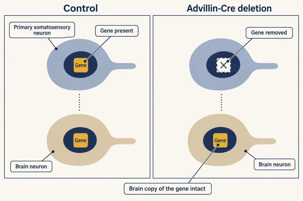
In mice, crossing Advillin-Cre to a floxed Mecp2 or Gabrb3 allele deletes the target in dorsal-root, trigeminal and sympathetic ganglion neurons. The diagram shows a representative somatosensory neuron and brain neuron. The sensory neuron's central terminal is also part of that targeted cell; resident brain and spinal-cord cells retain the gene. AI-generated illustration -
Build the terminal mechanism as an evidence ladder
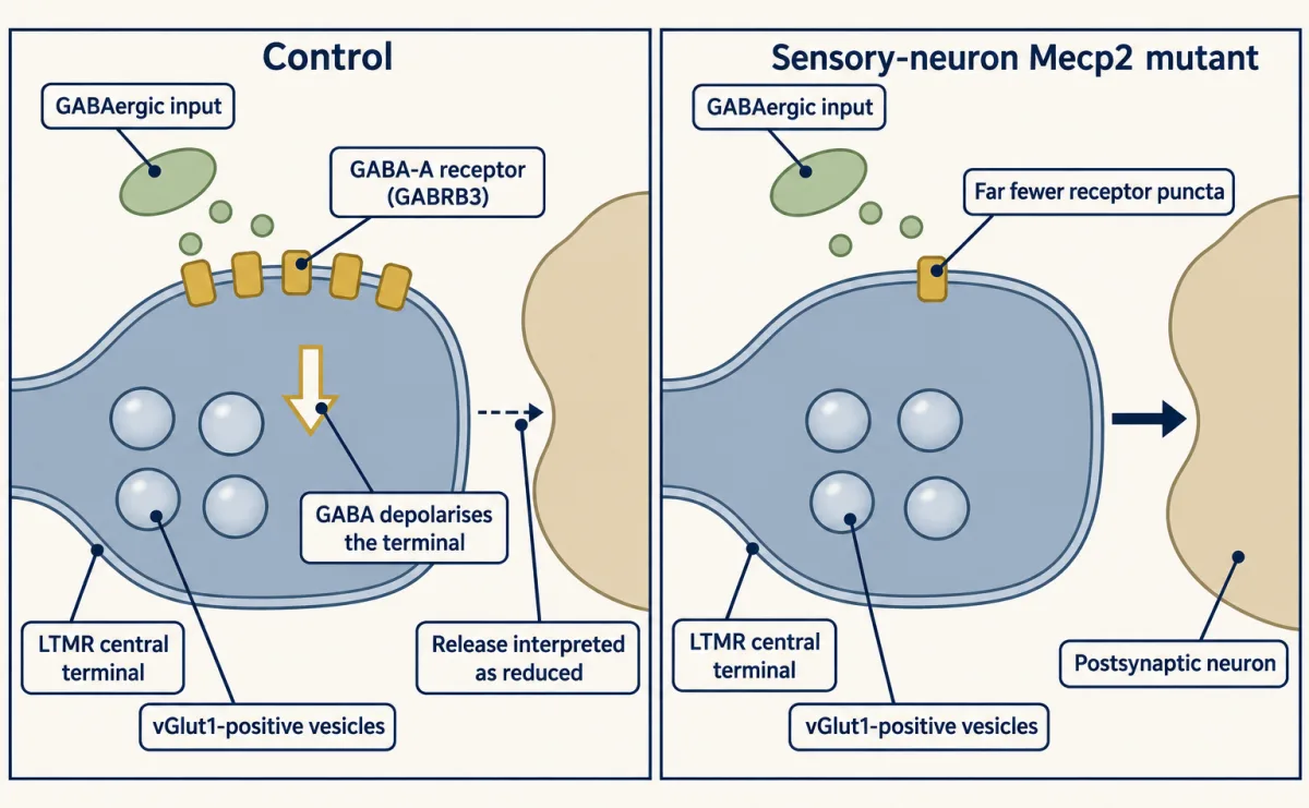
In mouse sensory-neuron Mecp2 conditional mutants, immunolabelled GABRB3 puncta associated with vGlut1-positive LTMR central terminals fell by about 70 per cent. That is a fixed-tissue puncta measurement. Separate synaptic and dorsal-root-potential recordings support the authors' presynaptic-inhibition account, which should not be read as settled physiology for all sensory afferents; the image itself does not measure conductance or release. AI-generated illustration -
What the route map did not show: the causal control
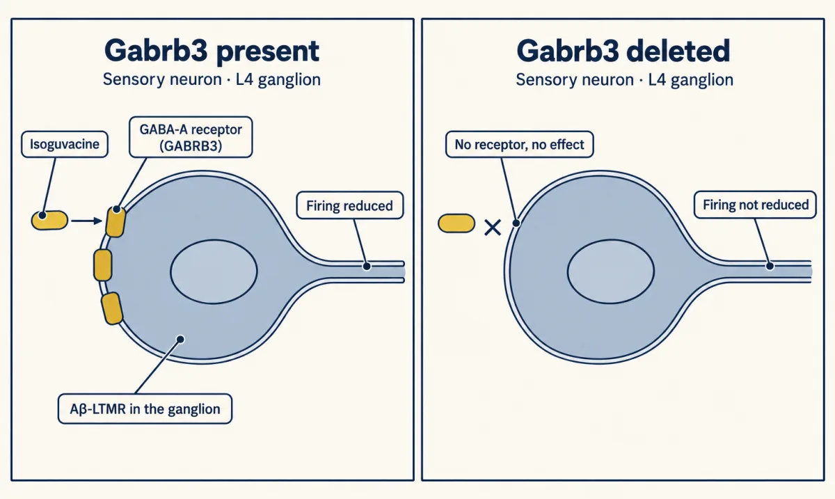
Isoguvacine reduced stimulus-evoked A-beta-LTMR firing in anaesthetised mice, but not after sensory-neuron Gabrb3 deletion. The loss of drug effect locates the required receptor subunit in those neurons. AI-generated illustration -
Timing requires two different designs
-
What this does and does not mean for people
-
Going deeper
Where this lesson fits
Before thisReview first
- The Low-Threshold Mechanoreceptors: One Touch, Several SignalsThis course · Lesson 4
The lesson works with A-beta and A-delta low-threshold mechanoreceptors, whose classes and responses are introduced there.
- Where Touch Is First Processed: Dorsal Horn and Dorsal Column NucleiThis course · Lesson 8
You need its account of presynaptic control of touch afferent terminals to follow the GABA-A inhibition mechanism here.
- Five Ways to Turn a Stimulus into a CurrentThis course · Lesson 1
The lesson assumes you know how a sensory neuron turns a stimulus into a current.
After thisStudy next
- When Pain PersistsThis course · Lesson 14
It applies the same locus, mechanism and timing questions to touch that turns painful after nerve injury.
- Spinal Circuits That Gate PainThis course · Lesson 12
It uses spinal inhibitory gate failure, the same kind of defect that lets touch signals escape control here.
Elsewhere on the siteFilms, simulations and method films in other courses
- What Can a Label Prove?Film · Developmental Neuroscience
It shows what a conditional Cre-based label or deletion can and cannot prove about where a gene acts.
- Conditional mouse builder: where does Cre act?Simulation · Developmental Neuroscience
It lets you see where Cre acts in a conditional mouse, the logic behind the peripheral-versus-brain deletion here.
Suggested by an AI model from the lessons themselves, then checked. How we use AI