Sensory Systems · Unit 3: Temperature, pain and itch
Spinal Circuits That Gate Pain
Light touch can start to hurt when a spinal gate fails. A gate can fail because the gatekeeper dies, or because the gatekeeper is still there and its signal has changed sign. In adult male rat lamina I, nerve injury was shown to do the second.
Pause when the reading reaches a film, and offer to play it. Saved on this device.
Watch the film now, or keep listening
Builds on:Where Touch Is First Processed: Dorsal Horn and Dorsal Column NucleiNociceptors and the Sodium Channels of PainThe Low-Threshold Mechanoreceptors: One Touch, Several SignalsWhy these?
 ▶ Watch Who Opens the Touch Gate? · 3:34
Read the transcript of “Who Opens the Touch Gate?”
Film transcript: Who Opens the Touch Gate?
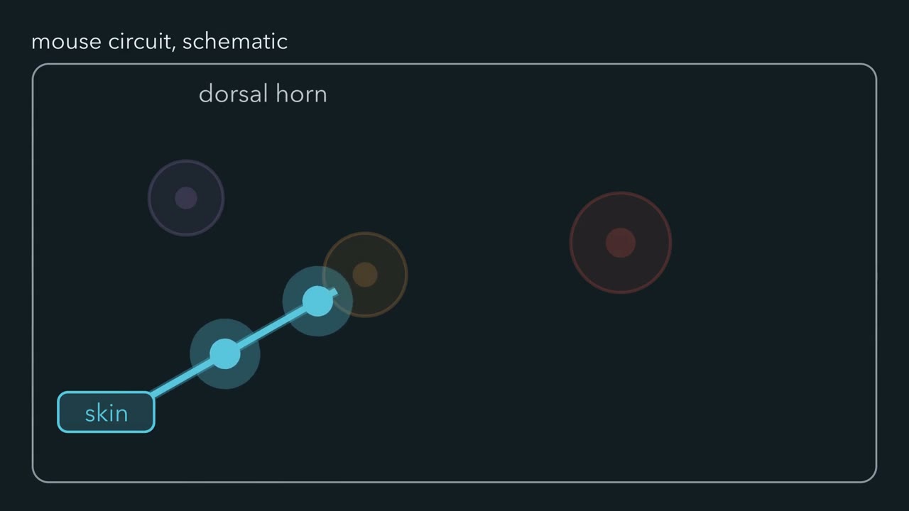
How can the same light touch recruit a pain pathway without changing the skin stimulus? Watch one blue touch signal enter the dorsal horn. It meets an orange relay, a purple inhibitory brake, and a red output route. First test the relay. In mice, removing the mostly excitatory somatostatin lineage weakens acute and injury-associated mechanical nocifensive behaviour. The result supports a relay role in the assays tested. It does not identify every route for mechanical pain. Now restore the relay and test its brake. Dynorphin-lineage inhibitory neurons restrict recruitment of superficial somatostatin neurons by large myelinated afferents. The blue input is unchanged.
Removing purple inhibition lets that input recruit many more orange cells. Predict the slice result. With the inhibitory population removed, does identical Aβ input recruit fewer superficial somatostatin cells, the same number, or more? Keep the incoming pulse fixed while you decide.
More. At the lamina II–III border, Aβ-evoked spiking rose from 5% to 40% percent of recorded neurons. The measurement locates a gated connection. The behaviour alone could not tell us where the circuit failed. Tools set the claim. Ablation removes captured cells, including cells marked by earlier expression. Tetanus toxin light chain can leave a neuron alive while blocking transmitter release. Activation asks whether added output can shift the measured behaviour. Opposite manipulations sharpen the relay claim. Somatostatin activation elicited nocifensive behaviour. Inhibition raised mechanical withdrawal thresholds and lengthened thermal withdrawal latency in naive mice.
Slice recordings confirmed that the light-driven neurons followed the stimulus trains. After spared nerve injury, permanent reporter counts of parvalbumin neurons did not fall significantly at 3, 5, or 8 weeks. Yet activating those neurons relieved injury-associated hypersensitivity.
The cells remained. The next question is what happened to their signal. Inhibitory populations are not interchangeable. Parvalbumin activation changed mechanical sensitivity without changing thermal sensitivity in the assays tested. Broader glycinergic manipulations changed mechanical, heat, and cold sensitivity together. One gate does not control every modality. So begin with behaviour, but do not stop there. Ask which cells were captured, what the tool changed, and which connection was measured. Behaviour identifies a failed gate.
Circuit measurements help locate the failure.
From the film
- In mice, inhibitory dorsal-horn neurons can keep touch input from recruiting mechanical-pain relays.
- Removing somatostatin-lineage relays reduces mechanical nocifensive behaviour in the assays tested.
- Ablation removes cells, while silencing can remove transmitter output from surviving cells.
- Behaviour identifies a failed gate; circuit measurements help locate the failure.
The first film established the circuit. The text begins where it stopped: what each intervention can actually prove.
Allodynia is pain caused by a stimulus that is normally innocuous, such as light touch. In animal experiments, the readout is described more cautiously as mechanical hypersensitivity or allodynia-like behaviour. Nocifensive behaviour is a defensive response to a noxious stimulus. Disinhibition means that inhibitory control has weakened.
In 1965, Melzack and Wall proposed that cutaneous input is modulated in the spinal cord before pain perception and response. Their diagram had no molecular identities for its cells. Modern experiments can target particular dorsal-horn populations, but the result remains bounded by the tool.
The guiding idea. Within the preparations tested, a spinal gate can fail because inhibitory output is absent or because its postsynaptic effect changes; behaviour alone cannot distinguish those mechanisms.
Scope: interpreting the matched behavioural, anatomical and electrophysiological endpoints in the dorsal-horn preparations tested here.
Touch and pain develops the original proposal, the spinothalamic crossing and central sensitization. Dorsal horn touch covers where low-threshold afferents end.
Build one circuit, then test the tool
Duan and colleagues (Cell, 2014) used intersectional mouse genetics to remove one genetically captured population at a time. Ablating the mostly excitatory somatostatin lineage impaired acute and injury-associated mechanical nocifensive behaviour. Reporter counts four weeks after diphtheria toxin confirmed depletion.
The slice experiment placed inhibition onto that relay. In dorsal-root-attached lumbar slices from young mice, inhibitory dynorphin-lineage neurons restricted recruitment of superficial somatostatin neurons by large myelinated A-beta afferents. At the lamina II–III border, removing those inhibitory neurons raised A-beta-evoked spiking from 5 to 40 percent of recorded neurons.
Read the lineage label carefully. A Cre lineage is a history of expression. The authors found detectable dynorphin messenger RNA in about three-quarters of labelled cells and cautioned that expression was probably transient in a minority. Killing every captured cell tests the population’s contribution, not the peptide’s action or only current dynorphin-expressing cells.
Reversible tools strengthen a different inference. Christensen and colleagues (Cell Reports, 2016) found that optogenetic activation of spinal somatostatin neurons elicited nocifensive behaviour in freely moving mice. Chemogenetic inhibition raised mechanical withdrawal thresholds and lengthened thermal withdrawal latency in naive mice. The channelrhodopsin-expressing neurons followed light trains in slices, a fidelity check that makes the driven spike train interpretable.
Worked problem: how complete was the ablation?
Duan and colleagues counted labelled somatostatin-lineage neurons per sampled lumbar hemisection four weeks after toxin treatment: 105 ± 4 in controls and 17 ± 2 after ablation. These are means ± SEM from 15 to 17 hemisections and three mice per group.
Predict. What depletion do the two means give, and what is the independent biological unit?
Check your prediction
Depletion = 100 × (105 − 17) / 105 = 100 × 88 / 105, which is about 84 percent. The paper reports 82 percent ablation; extra decimal places would claim more than the rounded means support.
The independent biological unit is the mouse, three per group, with hemisections nested within each animal. The section-level standard errors cannot supply animal-level uncertainty without the per-mouse values. Seventeen cells per hemisection remain, roughly one-sixth of the control mean. This is a percentage of labelled cells removed, not a percentage of pain removed.
More than one excitatory route
Peirs and colleagues (Neuron, 2015) identified another route. Deleting VGLUT3 from dorsal-horn neurons raised mechanical withdrawal thresholds in mice, in an acute assay and after carrageenan or nerve injury. Dorsal-horn VGLUT3 immunoreactivity appeared around postnatal day 5, peaked around days 10 to 12 and was much reduced by about day 20.
Keep those claims separate: one concerns a developmental requirement for the transporter; the other describes when the protein was present. Chemogenetic activation of the transient-VGLUT3 lineage produced allodynia-like behaviour, as did activation of adult lamina II calretinin neurons. The adult calretinin experiment is the cleaner adult sufficiency test because the VGLUT3-lineage vector was injected at postnatal day 10 and behaviour tested three weeks later.
The same group later reported (Neuron, 2021) that lamina II inner calretinin neurons contributed to inflammatory mechanical allodynia, PKC-gamma neurons to neuropathic allodynia, and deeper laminae III to IV cholecystokinin (CCK) neurons to both, in male and female mice. There is no invariant allodynia chain; the population required depends on the injury.
Inhibitory populations are not interchangeable
Petitjean and colleagues (Cell Reports, 2015) worked in male mice. Ablating spinal parvalbumin neurons produced mechanical allodynia associated with disinhibition of PKC-gamma interneurons. Activating parvalbumin neurons alleviated nerve-injury-associated mechanical hypersensitivity.
Boyle and colleagues (Cell Reports, 2019) refined the circuit. Parvalbumin interneurons act largely through presynaptic inhibition of myelinated low-threshold afferent terminals and inhibition of lamina II excitatory interneurons feeding lamina I. Activation filtered mechanical input without changing thermal sensitivity in the assays tested.
Foster and colleagues (Neuron, 2015) targeted a broader glycinergic population in male GlyT2-Cre mice. Local ablation caused mechanical, heat and cold hypersensitivity. So did tetanus-toxin-light-chain silencing, which blocks vesicular transmitter release while leaving the neuron alive. The block is persistent, not a rapid reversible interruption of firing. Chemogenetic activation ameliorated neuropathic hyperalgesia after chronic constriction injury.
Predict. Tetanus toxin light chain leaves the targeted neuron alive, but its block is persistent. Can this experiment show that the neuron’s output is required at the precise moment of testing?
Check your prediction
No. It shows that loss of transmitter output across the persistent intervention changes the behaviour. It is not a rapid, reversible interruption, so it cannot isolate the precise moment when the output is needed.
The population comparison answers a different question. Parvalbumin activation changed mechanical but not thermal sensitivity in the assays tested. Manipulating the broader glycinergic population moved mechanical, heat and cold sensitivity together. The populations differ in the modalities they control, so there is no all-purpose inhibitory gate.
Now change the question from necessity to mechanism. Permanent reporter counts of parvalbumin interneurons did not fall significantly at 3, 5 or 8 weeks after spared nerve injury. Petitjean and colleagues reported fewer parvalbumin terminal contacts on PKC-gamma cell bodies, while noting that the contribution of detachment to allodynia was unknown. Boyle and colleagues did not find contact loss and instead reported reduced parvalbumin-cell excitability. Surviving cells can signal too weakly.
Stable gatekeepers force the next question: can the target change what their signal does?
Read the transcript of “When Inhibition Changes Sign”
Film transcript: When Inhibition Changes Sign
Can the gate from chapter one fail while every gatekeeper remains present? The purple inhibitory neuron, its ending, and the red target all remain. This time, the change occurs inside the target neuron. An inhibitory synapse opens channels permeable to chloride and bicarbonate. Its current depends on the difference between membrane potential and anion reversal potential. Teal KCC2 helps keep intracellular chloride low in these spinal neurons. In adult male rats, nerve injury reduced dorsal-horn KCC2. In acute lamina I slices, anion reversal shifted in the depolarizing direction. The same synaptic conductance now produced current with a different sign.
Use one computed case at a membrane potential of minus −65 mV. Reversal at minus −75 gives plus +10 mV of driving force. Move reversal to minus −50 and driving force becomes minus −15 Conductance stayed fixed. Current direction changed. Current reversal is not the whole claim. An open anion conductance can still shunt other current. A depolarizing response becomes excitatory only if its net effect raises firing. The rat experiments measured increased excitability too. Now predict the diagnostic pattern.
Cell counts remain stable after injury. Which added measurement can reveal a changed postsynaptic effect: more counting, an evoked output recording, or anion reversal measured without replacing chloride? Measure anion reversal with a gramicidin perforated patch. It gives electrical access while preserving the cell's chloride.
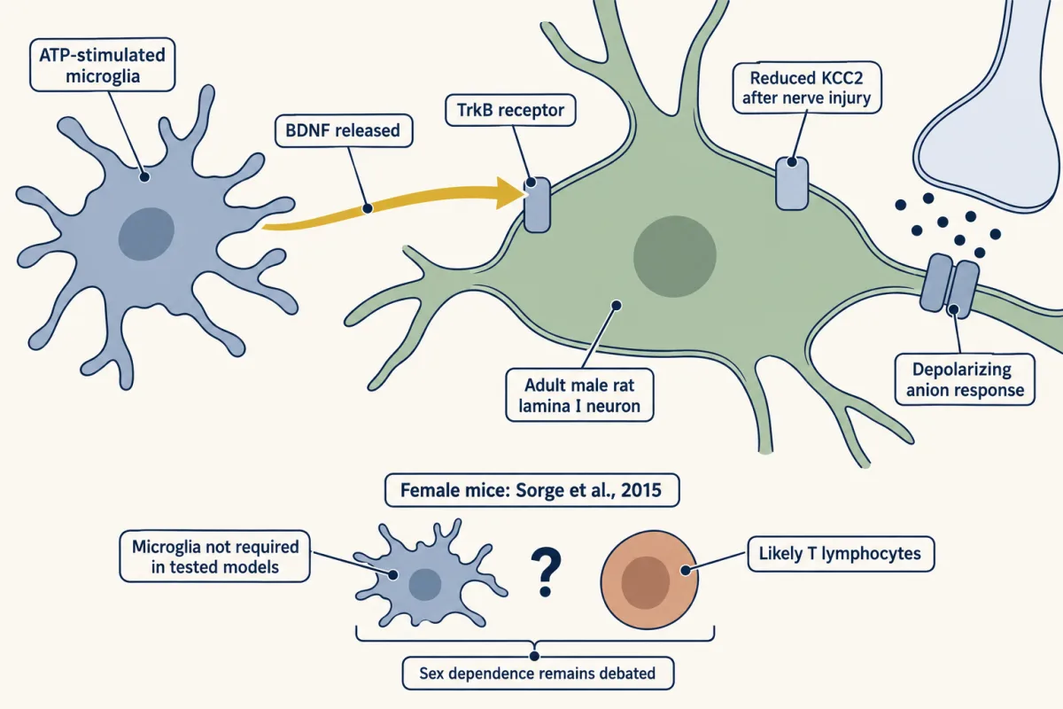
Stable counts test survival. Evoked inhibitory events test output. Reversal tests the target's response to that output. The rat studies also traced the signal upstream. ATP-stimulated microglia released BDNF. Gold BDNF blocks then reached TrkB on the red target. Gold BDNF then moved the reversal marker in the target. These rat transfer experiments did not measure KCC2 afterwards. The purple synapse remained present throughout the shift. Compare three pure patterns. Cell death lowers the count and evoked events.
Lost output leaves counts stable but reduces events. A changed postsynaptic effect leaves counts stable and events present while reversal moves. Real mechanisms can coexist. This route was established in adult male rats. It is not automatically obligatory across species or sexes. Keep the experimental rule instead. Count cells, test output, then measure reversal in the target neuron.
From the film
- In adult male rats, nerve injury lowered KCC2 and shifted lamina I anion reversal.
- A synapse can remain present while its postsynaptic effect changes.
- A depolarizing anionic response is not automatically excitatory because it can still shunt current.
- Cell counts, evoked output and gramicidin recordings test different failure points.
The causal tests the second film did not show
The records behind the moving gradient came from acute parasagittal lamina I slices. Coull and colleagues (Nature, 2003) used adult male Sprague-Dawley rats with a sciatic nerve cuff. Peripheral nerve injury reduced dorsal-horn expression of potassium-chloride cotransporter 2 (KCC2), the potassium-chloride exporter that keeps intracellular chloride low in these neurons, and the shifted anion gradient made normally inhibitory anionic synaptic currents excitatory. They measured increased excitability rather than inferring it from a depolarizing current alone. Synaptic integration develops why an open conductance can also shunt current.
The next step was sufficiency.
Predict. If lowering KCC2 is sufficient, what should spinal KCC2 blockade or knockdown do in otherwise intact rats?
Check your prediction
It should lower nociceptive thresholds without requiring the nerve injury. That is what the experiments found, making the chloride change sufficient in this preparation rather than merely correlated with injury.
The 2005 Nature paper then tested the upstream chain pictured in the chapter. Intrathecal transfer of ATP-stimulated rat microglia into uninjured adult male rats reproduced the depolarizing anion-reversal shift and behavioural change. Pretreating the cells with BDNF-directed interfering RNA inhibited both effects. Blocking BDNF-TrkB signalling reversed nerve-injury-associated allodynia and the anion-potential shift in injured rats, tying the pathway to injury rather than only transfer. Trk and p75 covers the receptor; microglia and complement pruning covers microglial signalling.
Two limits travel with this mechanism: it was established in adult male rats. Sorge and colleagues (Nature Neuroscience, 2015) found that microglia were not required for mechanical hypersensitivity in immunocompetent female mice in the models tested and implicated adaptive immune cells, likely T lymphocytes. Whether the contribution differs by sex remains debated because other studies find that blocking microglial signalling relieves nerve-injury pain in both sexes. A mechanism can be real in one preparation without being obligatory across species or sexes.
- Driving force (Vm − reversal)
- +5 mV
- Sign
- positive
| Question | What answers it | In this state |
|---|---|---|
| 1. Is the gatekeeper’s output there? | A cell count, or evoked inhibitory events | |
| 2. Which way does that output push? | The anion reversal potential, by gramicidin-perforated patch |
In this sketch: one compartment, one anionic synapse, driving force only, no spikes. The voltages are illustrative, not a measured reversal potential.
An inhibitory synapse has no fixed effect: the current depends on the driving force, membrane potential minus anion reversal potential, so a gate can fail because the gatekeeper’s output is gone or because the sign has changed. A cell count answers the first and says nothing about the second.
The output neurons and the denominator trap
Projection neurons carry the dorsal horn’s result onward, and they are uncommon. Cameron and colleagues (Pain, 2015) estimated that they make up about 5 percent of mouse lamina I neurons in lumbar segment L4, using fixed tissue from adults of either sex after retrograde tracing. About 90 percent of traced lamina I spinoparabrachial neurons expressed NK1r, the substance-P receptor encoded by Tacr1.
Predict. Does that 90 percent mean that most NK1r-positive lamina I cells project to the parabrachial nucleus?
Check your prediction
No. It is the fraction of traced projection neurons that express NK1r, not the fraction of NK1r-positive cells that project. Sheahan and colleagues (Journal of Neuroscience, 2020) found that most genetically targeted superficial Tacr1-CreER dorsal-horn neurons were interneurons. The denominators differ, so the results are compatible.
Roome and colleagues (Cell Reports, 2020) supplied a developmental handle. Mouse neurons with embryonic Phox2a expression included adult anterolateral-system projection neurons. In adults of both sexes aged 6 to 19 weeks, intersectional tracing showed Phox2a-lineage neurons reaching parabrachial and thalamic targets. Human fetal spinal cords at gestational weeks 7.3 to 8.4 also showed Phox2a neuronal molecular identity by immunohistochemistry: a fixed-tissue correspondence, not adult human physiology.
Predict. Developmental Phox2a loss reduces supraspinal nocifensive responses while measured spinal nociceptive reflexes remain. Does that establish an acute adult function?
Check your prediction
No. The pattern fits a thinned ascending limb with local reflex circuitry left intact, but the deletion acts during development. It cannot measure what an acute interruption of Phox2a-lineage neurons would do in an adult.
Do not turn the two projection targets into mutually exclusive sensation and affect channels. The tracing shows that the lineage reaches both. It does not assign exclusive roles. Lee and colleagues (Nature Communications, 2025) found Calcrl-positive spinal projection neurons carrying cool-specific signals to the lateral parabrachial nucleus in mice, reminding us that the parabrachial route is not pain-only. Chronic pain follows the output; itch uses partly separate populations in the same circuitry.
Design problem: where did inhibition fail?
Remember the short version: behaviour shows failure; matched endpoints locate it. The second film supplied the three diagnostic patterns. Now design a test robust enough to distinguish them after nerve injury.
One way to do it
Survival. Mark the inhibitory population permanently before injury and count labelled cells later. A permanent reporter matters because injury-driven loss of marker expression could mimic cell death.
Output. Activate the parvalbumin population optogenetically and record evoked inhibitory postsynaptic currents in PKC-gamma interneurons or a lamina I projection neuron. Behaviour and this recording alone cannot separate a dead cell from a surviving cell that lost its synapses; the survival count can.
Postsynaptic effect. Measure anion reversal in the target with a gramicidin-perforated patch, which preserves intracellular chloride. A conventional whole-cell pipette replaces the cell’s chloride and erases the quantity at issue.
Use acute, reversible silencing in adults rather than lineage ablation to test present function. Measure behaviour during perturbation and include a motor-control task, because impaired paw placement can resemble hypersensitivity.
Collect all three endpoints in matched preparations because mechanisms can coexist. Current amplitude at one holding potential will also change when driving force changes, so do not mistake amplitude alone for lost release.
Going deeper
Stimulation, dosing and control details
Somatostatin neurons expressing channelrhodopsin followed light trains at 1 to 10 Hz with 10 ms pulses in slices, although spike probability fell at higher frequencies. Petitjean and colleagues used male mice aged 8 to 14 weeks. Foster and colleagues used GlyT2-Cre mice aged 7 to 8 weeks at lumbar injection and clozapine-N-oxide at 1 mg/kg intraperitoneally.
For chemogenetics, include receptor-free animals given the same ligand. Clozapine-N-oxide is converted to clozapine, which occupies these receptors with higher affinity in rodent experiments. Saline in receptor-expressing animals does not test those off-target drug effects.
Light-driven silencing has its own threat here. Chloride-pumping silencers can leave an after-effect on reversal potential, reported in rat hippocampal slices and absent with a proton pump. That side effect changes the very endpoint this design aims to measure.
Your notes
Select any text in the lesson to highlight it or add a note to it. Your highlights and notes are saved in this browser and collected in your notebook.
This browser is not letting the site save anything (a private window or strict privacy settings), so highlights and notes will be lost when you leave the page.
How we measure it
Intersectional Cre/Flp toxin ablation
Two genetic drivers restrict a diphtheria-toxin or toxin-receptor allele to cells that expressed both, so one genetically captured dorsal-horn population can be removed and the animal's behaviour measured weeks later. It tests necessity for the assay used, and the label is a lineage: cells that expressed the driver at some time, not cells expressing the marker now.
Tetanus toxin light chain silencing
A viral vector delivers tetanus toxin light chain, which prevents vesicular transmitter release from the targeted neurons. The cell survives, which separates loss of output from loss of the cell, but the block is persistent rather than a rapid reversible interruption of firing.
Chemogenetic activation and inhibition
An engineered receptor such as hM3Dq or hM4D is expressed in a defined population and driven by a systemic ligand, usually clozapine-N-oxide. Because CNO is converted to clozapine, which occupies these receptors with higher affinity in rodent experiments, a receptor-free ligand-treated control is part of the method rather than an extra.
Gramicidin-perforated-patch recording
A pore-forming antibiotic gives electrical access to a neuron while leaving its own chloride concentration intact, so the anion reversal potential measured is the cell's and not the pipette's. It is the endpoint that turns "inhibition failed" into a number.
Practice questions on this lesson (3) → MCAT-style, original, each with an explanation
Where this lesson fits
Before thisReview first
- Where Touch Is First Processed: Dorsal Horn and Dorsal Column NucleiThis course · Lesson 8
It shows where low-threshold touch afferents end in the dorsal horn, which is the input this gate controls.
- Nociceptors and the Sodium Channels of PainThis course · Lesson 11
It covers the nociceptor input that touch must be kept from recruiting when the gate works.
- The Low-Threshold Mechanoreceptors: One Touch, Several SignalsThis course · Lesson 4
The A-beta touch afferents that the inhibitory neurons restrict are described there.
After thisStudy next
- When Pain PersistsThis course · Lesson 14
It takes disinhibition and a changed sign of inhibition further, to pain that persists after nerve injury.
- Itch: A Sense of Its OwnThis course · Lesson 13
Itch uses partly separate dorsal-horn populations, so the cell-type logic of gating carries over to it.
Elsewhere on the siteFilms, simulations and method films in other courses
- Why Capsaicin Feels HotFilm · Neuroscience
It shows how spinal pathways carry touch and pain, the circuit that is gated here.
- One More RopeFilm · Neuroscience
It shows how inhibition can shunt a cell without changing its voltage, as with depolarizing anionic responses here.
- The StandoffFilm · Neuroscience
It gives the resting balance of ion currents that a shifted anion reversal potential disturbs.
Suggested by an AI model from the lessons themselves, then checked. How we use AI
Finish line
Test yourself
8 questions
Practice and MCAT-style, with explained answers.
Next · Lesson 13
Itch: A Sense of Its Own
A mouse missing one cluster of Mrgpr genes scratches normally to histamine but much less to chloroquine. Which parts of the itch pathway are shared with pain, and which are not?
Continue →