Sensory Systems · Unit 3: Temperature, pain and itch
When Pain Persists
Rewritten on : new text, flashcards and film. Your notes and highlights stay, but the text around them may have moved.
After a nerve injury, a touch the animal used to ignore makes it pull away, and it keeps doing so for weeks. Is persistent pain an alarm that never switched off, or a nervous system that has changed?
Pause when the reading reaches a film, and offer to play it. Saved on this device.
Watch the film now, or keep listening
Builds on:Spinal Circuits That Gate PainNociceptors and the Sodium Channels of PainWhere Touch Is First Processed: Dorsal Horn and Dorsal Column NucleiWhy these?
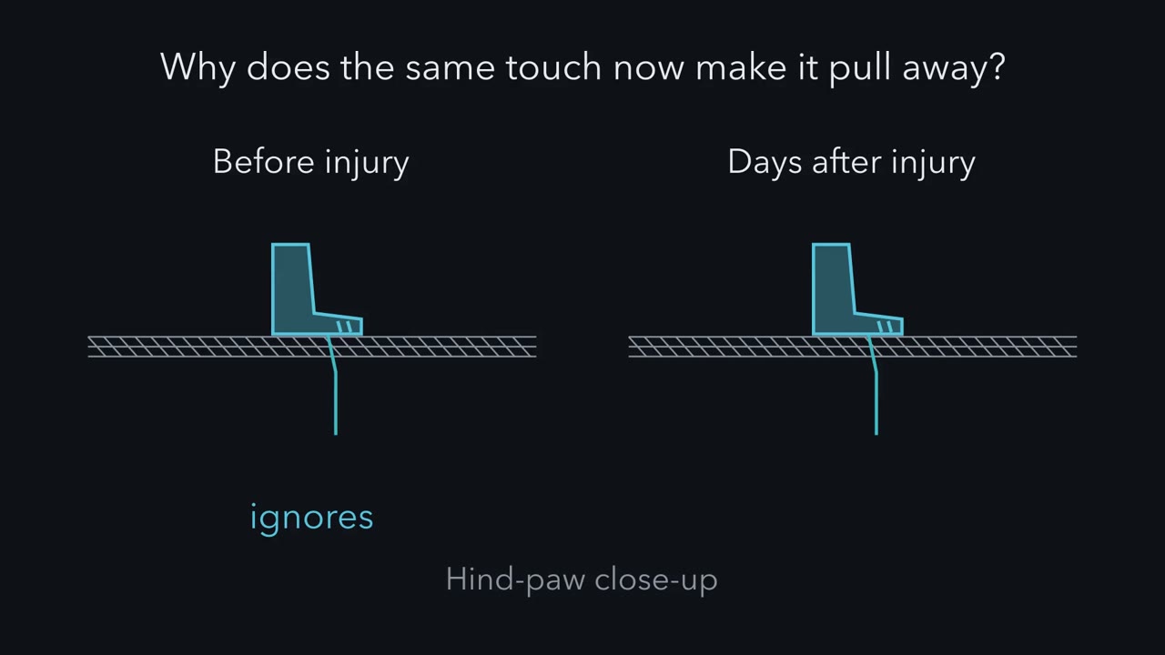 ▶ Watch A Touch That Hurts · 2:48
Read the transcript of “A Touch That Hurts”
Film transcript: A Touch That Hurts
A rat ignores a light touch on its paw. Days after a nerve injury, the same touch makes it pull away. Why? The rat stands on a wire-mesh floor. A thin filament presses up against its hind paw. The paw stays down. Then comes a spared nerve injury. Two branches of the nerve to the paw are cut, and one is spared. Days later, the same filament makes the paw lift.
Allodynia means pain from a stimulus that normally does not hurt. A rat cannot tell us, so the paw lift stands in for it. The touches come from filaments of rising stiffness, pressed over repeated trials. Many labs take the force that lifts the paw half the time. That is the withdrawal threshold. Plot that threshold over time. In sham-operated rats it stays flat. In injured rats it falls within days, and stays low. When a treatment is said to reverse allodynia, it pushes that curve back up, often only for a while.
Two other words matter. Hyperalgesia is more pain to a stimulus that already hurts. Spontaneous pain comes with no applied stimulus. Notice what is measured. Most rodent papers on allodynia report a withdrawal threshold, which is a reflex. Hold on to that. So is persistent pain an alarm that never switched off, or a changed nervous system? Which would you pick?
The answer is both. After injury, some sensory neurons fire with no stimulus. So the alarm wiring itself has changed. Here is the one idea to carry through. After a nerve injury, pain persists because the rules of the system have changed. First, sensory neurons make signals with no stimulus. Second, in rats, the dorsal horn loses inhibition. In a simple model, a light touch that used to fall short can then fire a pain-signalling neuron. Third, in rodents, immune cells help hold that change, by routes that differ with sex and compartment. Fourth, separate brain circuits set how unpleasant the result is, apart from the reflex.
Most of this comes from rodent nerve-injury models. In people, the evidence is association. The takeaway: the alarm wiring changed, and so did the rules for what counts as a pain signal.
From the film
- After a nerve injury, pain can persist because the rules of the system change, and the alarm wiring is part of what changes.
- In rodent models, the rodent sign of allodynia is a withdrawal threshold that falls and stays low.
- Most of this comes from rodent nerve-injury models; in people the evidence is association.
A rat stands on a wire-mesh floor, and a thin filament presses up against its hind paw. The paw stays down. Days after a spared nerve injury, in which two branches of the nerve to the paw are cut and one is spared, the same filament makes the paw lift. Why does a touch the animal used to ignore now make it pull away?
First, name three things that can go wrong after an injury like this.
- Allodynia is pain from a stimulus that does not normally provoke pain. A rat cannot say that a touch hurts, so rodent papers measure a stand-in: the paw withdraws from a touch it used to ignore. The touches come from calibrated filaments of increasing stiffness, called von Frey filaments. The withdrawal threshold is the force that makes the paw lift, estimated over repeated trials (many labs use the force that lifts the paw half the time). A fall in that threshold is the rodent sign of allodynia.
- Hyperalgesia is more pain to a stimulus that already hurts.
- Spontaneous pain is pain with no applied stimulus. It can still have a neural cause, such as nerve fibres firing on their own. It is hard to measure in an animal.
Most rodent papers that report “tactile allodynia” or “mechanical hypersensitivity” are reporting a withdrawal threshold of this kind, which is a reflex; some count responses or score other behaviours, so check the assay. A later experiment shows why that matters.
So is persistent pain an alarm that never switched off, or a nervous system that has changed? Both: the alarm wiring itself changes, and further in, so do the rules for what counts as a pain signal.
Touch and pain taught sensitization, gate control and descending control, and spinal pain circuits taught the dorsal-horn cell types and the potassium-chloride cotransporter 2 (KCC2) shift. This lesson starts past both.
The one idea to carry through this lesson. After a nerve injury, pain persists because the rules of the system have changed, and the alarm wiring is part of what changed. The change sits at four levels:
- Sensory neurons make signals with no stimulus.
- The dorsal horn loses inhibition, so input that used to fall short of firing a pain-signalling neuron can now fire it. A light touch is one such input. This step is a model, built below.
- Immune cells help hold that change, by routes that differ with sex and compartment.
- Separate brain circuits set how unpleasant the result is, apart from the reflex.
Nearly all of this comes from rodent nerve-injury (neuropathic) models; in people it is supported only by association, from surgically removed ganglia and from a brain-imaging cohort. These studies do not establish the chain in nociplastic pain (changed processing with no tissue damage or nerve lesion to explain it) or in pain driven by ongoing tissue damage, such as arthritis, where ongoing input and changed processing can coexist. The papers below also use different nerve-injury models, so the chain is assembled across them.
The signature: a threshold that falls and stays down
Picture the withdrawal threshold plotted against time, for injured rats and for sham-operated rats that had the same surgery without the nerve cut. The table gives the typical shape, not the data of one study.
| Time | Sham-operated | Nerve-injured |
|---|---|---|
| Before surgery | high threshold: light filaments leave the paw down | the same |
| Days after | unchanged | threshold falls: light filaments now make the paw lift |
| Weeks after | unchanged | threshold stays low |
| After a treatment said to reverse allodynia | — | threshold rises back toward baseline, often only for a while |
That last row is what every “reversed the allodynia” in this lesson means: the curve was pushed back up. The rest of the lesson asks which cells hold the curve down.
The ganglion that will not stop firing
Start with the first neuron. Rat models show that nerve injury changes both injured afferents and their uninjured neighbours. After the L5 spinal nerve was ligated and cut, about half of the C-fibre nociceptors in the uninjured L4 nerve developed spontaneous activity within a day (Wu and colleagues, 2001). That is ongoing peripheral input with no stimulus: the alarm wiring itself making signal.
Channel expression shifts too. Li and colleagues ligated the L5 spinal nerve in rats and measured Nav1.7, a sodium channel that helps nociceptors start firing, in the L4 and L5 ganglia seven days later. Nav1.7 mRNA and protein fell in the injured L5 ganglion and rose in the adjacent, uninjured L4 ganglion. Read that twice, because “injury raises Nav1.7” is a tempting summary and it is wrong. In this model the sign of the change depends on which ganglion you sample. In a second nerve-injury model Li and colleagues saw Nav1.7 rise in both ganglia, and an expression change is not the same as a change in firing. The sodium channel lesson covers what Nav1.7 does when it is present or absent.
Does anything like this happen in people? North and colleagues studied dorsal root ganglia surgically excised from human donors who were undergoing tumour surgery, some with radicular or neuropathic pain and some without. Some of the cultured neurons fired spontaneously, with no applied stimulus, and that firing was associated with the donors’ pain. Two limits matter. It is an association, and dissociation cuts every axon and the cells spent more than a day in culture, so firing in the dish is only a proxy for ectopic firing in the body.
Notice what just happened: a rat result, then a human association, each measured in its own preparation, with its own readout and limits. Keeping each result at its own level is the skill this lesson trains.
How a touch could now fire a pain neuron
Read the transcript of “How Microglia Let a Touch Through”
Film transcript: How Microglia Let a Touch Through
After a nerve injury, a light touch can make a rat pull away. What changed in the spinal cord, and who changed it? Meet a lamina I neuron, in the outermost layer of the dorsal horn. It sends signals toward the brain. In a simple picture, a light touch excites it a little. Inhibitory synapses hold it below threshold. Those synapses open channels that pass chloride. They pull the voltage toward the anion reversal potential. That potential stays low because a transporter, KCC2, carries potassium and chloride out of the cell. In 2003, Coull, De Koninck and colleagues studied rats after nerve injury. Their lamina I neurons had less KCC2. Their anion reversal potential had shifted up.
That does not mean the neuron itself depolarized. Predict. As that potential rises toward threshold, what happens to the inhibitory synapse's grip? Recall the balance-point rule. The synapse can only pull the voltage toward its own reversal potential. So as that potential rises, the pull weakens. Less excitation is needed to fire the cell. In a passive model with made-up values, this touch leaves the steady voltage below threshold in the healthy cell. With the reversal potential at minus −55 mV, it sits exactly at the threshold boundary. At minus −50 mV, it sits above it. Notice what did not happen. Inhibition did not have to turn into excitation.
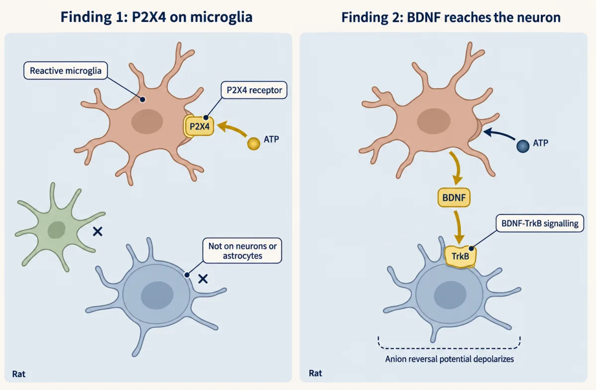
It only had to weaken. So what changed the neuron? Not a neuron. Tsuda and colleagues, in rats, found P2X4 receptors on reactive microglia. Microglia are the resident immune cells of the cord. They ran two tests. Lowering P2X4 suppressed the allodynia. So the receptor was needed. Microglia stimulated with ATP were transferred into rats never injured. Those rats developed allodynia. So stimulated microglia were enough. The messenger came in 2005. Coull and colleagues showed that stimulated microglia release BDNF. BDNF acts through TrkB receptors. It shifts the anion reversal potential up, the same change seen after injury. Block that signalling, and both the shift and the allodynia reverse.
Three papers, different links, all in rats. The behavioural readout was always a withdrawal threshold. The last link, from weaker inhibition to a touch that fires, is a model. Now the twist. Sorge, Mapplebeck and colleagues gave male and female mice a nerve injury. The hypersensitivity was comparable. In males, a microglial inhibitor reversed the hypersensitivity. In females, under the conditions tested, it did not. Predict. Intact females seemed to rely on adaptive immune cells. What does the inhibitor do in females lacking those cells?
It works. The route can switch. A later study found the same pattern in rats. So it is not a quirk of mice, but it stays conditional. Macrophages in the dorsal root ganglion add another route. In mice, depleting macrophages throughout the body reduced hypersensitivity in both sexes. The authors favoured the ganglion macrophages, but skin dendritic cells were depleted too. So ask of any immune result: which cell, in which sex, in which compartment?
From the film
- In rats, nerve injury lowers KCC2 in lamina I neurons and shifts the anion reversal potential up, which weakens inhibition without turning it into excitation.
- In rats, a chain of tested links runs from microglial P2X4 to BDNF, TrkB and that shifted potential.
- In mice, the immune route to hypersensitivity can differ with sex, so ask which cell, in which sex, in which compartment.
Now the cord, and the cell that has to change. A lamina I neuron, in the outermost layer of the dorsal horn, sends signals toward the brain. In the simple picture used here, a light touch excites it a little before injury, but inhibitory synapses that use GABA and glycine hold it below threshold (the real touch route runs through dorsal-horn interneurons). Those synapses open channels that pass chloride, and they pull the voltage toward the chloride reversal potential (strictly the anion reversal potential, Eanion, because the channels also pass some bicarbonate). That potential stays low because a transporter, potassium-chloride cotransporter 2 (KCC2), carries potassium and chloride out of the cell together.
In 2003 Coull, De Koninck and colleagues found that after a nerve injury in rats, lamina I neurons had less KCC2, and their Eanion had shifted in a depolarizing direction. That phrase is easy to misread. It means the reversal potential moved up, not that the neuron itself depolarized. Why would it let a touch through?
Predict. Use the balance-point rule from the action potential: at steady state, with no current injected, the voltage sits at the conductance-weighted average of the reversal potentials. Suppose an inhibitory synapse is open, and Eanion moves from −70 mV up toward −50 mV, the threshold. What happens to the synapse’s power to keep the cell below threshold? Does it have to start exciting the cell for a touch to get through?
Check your prediction
The synapse can only pull the voltage toward Eanion. As Eanion rises toward threshold, that pull weakens, so less excitation is needed to reach threshold. Here is a passive model with illustrative values, not lamina I measurements. Passive means fixed conductances and no spike mechanism, so the model gives only the steady voltage. The values: a leak conductance of 1 at −65 mV, a threshold of −50 mV, an inhibitory conductance of 1 at Eanion, and an excitatory touch conductance driving toward 0 mV.
| Eanion | Excitation that puts the steady voltage at threshold | Steady voltage with a touch input of 0.4 |
|---|---|---|
| no inhibition | 0.3 | — |
| −70 mV (healthy) | 0.7 | −56.2 mV: below threshold |
| −60 mV | 0.5 | −52.1 mV: below threshold |
| −55 mV | 0.4 | −50.0 mV: exactly at threshold, the boundary |
| −50 mV | 0.3, the same as with no inhibition | −47.9 mV: above threshold |
For the healthy row, the balance point is (1 × −65 + 0.4 × 0 + 1 × −70) / (1 + 0.4 + 1) = −56.2 mV. With the same touch, the steady voltage sits well below threshold in the healthy cell, exactly at the boundary at −55 mV and above it at −50 mV. Starting below threshold, the voltage crosses in real time only if the steady value lies above threshold and the touch lasts long enough.
So the answer to the second question is no. At −55 mV the inhibitory synapse, acting alone, depolarizes the cell from −65 mV to −60 mV, yet it still raises the excitation needed to reach threshold (0.4 against 0.3 with no inhibition), because it pulls toward a value below threshold. In this steady-state model the synapse adds to the excitation needed whenever Eanion is below threshold, and lowers it only once Eanion is above. Real neurons are less tidy: a depolarizing GABA input can sometimes help a cell fire with Eanion below threshold, depending on its timing, its location on the cell and active channels. “Lost inhibition” is the safe summary; “GABA becomes excitatory” needs more evidence than a shifted Eanion.
The chloride arithmetic shows why the transporter matters. From the Nernst equation at 37 °C, with outside chloride fixed, each doubling of chloride inside the cell makes the chloride reversal potential about 18.5 mV more positive (26.7 mV × ln 2). So a cell that exports less chloride can lose much of its inhibition. How fast depends on chloride fluxes, and bicarbonate also shifts Eanion.
That explains how the neuron changes. It does not say what changed it, and the answer was not a neuron. Tsuda and colleagues, working in rats after nerve injury, found that induced P2X4 receptors, channels opened by ATP, sat on reactive spinal microglia, the resident immune cells of the cord, and not on neurons or astrocytes. The authors ran two tests, and they answer different questions.
Predict. If microglial P2X4 is part of the cause, what should happen to allodynia when P2X4 levels are lowered? And what should happen if microglia stimulated with ATP are transferred into a rat that was never injured?
Check your prediction
Lowering P2X4 levels suppressed the tactile allodynia, so the receptor was needed. Microglia stimulated with ATP in culture and transferred to naive rats produced allodynia, so stimulated microglia were enough. The test of need was on a receptor, and the test of enough was on whole cells; neither shows that the receptor alone is enough. Together they put microglia in the mechanism. They still did not show how a microglial cell reaches the neuron.
The link came in 2005. Coull and colleagues, working in rats, showed that ATP-stimulated microglia release brain-derived neurotrophic factor (BDNF), and that BDNF acting on TrkB receptors depolarizes the anion reversal potential of lamina I neurons in spinal slices. That is the same change found after injury.
Predict. If BDNF is the messenger, what should blocking BDNF-TrkB signalling do to the shift, and to the allodynia?
Check your prediction
It should reverse both, and it did. The chain now has tested links: nerve injury, P2X4 in microglia, BDNF release, TrkB, a depolarized Eanion, weaker inhibition. On the balance-point picture, touch input can then fire a neuron it used to fall short of; that last step is an inference from the model and the definition of allodynia, not a recording in these papers.
Each link comes from a different experiment, which is why the 2003 P2X4 paper and the 2005 BDNF paper should not be merged into one discovery. Every tested link was shown in rats. The molecular and cellular links were measured directly, and every behavioural readout was a withdrawal threshold.
The spinal pain circuits lesson covers the dorsal-horn cell types in which this happens, and Trk and p75 covers the receptor.
A worked problem: the other half of the balance
Lost inhibition is one half of the balance. The other half is excitation: does more of it arrive after injury? Chen and colleagues recorded in vivo, by whole-cell patch clamp, from lamina I and II neurons of anaesthetized rats after nerve injury. The mean spontaneous excitatory postsynaptic current (EPSC) frequency was 9.0 Hz (SEM 1.3, n = 20 neurons) in sham-operated rats and 15.9 Hz (SEM 1.6, n = 20 neurons) seven days after spared nerve injury.
- Absolute change: 15.9 Hz minus 9.0 Hz is 6.9 Hz.
- Ratio: 15.9 divided by 9.0 is 1.77, a 1.77-fold rise.
- Percentage increase: 100 times 6.9 divided by 9.0 is 76.7 percent. The base is the sham value, so 1.77-fold and a 76.7 percent increase are the same fact.
- Spread: neurons vary widely, so the groups overlap even though the means differ, and neurons recorded from the same animal are not independent.
So, putting two rat studies side by side, more excitation reaches dorsal-horn neurons after injury while less inhibition opposes it. These EPSC measurements do not show a loss of inhibition in the same neurons: Chen’s separate inhibitory-current recordings tested anterior cingulate cortex (ACC) stimulation, and the KCC2 and anion-gradient findings came from Coull and colleagues in other rats. What the 76.7 percent is: a rise in the frequency of synaptic events onto these neurons. What it is not: a 76.7 percent rise in how unpleasant the animal finds anything. The amygdala experiment below says why those must stay apart.
The twist: the chain depends on sex and compartment
Those links come from rats. Sorge, Mapplebeck and colleagues asked whether the microglial route holds in both sexes. They gave male and female mice a nerve injury, which produced comparable hypersensitivity in the two sexes, then a microglial inhibitor, in intact mice and in female mice lacking adaptive immune cells.
Predict. Here are the first two rows. Predict the third: if intact females rely on adaptive immune cells instead of microglia, what should the inhibitor do in females that lack those cells?
| Mice | Microglial inhibitor reverses the hypersensitivity? |
|---|---|
| Male, intact | yes |
| Female, intact | no, under the conditions tested |
| Female, lacking adaptive immune cells | ? |
Check your prediction
Yes. Female mice lacking adaptive immune cells became sensitive to the glial inhibitor, so the pathway can switch. Intact females appeared to use adaptive immune cells, likely T lymphocytes. Note the wording of the second row: “no, under the conditions tested” is narrower than saying females do not use microglia.
The result is conditional and in mice. The authors themselves raise the idea of treating men and women differently, but they tested no people. It does not say that women use T cells and men use microglia.
Mapplebeck, Salter and colleagues later found the same pattern in rats: inhibiting spinal microglia or blocking P2X4 reversed allodynia in males but not in females. So the difference is not a quirk of mice, but it remains conditional on model and assay.
Compartment matters as well. Yu and colleagues (Nature Communications, 2020) used mice and a nerve injury that spared one branch. Depleting macrophages throughout the body reduced mechanical hypersensitivity in both males and females, and macrophages in the dorsal root ganglion expanded after injury. Deleting CSF1 from sensory neurons reduced that expansion in males but not in females.
The depletion has one caveat. Because it was systemic, it could not exclude a contribution from skin dendritic cells depleted at the same time, though the authors favoured the ganglion macrophages as the main contributor. File the ganglion as a second immune compartment, where what differed by sex was the CSF1 dependence of the expansion, not the effect of depletion.
So “immune cells drive chronic pain” splits into spinal microglia, ganglion macrophages and adaptive cells, each with its own sex and context dependence. The question to ask of any immune result is: which cell, in which sex, in which compartment? Testing this in people would need cell-type-specific tests in human tissue or a human intervention, and none of the mouse data support a treatment claim.
A reflex is not the hurt
Read the transcript of “A Reflex Is Not the Hurt”
Film transcript: A Reflex Is Not the Hurt
A paw pulls away from a touch. Does that tell you how much it hurts? Every behavioural result so far was a withdrawal threshold. A reflex is easy to mistake for pain. Corder and colleagues imaged neurons in the basolateral amygdala of male mice, across repeated sessions. One ensemble responded to noxious stimuli and encoded their unpleasantness. After a nerve injury, innocuous stimuli recruited the same ensemble.
Then they tagged those neurons genetically, and silenced them. Predict. With the ensemble silenced, does the paw still pull away? Yes. The withdrawal reflexes were preserved. But escape and avoidance fell. The reflex and the affective behaviour are two measurements, and they can come apart. This was male mice, in one model. In people, these cell-type manipulations cannot be done. So the human study here asks a different question: prediction. Baliki and colleagues scanned adults whose back pain was still subacute. A year later, they asked who still had pain.
Recovering meant a set drop in pain intensity. It did not mean pain-free. Stronger baseline connectivity between medial prefrontal cortex and nucleus accumbens predicted who persisted. That area is a pair-ordering probability. It is not a cause, and not one person's risk. The takeaway: in rodent models, the wiring, the cord and the immune cells all change. In mice, affective behaviour can be separated from the reflex. In people, we have association and prediction.
From the film
- A withdrawal reflex is easy to mistake for pain, because the reflex and the affective behaviour are two measurements that can come apart.
- In male mice, silencing a basolateral amygdala ensemble reduced escape and avoidance while withdrawal reflexes were preserved.
- In people, the evidence is prediction and association: baseline prefrontal-accumbens connectivity predicted persistence, which is not a cause.
Every behavioural result so far was a withdrawal threshold; the rest were recordings, expression measures and immune-cell assays. A withdrawal reflex is cheap to measure and easy to mistake for pain. Corder, Ahanonu and colleagues asked whether affective-motivational behaviour, such as escape and avoidance, has its own circuit. In male mice they imaged basolateral amygdala (BLA) principal neurons in repeated sessions and identified an ensemble that encoded the negative valence, or unpleasantness, of noxious stimuli. After a nerve injury, innocuous stimuli recruited the same ensemble. They then used activity-dependent genetic tagging to silence that ensemble.
Predict. If the ensemble were needed for the withdrawal response, what would happen to paw withdrawal when it is silenced? If it carries only how bad the stimulus is, what would you see instead?
Check your prediction
If the ensemble were needed for withdrawal, withdrawal would fall too. It did not. Affective-motivational pain behaviours fell, while the measured withdrawal reflexes were preserved. The reflex and the behaviours that signal unpleasantness are two measurements, and they can come apart. Whether the mouse still detects the touch would need its own test.
Scope matters: this is male mice, a nerve-injury model, and the behaviours the authors chose. The mouse gives no verbal report, and the result does not locate a place where unpleasantness is made. The emotion and stress lesson places the amygdala in the wider circuit.
One more brain result bears on the cord. In the same 2018 study as the cord recordings in the worked problem, Chen and colleagues found that stimulation of the anterior cingulate cortex (ACC) potentiated spinal excitatory synaptic transmission in the rodent experiments, that the facilitation persisted independently of the rostral ventromedial medulla, and that optogenetic activation of ACC neurons projecting to the spinal cord sensitized pain-related behaviour in mice. So the brain can turn up excitatory transmission in the cord: descending control is not only the inhibitory brainstem route of touch and pain.
Predicting who stays in pain
Prediction is the human question here, because the cell-type-specific manipulations above, such as genetic tagging and silencing, cannot be done in people. Baliki and colleagues followed adults with subacute back pain for a year with fMRI. In the main completed cohort, 20 patients were classed as recovering and 19 as persisting, using a 20 percent drop in pain intensity as the cutoff, so recovering did not mean pain-free. Greater baseline functional connectivity between medial prefrontal cortex and nucleus accumbens predicted persistence, with an ROC area of 0.83, and 13 further patients were used for validation. The corticostriatal circuit is the one met in motivation and reward, which fits the idea that reward and motivation circuits take part in chronification.
- Area under the ROC curve
- 0.83
- Separation
- 1.35
- Cutoff
- 0.70
| Expected | Drawn | |
|---|---|---|
| Persisting patients above the cutoff (n = 19) | ||
| Recovering patients above the cutoff (n = 20) | ||
| Pairs of 380 with the persisting patient higher | ||
| Area from this draw (pairs ÷ 380) |
In this sketch: two normal shapes of equal spread (1 relative unit), independent draws, and a separation set by you, with the default chosen to give the area of 0.83 that the lesson reports; the study’s individual scores are not shown, and only the group sizes (19 persisted, 20 recovered) and the reported area are its numbers. Rule to keep: the area is a probability about pairs of patients, not an accuracy and not one patient’s risk.
Four words for pain, now that they mean something
The International Association for the Study of Pain (IASP) names three mechanistic descriptors. Nociceptive pain arises from actual or threatened non-neural tissue damage through nociceptor activation. Neuropathic pain is caused by a lesion or disease of the somatosensory nervous system. Nociplastic pain arises from altered nociception without clear evidence of either of those causes. The descriptors can coexist in one patient: nociceptive and nociplastic pain, for instance, can both be present. When a paper says “neuropathic” it should mean a documented lesion or disease. When it says “nociplastic” it is making a claim that rests partly on exclusion, and the proposed clinical criteria also ask for signs of pain hypersensitivity in the painful region. A fourth word, inflammatory, comes from a different classification: Woolf describes inflammatory pain as pain caused by immune-system activation after tissue injury or infection. It names a process, not a fourth IASP descriptor, so one patient can fit a process label and a mechanistic descriptor at once. “Chronic” is a statement about duration, and none of these words is.
The animal models here are nerve-injury models, so they speak most directly to neuropathic mechanisms; extending them to the other descriptors is a hypothesis. In one line: in rodent nerve-injury models the wiring, the cord and the immune cells change, and a separate circuit carries how bad it feels; in people the evidence is association and prediction.
Design problem
Baliki’s baseline connectivity predicted persistence of back pain at one year with an ROC area of 0.83. What does that support, and what study would go further?
Saved in this browser only.
One way to do it
What it supports. The measurement came before the outcome, so connectivity carries information about who will persist. The area under the ROC curve, 0.83, means that if you pick one patient who persisted and one who recovered at random, the persisting patient has the higher connectivity about 83 percent of the time. It is a discrimination index.
What it does not support. It is not 83 percent accuracy, and it is not an individual’s chance of lasting pain, which would need a calibrated model and would depend on how common persistence is. Accuracy, by contrast, depends on the cutoff chosen. The design is observational, so it does not show that the circuit causes chronification. Sample size is also small: 39 patients in the main cohort.
A study that goes further. First, a prospective replication in an independent, larger cohort, with calibration reported (do predicted risks match observed outcomes?) and the cutoff fixed in advance. Second, for cause, a manipulation: an intervention that changes connectivity or the circuit in a randomized design, with persistence as the outcome. Third, report who dropped out, since persisting and recovering patients may leave a study at different rates.
Going deeper
Sampling in the human ganglion study
North and colleagues recorded the cultured neurons by patch clamp and sequenced RNA from the same tissue. Spontaneous action potentials were recorded in 20 of 149 sampled neurons. The neurons came from 17 patients with spinal tumours, 10 of whom contributed a spontaneously active cell, so 20 of 149 is not the fraction of human sensory neurons that misfire.
Animals, ages and spread in the cord recording
Chen and colleagues (Nature Communications, 2018) recorded from anaesthetized male Sprague-Dawley rats aged 5 to 6 weeks. Spread: an SEM of 1.3 Hz with n = 20 means a standard deviation of about 1.3 times the square root of 20 (4.47), or 5.8 Hz, and 1.6 gives about 7.2 Hz. As a rough size of the difference, the standard error of the difference is the square root of 1.3 squared plus 1.6 squared, or 2.06 Hz, and 6.9 divided by 2.06 is about 3.3. That is a back-of-envelope figure, not the authors’ test.
How much do the groups overlap? The pooled standard deviation is about 6.5 Hz, so the means differ by about 1.06 pooled standard deviations. If both groups were normal with equal spread, that separation would correspond to an area under the curve of about 0.77, and the two distributions would overlap by about 60 percent. For comparison, Baliki’s 0.83 corresponds to a separation of about 1.35 standard deviations. The pair-ordering simulation in the prediction section lets you set such separations yourself. Because neurons from one animal are not independent, this is an illustration, not an analysis.
Li and colleagues used male Sprague-Dawley rats. Sorge, Mapplebeck and colleagues used mice mostly 7 to 12 weeks old. Yu and colleagues used mice aged 4 to 12 weeks.
How the anterior cingulate codes noxious stimuli
Acuña and colleagues imaged CaMKIIa-expressing anterior cingulate cortex (ACC) neurons in male mice with a head-mounted microscope, before and after nerve injury. Population activity discriminated noxious stimuli from other sensory stimuli better than any single cell’s response identity did, and the population representation stayed stable even as single-cell selectivity changed. Nerve injury impaired the sampled population’s classification of sensory events. In the sampled CaMKIIa population the authors found no nociception-specific neurons, so in this region a search for a single “pain neuron” asks the wrong question.
The back-pain cohort in numbers
In Baliki’s main completed cohort the mean age was 40.9 years (SEM 2.3), and 20 of the 39 patients were women.
Tolerance is a separate topic
Tolerance concerns drugs, not chronification. Opioid receptors signal through Gαi/o: that inhibits adenylyl cyclase, presynaptic Gβγ inhibits the opening of voltage-gated calcium channels, and postsynaptic Gβγ activates G protein-gated inwardly rectifying potassium (GIRK) channels, so both reduce transmission (a review by Corder, Castro, Bruchas and Scherrer, 2018). In knock-in mice whose receptors lacked carboxyl-terminal phosphorylation sites, Kliewer and colleagues found much less analgesic tolerance, yet tolerance to morphine persisted even when every tested site was removed. Phosphorylation contributes to tolerance and is not the whole account. A drug that works by a different mechanism, suzetrigine, an inhibitor of the NaV1.8 sodium channel, was approved by the FDA on 30 January 2025 for adults with moderate to severe acute pain. That approval does not establish efficacy in chronic pain.
Your notes
Select any text in the lesson to highlight it or add a note to it. Your highlights and notes are saved in this browser and collected in your notebook.
This browser is not letting the site save anything (a private window or strict privacy settings), so highlights and notes will be lost when you leave the page.
How we measure it
In vivo whole-cell patch clamp
A glass electrode forms a seal on one spinal neuron in an anaesthetized animal and records the synaptic currents it receives. The frequency of spontaneous excitatory currents is a readout of how often excitatory synaptic events reach the cell, not of any sensation.
Activity-dependent genetic tagging and silencing
Neurons that were active during a chosen experience are made to express an inhibitory tool, so that the same cells can be silenced later while behaviour is measured. It tests whether that ensemble is needed for the behaviours measured, in that animal and assay.
Longitudinal in vivo calcium imaging
A head-mounted miniature microscope records a fluorescent calcium indicator in neurons of a freely moving mouse in sessions on different days, before and after injury. It shows which neurons and populations respond within a session; matching the same cells from one day to the next is harder.
Functional MRI connectivity with ROC analysis
Correlated activity between brain regions is measured in people without an external stimulus (in Baliki's study, while they rated their ongoing back pain), and a receiver operating characteristic curve asks how well that measure separates later outcomes. The area under the curve is a discrimination index, and the design is observational.
Method films · the methods library
Practice questions on this lesson (3) → MCAT-style, original, each with an explanation
Where this lesson fits
Before thisReview first
- Spinal Circuits That Gate PainThis course · Lesson 12
It teaches the dorsal-horn cell types and the KCC2 shift that this lesson builds its chain on.
- Nociceptors and the Sodium Channels of PainThis course · Lesson 11
You need to know what Nav1.7 does in nociceptors to read the ganglion expression changes here.
- Where Touch Is First Processed: Dorsal Horn and Dorsal Column NucleiThis course · Lesson 8
It explains how touch input is first processed in the dorsal horn, the route a light touch takes to a pain neuron.
After thisStudy next
- Proprioception: Sensing the Body's Own PositionThis course · Lesson 15
It continues the unit's study of how body signals are sensed, now for body position rather than pain.
- Combining Senses and Predicting ThemThis course · Lesson 29
It takes up how the brain predicts and weighs sensory reports, which bears on pain being more than reflex withdrawal.
Elsewhere on the siteFilms, simulations and method films in other courses
- One More RopeFilm · Neuroscience
It shows how an inhibitory synapse pulls voltage toward its reversal potential, the rule used here for the shifted anion reversal.
Suggested by an AI model from the lessons themselves, then checked. How we use AI
Finish line
Test yourself
9 questions
Practice and MCAT-style, with explained answers.
Next · Lesson 15
Proprioception: Sensing the Body's Own Position
Vibrate a tendon at 100 Hz and a person with their eyes closed misjudges the position of the elbow, by as much as 40 degrees when the muscle is contracting.
Continue →