Developmental Neuroscience · Unit 5: Cells on the move
Radial Migration: Climbing the Glial Ladder
Rewritten on : new text, figures, flashcards and film. Your notes and highlights stay, but the text around them may have moved.
How does a newborn neuron cross a thick, crowded wall to reach its layer, and what brings its nucleus along?
Pause when the reading reaches a film, and offer to play it. Saved on this device.
Watch the film now, or keep listening
Builds on:The Ventricular Zone: Radial Glia as Stem CellsBirth Order and Fate: Temporal Identity and the Inside-Out CortexThe Growth Cone: The Tip That Steers the AxonWhy these?
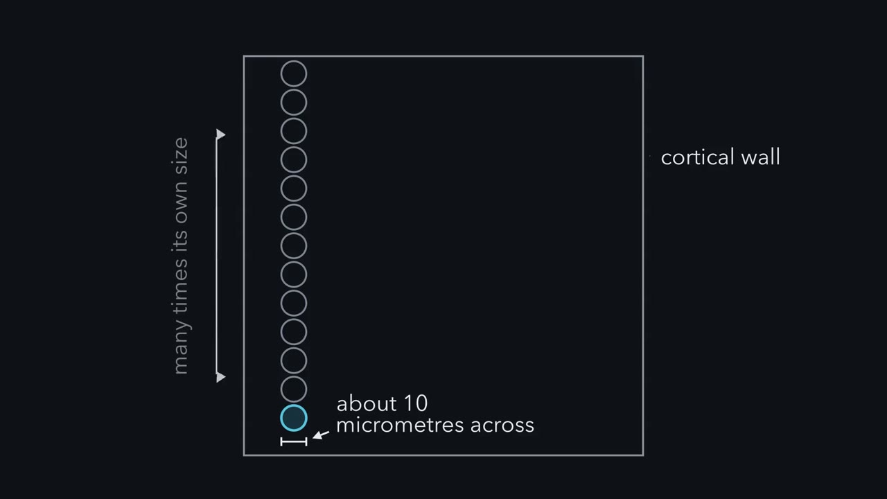 ▶ Watch The Front Goes First · 2:40
Read the transcript of “The Front Goes First”
Film transcript: The Front Goes First
A newborn neuron must cross a wall many times its own size. It has no legs. How does it get across? The wall has layers. Neurons are born in the ventricular zone, on the inside. Next come the subventricular and intermediate zones. Then the cortical plate, where neurons settle. The marginal zone sits on top, just under the surface. A neuron's birth date predicts its layer. Early-born neurons settle deep, later-born ones nearer the surface. So every neuron must travel. A migrating neuron has a leading process, one long arm pointing the way. Behind it is the cell body, which holds the nucleus. Near the nucleus sits the centrosome, the cell's main organizing centre for microtubules.
In 1972, Pasko Rakic rebuilt young neurons from fetal monkey cortex. He found them pressed along fibres of radial glia, long cells spanning the wall. But fixed tissue shows contact. It cannot show which cell moved, or how. In 1987, James Edmondson and Mary Hatten filmed living neurons moving on glial fibres in a dish. The neurons moved in a stop-and-go rhythm. The tip reached forward and pulled back, out of step with the cell body. Bruce Schaar and Susan McConnell then watched single cycles in neuroblasts, young migrating neurons, from newborn rats. These are not cortical neurons. A swelling forms in the leading process, ahead of the nucleus.
Which moves into it first, the centrosome or the nucleus? The centrosome goes first. Cells fixed at this moment show it ahead, and later movies of cortical neurons tracked it. Then the nucleus jumps forward. Here is the one idea. In the typical bipolar climb, the front goes first. The cell has moved only when its nucleus catches up. A tip can keep growing while the nucleus stays put. So track the nucleus against the tissue, not the tip. Count the steps of the nucleus. 6 micrometres a step, 4 steps an hour, gives 24 micrometres an hour.
Follow the nucleus, not the tip.
From the film
- In the typical bipolar climb the front goes first, and the cell has moved only when its nucleus catches up.
- A tip can keep growing while the nucleus stays put, so migration is measured at the nucleus relative to the tissue, not at the tip.
- Each step has two strokes: the centrosome moves ahead into a swelling, then the nucleus is brought forward after it.
Knock down one protein in a migrating cortical neuron, and its front keeps reaching toward the surface while its cell body, nucleus and all, barely moves.
The protein is LIS1, and losing one copy of its gene is enough to give a child a cortex with reduced or absent folding. To see why, you need to know how a neuron moves at all. A newborn cortical neuron is about ten micrometres across, and the wall it has to cross can be many times that thick. It makes the trip with no legs, commonly climbing a radial glial fibre, and it moves its own nucleus forward one stroke at a time.
Across the wall
You have seen in the lesson on birth order and layers that a neuron’s birth date predicts its layer: early-born neurons settle deep, and later-born neurons settle in more superficial layers. That rule needs travel.
The main zones, from the inside out (the subplate, between the intermediate zone and the cortical plate, is left out):
| Zone | Where it lies | What happens there |
|---|---|---|
| Ventricular zone | the inner surface, beside the fluid-filled ventricle | progenitors divide; excitatory neurons are born |
| Subventricular zone | just outside the ventricular zone | more neurons are born; many newborn neurons linger here as multipolar cells |
| Intermediate zone | the middle of the wall | axons run; migrating neurons cross it |
| Cortical plate | near the outer surface, which the pia, a thin membrane, covers | neurons settle into their layers |
| Marginal zone | above the cortical plate, just under the pia | the leading process of a locomoting neuron can reach it, and some cells then finish with a terminal translocation |
In a mouse the wall is thin at first; in a primate late in gestation it is far thicker. Either way the problem is the same: how does a cell move its bulky nucleus outward through a crowded wall?
During the climb, a migrating neuron has one leading process, a long extension pointing the way it is going, and a cell body, or soma, that holds the nucleus. The centrosome, the cell’s main organizing centre for microtubules, sits near the nucleus or a little ahead of it in the leading process.
Predict. You film a neuron in a slice. Over an hour the tip of its leading process advances steadily toward the cortical plate. Has the neuron migrated? What would you need to see before you said yes?
Check your prediction
Not necessarily. A process can grow while the cell body stays put, which is exactly what the LIS1 knockdown in the opening does. You would need to see the soma, and so its nucleus, move relative to the surrounding tissue.
The one idea to carry through this lesson. The front goes first, and the cell has moved only when its nucleus catches up. Migration is measured at the nucleus, relative to the tissue, not at the tip.
What the rule covers.
- Which cells. Bipolar neurons that advance in stop-and-go steps: cortical neurons climbing glia, cerebellar granule neurons on glia, and subventricular-zone neuroblasts (young migrating neurons) and interneurons, which can move the same way without glia. Somal translocation appears fairly smooth; the two-stroke description was worked out for stop-and-go locomotion. Cells in the multipolar stage (several short processes, met later) move differently.
- Which claim. The order of events, and where to measure them. It does not say where the force comes from: whether myosin II squeezes from behind the nucleus or pulls from the front is still argued, so the rule says “catches up” and names no force.
- Typical, not universal. In some cerebellar granule neurons the nucleus even passes the centrosome.
Two ways up
In 2001 Bagirathy Nadarajah, Alan Pearlman, Rachel Wong and colleagues filmed neurons in acute slices of embryonic mouse cortex and named the two ways they climb.
| Stage | Leading process | How the soma moves | Name |
|---|---|---|---|
| Early, when the wall is thin | in many cells, long and already attached near the pial surface | moves up within the process as it shortens, fairly smoothly | somal translocation |
| Later, when the wall is thick | shorter, ending freely | advances in jumps; from the cells’ shape, the authors inferred that radial glia guide it | locomotion |
| The final approach | reaches the marginal zone, above the cortical plate | in some cells, switches to a final translocation | terminal translocation |
Picture translocation as a person on a rope that is already tied to the ceiling, simply reeling in. Locomotion, the harder case, is where the front and the nucleus come apart, and most of this lesson is about it.
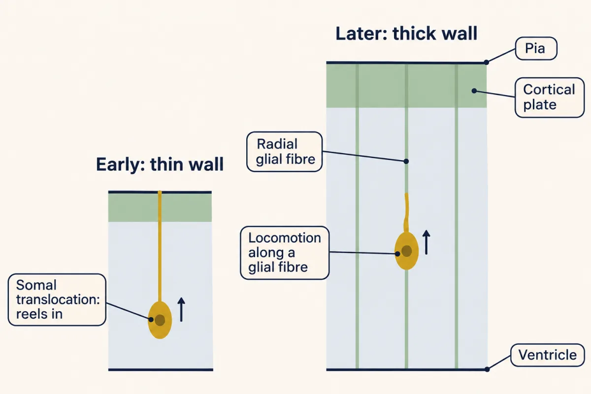
The glial ladder: from an inference to a movie
The fibres the neurons climb belong to radial glia. As the lesson on radial glia explains, these same cells are the main progenitors of the cortex. Each one that still touches the ventricle keeps a process running to the pia, so the wall is strung with parallel fibres.
Pasko Rakic proposed in 1972, for the cerebral cortex, that these fibres are the guide; he had made the same case for the cerebellum in 1971. He rebuilt young neurons from serial electron micrographs of fetal rhesus monkey cortex and found them pressed along radial glial fibres at every depth of the intermediate zone. That was fixed tissue. It showed contact, and it suggested a climb, but it could not show which cell moved, or how.
Predict. Suppose you could film living neurons and glia together in a dish. What could such a movie show that Rakic’s sections could not? And what would it still leave open about the living brain?
Check your prediction
In 1987 James Edmondson and Mary Hatten supplied the movie, in a different animal and a different brain region. They purified granule neurons and astroglia from the cerebella of early postnatal mice, recombined them on a coverslip, and filmed them. Neurons moved along glial processes in a stop-and-go rhythm, with the cell body closely apposed to the fibre and a leading tip that extended and retracted out of step with the soma. A later antibody stain confirmed that the guides were glial.
The experiment showed that a neuron can migrate on a glial fibre and how it moves when it does. It did not show that glia are required in the living brain, or what tells a neuron to get off.
Notice “out of step with the soma”: even in this early movie, the tip and the cell body kept separate schedules.
The front goes first
Nucleokinesis, the forward movement of the nucleus, works as a “two stroke” cycle.
- Stroke one. The leading process extends. A swelling forms in it, ahead of the nucleus, and the centrosome moves forward into the swelling.
- Stroke two. The nucleus is brought forward into the swelling. The trailing part of the cell follows.
Then the cycle repeats. The analogy is an inchworm: the front reaches and grips, then the back catches up.
Bruce Schaar and Susan McConnell watched this cycle in neurons from the anterior subventricular zone of newborn rats, migrating in a gel; they filmed the swelling and the nucleus, and located the centrosome in cells fixed mid-cycle. These are not cortical neurons but precursors of olfactory bulb interneurons, which normally travel without radial glia. Later imaging of rat cortical slices found the same order in radially migrating cells: centrosomes moving ahead of nuclei, and dynein concentrated at a swelling.
Predict. A swelling has just formed in the leading process, a short way ahead of the nucleus. Where will the nucleus be after its next jump? And if you fixed the cell at that moment and looked inside, where would you expect to find the centrosome?
Check your prediction
In Schaar and McConnell’s cells, the swelling predicted where the nucleus would go next. Electron micrographs of cells fixed mid-cycle showed the centrosome ahead of the nucleus and bundles of microtubules wrapped around the nucleus like a cage, with the microtubules running forward toward the centrosome.
So the proposed order is front, then centrosome, then nucleus.
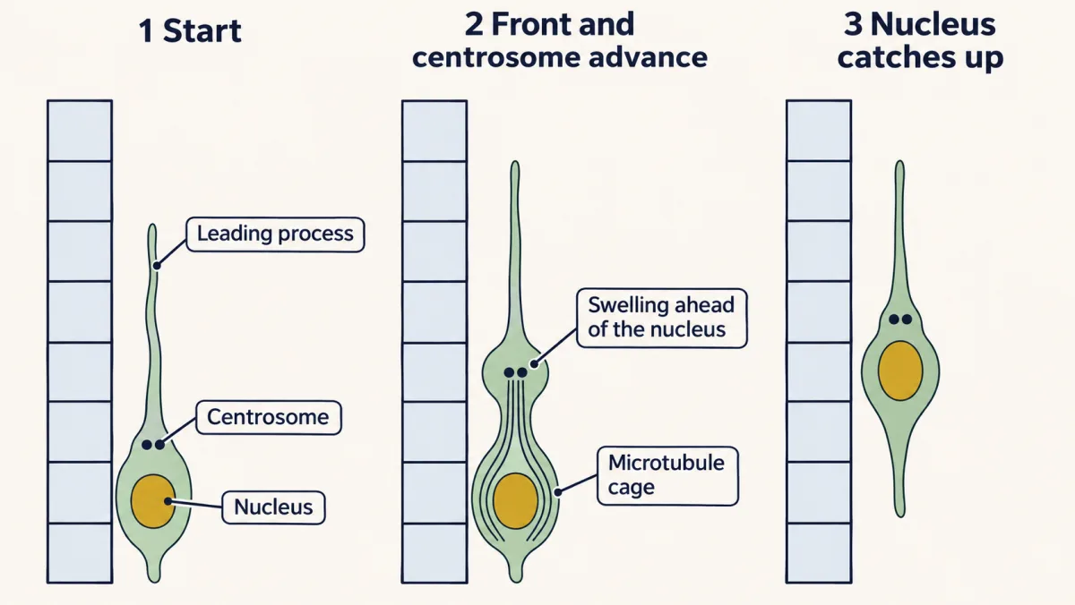
The engine: what brings the nucleus up
Read the transcript of “What Brings the Nucleus Up”
Film transcript: What Brings the Nucleus Up
Each step has two strokes. The centrosome moves ahead, then the nucleus catches up. But what makes it catch up? Inside the cell, bundles of microtubules wrap the nucleus like a cage. They run forward to the centrosome. Microtubules have a direction. In the usual picture, their minus ends gather at the centrosome. Their plus ends point away from it. The motor is dynein. It walks toward minus ends, that is, toward the centrosome. What dynein moves depends on what holds it. If dynein is held in place, it cannot walk. So it drags the microtubule instead, and the centrosome comes toward it. If dynein rides on something that can move, it carries that thing toward the centrosome.
In the usual model, a neuron uses both. In stroke one, dynein in the swelling pulls the microtubules forward. The centrosome comes with them. In stroke two, dynein on the nucleus walks along the cage. It carries the nucleus toward the centrosome, which now sits ahead. LIS1 binds dynein and regulates it in both places. It is not the motor itself. A second force comes from actin and myosin II. A drug called blebbistatin blocks myosin II. Now the experiment, in slices of embryonic rat cortex. Label the centrosome and the nucleus, and film the neurons. Knock down LIS1, or block myosin II. What do the tip, the centrosome and the nucleus each do?
Knock down LIS1 or dynein, and the centrosome stalls. The nucleus stalls too. But the tip keeps growing. Block myosin II, and the centrosome keeps moving. The nucleus stops, so the gap between them grows. Dynein's pull on the centrosome is still there. But the nucleus has lost a force it needs.
Exactly where myosin II acts, behind the nucleus or at the front, is still argued. So a cell can keep reaching without arriving. Measure migration at the nucleus.
From the film
- In the usual model, dynein moves the centrosome in stroke one and the nucleus in stroke two, toward the microtubule minus ends at the centrosome.
- LIS1 binds and regulates dynein; it is not the motor itself.
- In rat cortical slices, knocking down LIS1 or dynein stalls both the centrosome and the nucleus, while blocking myosin II stops only the nucleus.
- A cell can keep reaching without arriving, so measure migration at the nucleus.
So what makes the nucleus catch up?
Start with the track, the microtubule cage. Microtubules have a direction. In the usual picture, drawn from imaging of radially migrating rat cortical neurons, their minus ends gather at the centrosome, and their plus ends point away from it, back around the nucleus and forward into the leading process. Some other neurons arrange them differently: in some cerebellar granule neurons the microtubules around the nucleus need not meet at the centrosome.
The motor is cytoplasmic dynein, which walks toward minus ends, that is, toward the centrosome. What it moves depends on what holds it.
- If dynein is held in place, it cannot walk anywhere, so it drags the microtubule instead, and the centrosome at the microtubule’s minus end comes toward the dynein.
- If dynein is attached to something that can move, it carries that thing along the microtubule toward the centrosome.
Think of hauling hand over hand on a rope. Tied to a post, you pull the far end of the rope toward you; free on a fixed rope, you climb toward its far end. In the usual model, a migrating neuron uses both cases, one in each stroke.
- Stroke one, at the swelling. Dynein concentrated in the swelling ahead of the nucleus pulls on microtubules whose minus ends sit at the centrosome. The microtubule network, and with it the centrosome, is drawn forward into the swelling.
- Stroke two, at the nucleus. Dynein on the surface of the nucleus walks along the cage and carries the nucleus toward the centrosome, which now sits ahead.
LIS1 binds dynein and regulates it in both places; it is not the motor itself.
A second force comes from actin and myosin II, the contractile system of the lesson on integrins and Rho GTPases. Blebbistatin is a drug that blocks myosin II. When Schaar and McConnell added it, leading processes could keep growing while the cell bodies stopped.
Predict. In slices of embryonic rat cortex, the centrosome and the nucleus of migrating neurons were labelled and filmed. For control neurons, neurons with LIS1 or dynein knocked down, and neurons given blebbistatin, what do you expect the leading-process tip, the centrosome and the nucleus to do?
Check your prediction
| Leading-process tip | Centrosome | Nucleus | |
|---|---|---|---|
| Control | extends | moves ahead into the swelling | follows |
| LIS1 or dynein knocked down | keeps extending | stalls | stalls |
| Blebbistatin (myosin II blocked) | keeps extending | keeps moving | stops, so the gap to the centrosome grows |
Knocking down LIS1 or dynein impairs both of dynein’s proposed jobs, and both the centrosome and the nucleus stall. Blebbistatin leaves centrosome movement in place but stops the nucleus, which is why the two are thought to depend on different forces. In every row the tip keeps growing.
That last column is the reason for the rule. A cell can keep reaching without arriving.
Measure migration at the nucleus
A movie gives speed directly, as long as you follow the nucleus. If each cycle moves the nucleus a fixed step,
mean speed = step per cycle × cycles per hour
A nucleus that advances 6 micrometres per cycle, with one cycle every 15 minutes, travels 6 × 4 = 24 micrometres an hour.
Because the movement is stop-and-go, the mean is lower than the speed in a burst. In Edmondson and Hatten’s culture the cell body advanced in bursts of about 56 micrometres an hour, but about 33 an hour overall. If a cell moved only at burst speed and stood still the rest of the time, it would be moving 33 ÷ 56, about 60 percent of the time.
Predict. Take a toy model. Over 2 hours, the tip of a control neuron and the tip of a LIS1 knockdown neuron each advance 24 micrometres an hour relative to the tissue. The control nucleus steps 6 micrometres every 15 minutes; the knockdown nucleus does not step. How far does each tip move, and each nucleus? If you tracked only the tips, what would you conclude?
Check your prediction
| Over 2 hours | Tip moves | Nucleus moves | Tip-to-nucleus distance |
|---|---|---|---|
| Control | 48 micrometres | 8 steps × 6 = 48 micrometres | no net change (it stretches and shrinks within each cycle) |
| LIS1 knocked down | 48 micrometres | 0 | 48 micrometres longer |
Tracked at the tip, the two cells look the same, and you would score the knockdown as a normal migrator. Tracked at the nucleus, one cell has crossed 48 micrometres of wall and the other has not moved.
When the engine breaks: lissencephaly
Read the transcript of “When the Engine Breaks”
Film transcript: When the Engine Breaks
What happens to a brain when the nucleus cannot catch up? Disrupt that machinery, and neurons can stall short of their layers. One result can be lissencephaly. The cortex has reduced or absent folding. In the classic form it is also thick and poorly layered. Losing one copy of LIS1 is enough to cause it. LIS1 is deleted in Miller-Dieker lissencephaly. That deletion also removes neighbouring genes, which contribute to a more severe syndrome. A mouse cortex is smooth to begin with. So in mice you look at migration speed and where neurons end up. A second gene, doublecortin, sits on the X chromosome. Its protein binds and stabilizes microtubules.
Boys with a mutation typically have lissencephaly. Now a girl carries one mutant copy. Each of her cells switches off one X at random. What would her cortex look like? She is a mosaic. Neurons using the normal copy migrate. Some using the mutant copy stall. The stalled neurons form a band of grey matter beneath the cortex. It is called double cortex.
A kinase called Cdk5 sits upstream. Its targets include doublecortin and a dynein regulator. Mice lacking it have disordered layers. The two-stroke climb is not the whole trip. In embryonic rat cortex, newborn neurons first move to the subventricular zone. There they become multipolar, with several short processes. They move slowly, change direction, and make little net progress for about a day or longer. Many then move back toward the ventricle for a time. Why some cells go back is not settled. Then they turn bipolar and begin the sustained climb to the cortical plate. Now the front goes first. LIS1 matters at this turn too. So one defect can stall cells at more than one step.
The two-stroke cycle explains one way migration can fail. When you film a migrating neuron, track the nucleus, not the tip.
From the film
- Disrupting the machinery that brings the nucleus up can stall neurons short of their layers, and one result can be lissencephaly.
- Losing one copy of LIS1 is enough to cause lissencephaly, a cortex with reduced or absent folding.
- Doublecortin stabilizes microtubules; girls with one mutant copy are mosaics, and neurons that stall can form a band called double cortex.
- The two-stroke climb is not the whole trip: in embryonic rat cortex, newborn neurons spend about a day or longer as multipolar cells with little net progress.
Disrupt the machinery that brings the nucleus up, and neurons can stall short of their layers. Each of the main genes below touches the two-stroke machinery, or sits upstream of it.
Lissencephaly means a cortex with reduced or absent folding; in the classic form the cortex is also abnormally thick and poorly layered, because neurons failed to reach their places.
LIS1, the regulator of dynein. Losing one copy of LIS1 is enough to cause lissencephaly. LIS1 is deleted in Miller-Dieker lissencephaly, but that deletion also removes neighbouring genes, among them YWHAE, which encodes 14-3-3ε, and these make the syndrome more severe than lissencephaly caused by LIS1 alone. In rat cortical neurons LIS1 knockdown stalls both strokes.
Predict. A mouse cortex is smooth to begin with. How could a mouse with a defect in this machinery show that something is wrong, if not by losing folds?
Check your prediction
Not by folds. In a mouse you look at how fast neurons move and where they end up, for example whether the cortical layers are disordered. The readout depends on the gene, the dose and the brain region: mice lacking doublecortin alone have a nearly normal neocortex, though their hippocampus is abnormal.
Doublecortin, a microtubule stabilizer. In 1998 two groups (Gleeson, Walsh and colleagues; des Portes, Chelly and colleagues) identified DCX on the X chromosome. It encodes a protein that binds and stabilizes microtubules, the tracks the engine runs on. Boys with a mutation typically have lissencephaly. Girls with one mutant copy are mosaics, because each cell switches off one of its two X chromosomes at random: neurons using the normal copy migrate, and some using the mutant copy stall. The stalled neurons form a band of grey matter in the white matter below the cortex, called subcortical band heterotopia or “double cortex”.
Cdk5, the kinase upstream. Cdk5 is a kinase (an enzyme that adds phosphate groups to proteins) of the cell-cycle family that, in neurons, is activated by p35 (or the related p39) instead of a cyclin. Mice lacking Cdk5 or p35 have disordered cortical layers. Among its targets are NDEL1, a dynein regulator that works with LIS1, and doublecortin, so Cdk5 sits upstream of the same nuclear machinery.
These patients and mice show what the motor system is for, but each gene also acts at other stages, as the next section shows.
Not a straight climb
The two-stroke climb is not the whole trip.
In 2004 Stephen Noctor, Arnold Kriegstein and colleagues labelled progenitors in embryonic rat cortex with a GFP retrovirus and filmed their daughters in slices. Newborn neurons first moved to the subventricular zone, then became multipolar, with several short processes, and spent about a day or longer there or in the lower intermediate zone with little net outward progress. Many then moved back toward the ventricle for a time before turning bipolar and migrating radially to the cortical plate. Only a movie could show this.
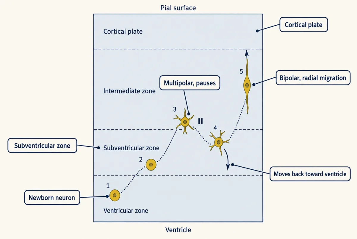
A multipolar cell is still migrating. It extends and retracts several processes and can move slowly, changing direction, between pauses. What it lacks is the one steady leading process of the bipolar climb, so the sustained two-stroke climb begins only after the cell turns bipolar. Why some cells first move back toward the ventricle is not settled.
LIS1 matters at this turn too. LIS1 knockdown in embryonic rat cortex blocks the nuclear movement of progenitors, the multipolar-to-bipolar change and the movement of the soma, so a single defect can stall cells at more than one step.
Putting it together
Each experiment below answered one question about the climb, and each left something open.
| Experiment | Preparation | Observed result | What it supports | What it does not establish |
|---|---|---|---|---|
| Serial-section electron microscopy (Rakic, 1972) | fixed fetal rhesus monkey cortex | young neurons pressed along radial glial fibres at every depth of the intermediate zone | contact, and a hypothesis that glia guide the climb | which cell moved, or how |
| Time-lapse culture (Edmondson and Hatten, 1987) | early postnatal mouse cerebellar granule neurons with astroglia | stop-and-go movement along glial processes; tip out of step with the soma | a neuron can migrate on a glial fibre; the movement is stop-and-go | that glia are required in the living brain; what tells a neuron to get off |
| Time-lapse slices (Nadarajah and colleagues, 2001) | acute slices of embryonic mouse cortex | translocation early, locomotion later, a final translocation at the end | the mode depends on stage | that locomotion is glia-guided, which was inferred from shape |
| Film, then fix and section (Schaar and McConnell, 2005) | anterior subventricular-zone neuroblasts of newborn rats, in a gel | filmed: a swelling forms ahead of the nucleus before it jumps; in cells fixed mid-cycle, the centrosome lies ahead and a microtubule cage surrounds the nucleus | a proposed order: front, centrosome, nucleus | the live timing of the centrosome, or that cortical neurons do the same; later slice imaging partly addressed both |
| Blebbistatin | the same neuroblasts; slices of embryonic rat cortex | processes keep growing; cell bodies stop; in slices the centrosome keeps moving | soma advance needs myosin II | where myosin II acts |
| LIS1 or dynein knockdown, with the centrosome and nucleus tracked | slices of embryonic rat cortex | centrosome and nucleus stall; the tip keeps extending | dynein, regulated by LIS1, is needed to move both | that every cell type gives the same readout |
| Retroviral labelling and time-lapse (Noctor and colleagues, 2004) | slices of embryonic rat cortex | a multipolar stage with little net progress; many cells move back toward the ventricle; then bipolar radial migration | the route is not a straight climb | why cells move back; that every neuron takes this route |
| Human genetics of LIS1 and DCX | patients | lissencephaly; band heterotopia in heterozygous girls | these genes are needed for normal cortical development | which single step fails, since each gene acts at several |
| Cdk5 or p35 knockout | mouse | disordered cortical layers | the kinase is needed for layering | which of its targets matters most |
Design problem
You image a migrating cortical neuron in a slice and see that its leading process keeps extending but the nucleus stays put. Name two molecular defects that could cause this and one experiment that tells them apart.
Saved in this browser only.
One way to do it
Defect one: the dynein system fails. Loss of LIS1 or of dynein function impairs dynein’s pull in both strokes.
Defect two: the actomyosin push or pull fails. Loss of myosin II activity stops the soma while the process extends.
The experiment. Label the centrosome (for example with a fluorescent centrin) and the nucleus, and film the cell. Track the leading process, the centrosome and the nucleus separately over time, and measure migration at the nucleus. As in the engine prediction, LIS1 or dynein knockdown stalls both the centrosome and the nucleus, while a myosin II block stops only the nucleus, so the centrosome-nucleus distance grows. As a control, add blebbistatin to normal cells and check that it reproduces the second pattern, and rescue the first with a knockdown-resistant LIS1.
Be careful with the interpretation. The two systems interact, and a long block in either may slow the other. The readout also depends on the cell type and on how much LIS1 is lost; see Going deeper.
Going deeper
Where the force is applied
Rho and its kinase ROCK switch on myosin II, and different preparations put actomyosin activity in different places. In Schaar and McConnell’s neuroblasts, once a swelling had formed, the nucleus still advanced after nocodazole disrupted microtubules, and blebbistatin blocked that step; microtubules and dynein remain essential in the knockdown work. The force distribution likely depends on cell type and stage.
The LIS1 readout depends on cell type and on how much LIS1 is lost. LIS1 or dynein knockdown stalls both the centrosome and the nucleus in rat cortical slices, but in cultured mouse cerebellar granule neurons with one copy of Lis1 the distance between the nucleus and the centrosome grows instead, and becomes more variable.
Which cells showed the swelling
Schaar and McConnell argued that the swelling is common to many kinds of migrating neuron. The Golgi’s move into the swelling was shown in interneurons from the ganglionic eminence.
Outer radial glia
In the large primate cortex, outer radial glia, which have no ventricular contact, add more fibres.
The longer list of mutants
The list of mutants runs from LIS1, NudE and Ndel (NDE1 and NDEL1 in mammals, dynein regulators that work with LIS1), doublecortin, Cdk5 and dynein to myosin II, ROCK and many more. Most of the names fit one of the two strokes.
Interkinetic nuclear migration in progenitors
Motor-driven nuclear movement also occurs earlier, in the progenitors. In interkinetic nuclear migration the nucleus of a radial glial cell moves away from the ventricle in G1 and back to it in G2, dividing at the ventricular surface. In embryonic rat cortex the kinesin Kif1a, a plus-end motor, carries it basally, and dynein carries it apically. So progenitors use two motors to move the nucleus in two directions, while a migrating neuron uses dynein, helped by myosin II, to move both its centrosome and its nucleus the same way.
How LIS1 was linked to dynein
In 1993 Orly Reiner, David Ledbetter, William Dobyns and colleagues isolated LIS1 from chromosome band 17p13.3 and showed it was deleted in Miller-Dieker lissencephaly. The clue to its job came from a fungus: in 1995 the laboratory of N. Ronald Morris found that nudF, a gene needed to spread nuclei along a filament of Aspergillus, encodes a protein similar to LIS1. Other genes from the same screen encode parts of the dynein system. By 2000 several groups had linked mammalian LIS1 to dynein and found LIS1, NDEL1 and dynein together in the nervous system.
Your notes
Select any text in the lesson to highlight it or add a note to it. Your highlights and notes are saved in this browser and collected in your notebook.
This browser is not letting the site save anything (a private window or strict privacy settings), so highlights and notes will be lost when you leave the page.
How we measure it
Serial-section electron microscopy
Fixed tissue is cut into a long series of very thin sections, each is photographed in the electron microscope, and a cell is traced from section to section to rebuild its shape in three dimensions. It shows which membranes touch, but because the tissue is dead it cannot show which cell moved.
Time-lapse imaging of slices and cultures
Living brain slices or cultured cells, labelled with a dye or a fluorescent protein, are photographed every few minutes for hours. The movie shows the order of events (process extends, swelling forms, nucleus jumps), but the dissection and the culture conditions can change speed and behaviour.
Retroviral and electroporation labelling
A retrovirus or a plasmid pushed into cells by an electric pulse delivers a gene for a fluorescent protein, and in the retrovirus case every daughter of the infected cell inherits it. The same delivery can carry an RNA that knocks down a gene, so a labelled cell with less of the protein of interest can be compared with control cells.
Acute drug inhibition
A small molecule that blocks one protein, such as blebbistatin for myosin II or nocodazole for microtubules, is added while a cell is being filmed. If one movement stops and another continues, the two depend on different machinery, although every drug can act on more than its intended target.
Practice questions on this lesson (3) → MCAT-style, original, each with an explanation
Where this lesson fits
Before thisReview first
- The Ventricular Zone: Radial Glia as Stem CellsThis course · Lesson 13
The fibres the neuron climbs belong to radial glia, whose role as progenitors and scaffold is taught there.
- Birth Order and Fate: Temporal Identity and the Inside-Out CortexThis course · Lesson 16
The rule that birth date predicts layer is the reason neurons must cross the wall, and this lesson builds on it.
- The Growth Cone: The Tip That Steers the AxonThis course · Lesson 3
You need the leading process and its actin and microtubule behavior from there to follow the front-first step.
After thisStudy next
- Reelin and the Reeler MouseThis course · Lesson 18
Reelin acts on migrating neurons in the cortical plate and decides where they stop and layer.
- Tangential Migration: Interneurons and the Long Way RoundThis course · Lesson 19
Interneurons use a similar leading-process and nucleus cycle but travel a different route into the cortex.
- When Building Goes Wrong: Microcephaly, Zika and LissencephalyThis course · Lesson 48
Lissencephaly from LIS1 and doublecortin, introduced here, is worked through there alongside other malformations.
Elsewhere on the siteFilms, simulations and method films in other courses
- Tight Ends, Loose EndsFilm · Cell Biology
Microtubule growth and shrinkage explains how doublecortin-stabilized microtubules form the cage around the nucleus.
- Grip, Swing, Let GoFilm · Cell Biology
It shows how dynein walks toward microtubule minus ends, the motor that pulls the centrosome and nucleus forward.
Suggested by an AI model from the lessons themselves, then checked. How we use AI
Finish line
Test yourself
8 questions
Practice and MCAT-style, with explained answers.
Next · Lesson 18
Reelin and the Reeler Mouse
Why does a mouse that cannot walk straight have its cortex built in the wrong order?
Continue →← Lesson 16 · Birth Order and Fate: Temporal Identity and the Inside-Out Cortex