Developmental Neuroscience · Skim version
Tangential Migration: Interneurons and the Long Way Round
Read the full lesson → Skim the whole course →
The guiding idea
In a mouse embryo, most of the neurons that will inhibit the cortex are born outside it.
The excitatory neurons of the cerebral cortex are made in the cortex's own wall, but in rodents most of its inhibitory interneurons are born in the ventral forebrain and travel sideways to get there.
From the film: Born Outside
- In rodents, most cortical inhibitory interneurons are born outside the cortex, in the ventral forebrain, and travel sideways into it.
- Excitatory cortical neurons are made in the cortex's own wall and climb out along radial glial fibres.
- A fault in migration can sit in the cell, which cannot answer a cue, or in the road, which lacks it.
- These results come from mice and rats.
From the film: Does the Defect Travel with the Cell?
- Following cells out, removing the source and swapping tissue are three ways to ask where a neuron came from.
- In mice lacking both Dlx1 and Dlx2, no migration from the ventral forebrain into the neocortex was detected and few cells contained GABA, while pyramidal neurons were still made.
- In graft experiments with slices, the defect travelled with the cells, so these genes act inside the travelling cells.
From the film: Staying in the Lane
- In mice, interneurons travel in two streams lined with the chemokine CXCL12, and they carry its receptor CXCR4.
- Without CXCR4 in mice, interneurons leave their streams and enter the cortical plate early, and they spread less widely.
- To leave a lane, a cell may stop answering the signal; that comparison of ages fits the idea but does not prove it.
-
Two populations, two birthplaces
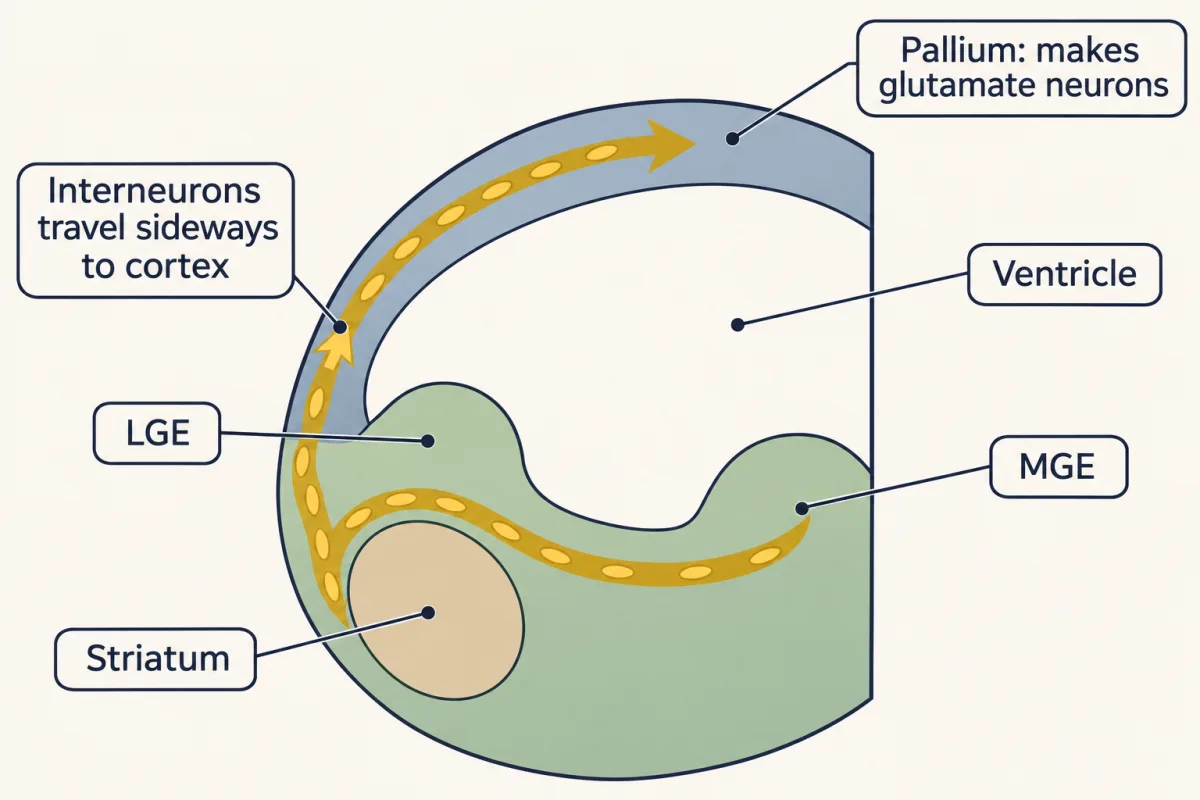
The pallium makes the cortex's excitatory, glutamate-releasing neurons. In rodents most of its inhibitory interneurons are born in the ganglionic eminences of the subpallium, mainly the MGE, and travel sideways around the striatum into the cortex. AI-generated illustration -
Follow, remove, swap
-
Moving with no fibre to climb
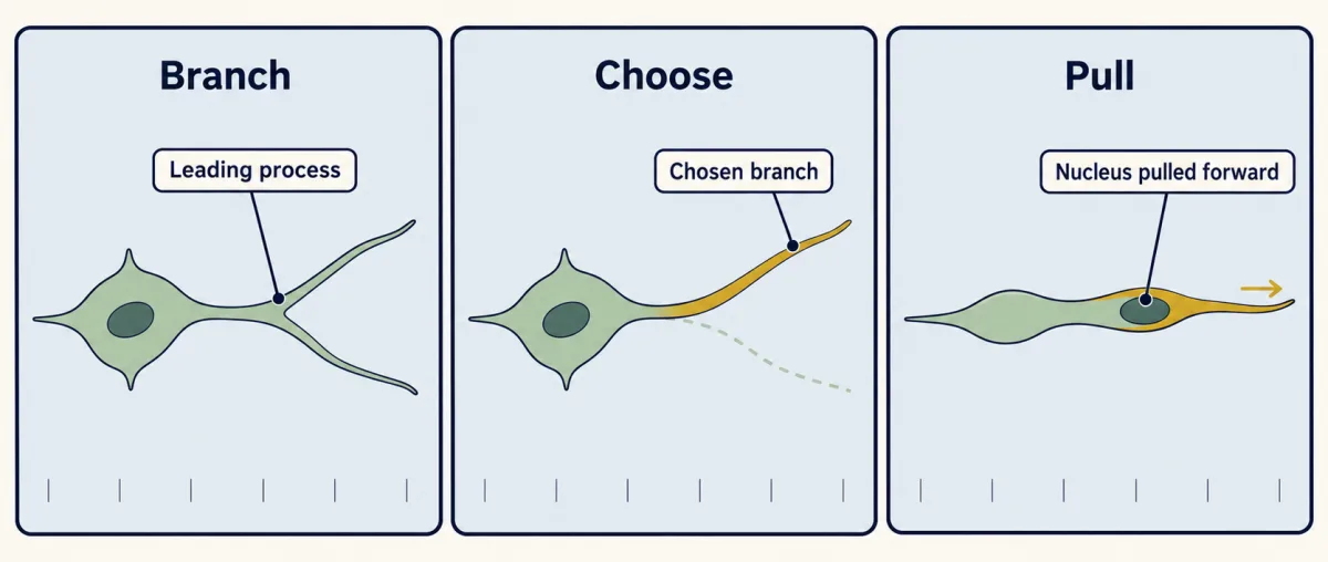
With no glial fibre to follow, the cell picks one branch of its leading process and moves its nucleus into it. AI-generated illustration -
A map of the cortical wall, and two streams
-
Staying in the lane: CXCL12 and CXCR4
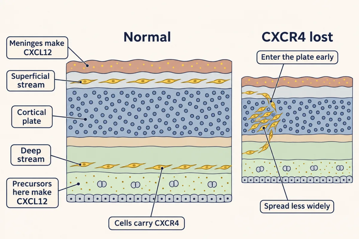
CXCL12 from the meninges and from precursors deep in the wall holds CXCR4-carrying interneurons in two streams. Without CXCR4 in mice, they enter the cortical plate early and spread less widely. AI-generated illustration -
Leaving the lane, and turning in
-
Where they settle
-
The rostral migratory stream: a tangential route for life
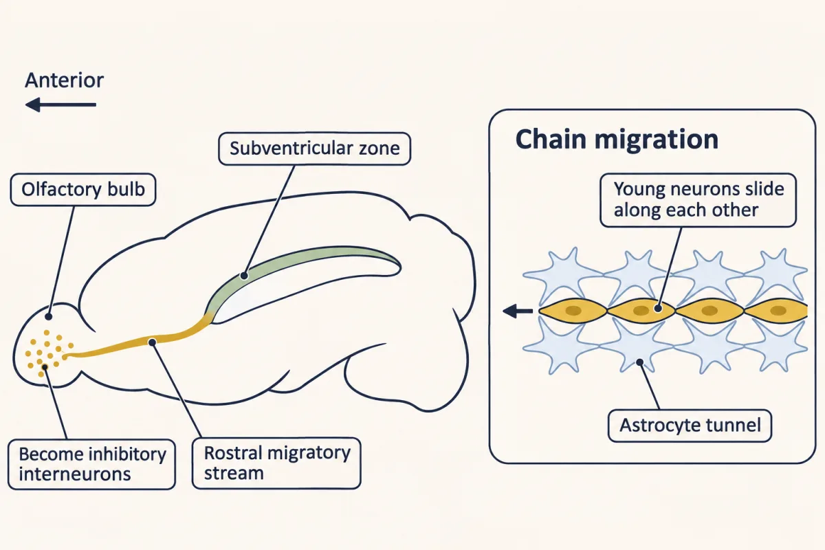
In adult rodents, young neurons from the subventricular zone travel forward along the rostral migratory stream to the olfactory bulb, sliding along one another in chains inside astrocyte tunnels, with no radial glia or axons to follow. AI-generated illustration -
Putting it together
-
Going deeper
Where this lesson fits
Before thisReview first
- Radial Migration: Climbing the Glial LadderThis course · Lesson 17
Pyramidal neurons climb glial fibres there, and this lesson contrasts that with interneurons moving sideways with no fibre.
- Ventral to Dorsal: Sonic Hedgehog and BMP in the Neural TubeThis course · Lesson 10
Sonic hedgehog patterning gives the ventral forebrain, including MGE identity through Nkx2.1, that these interneurons come from.
- The Ventricular Zone: Radial Glia as Stem CellsThis course · Lesson 13
You need radial glia as the cortex's own progenitors to see why excitatory neurons are made in its wall.
After thisStudy next
- Too Many Neurons: Naturally Occurring Cell DeathThis course · Lesson 28
Interneurons arrive in excess, and this lesson takes up the cell death that trims their numbers afterwards.
- Two Places the Adult Brain Still Makes NeuronsThis course · Lesson 47
It takes up the rostral migratory stream and asks whether adult humans add neurons.
- Opening and Closing a Critical PeriodThis course · Lesson 40
Once interneurons settle in cortex, GABA circuits help open and close the critical period.
Suggested by an AI model from the lessons themselves, then checked. How we use AI