Developmental Neuroscience · Unit 5: Cells on the move
Reelin and the Reeler Mouse
Rewritten on : new text, figures, flashcards and film. Your notes and highlights stay, but the text around them may have moved.
Why does a mouse that cannot walk straight have its cortex built in the wrong order?
Pause when the reading reaches a film, and offer to play it. Saved on this device.
Watch the film now, or keep listening
Builds on:Birth Order and Fate: Temporal Identity and the Inside-Out CortexRadial Migration: Climbing the Glial LadderAdaptors, Docking Proteins and ScaffoldsWhy these?
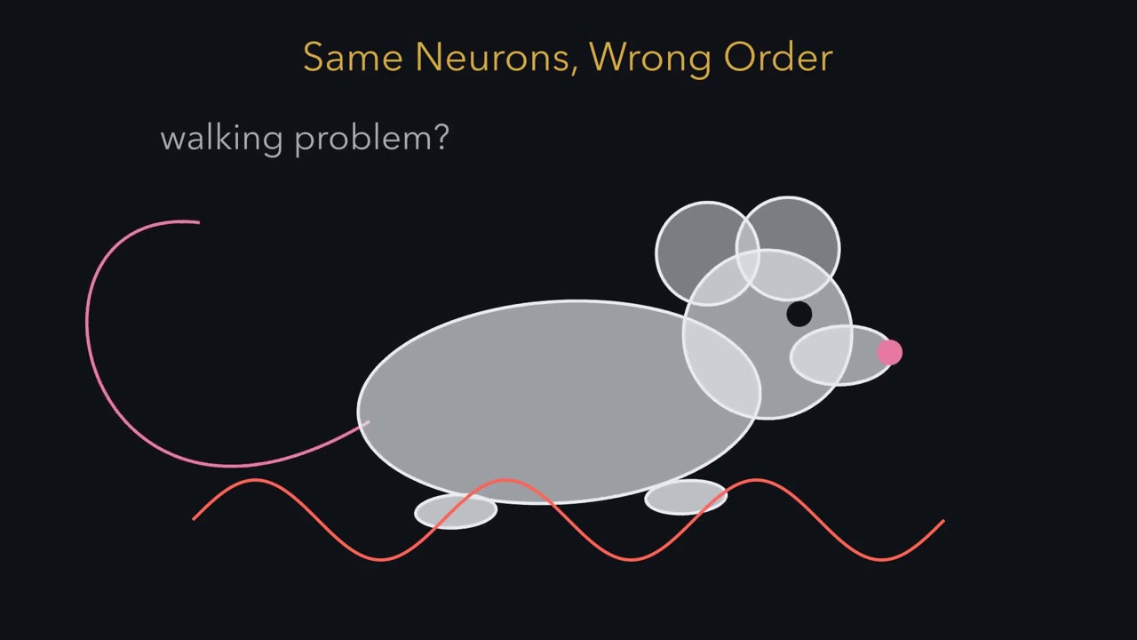 ▶ Watch Same Neurons, Wrong Order · 2:17
Read the transcript of “Same Neurons, Wrong Order”
Film transcript: Same Neurons, Wrong Order
A mouse reels as it walks. Is that a walking problem, or a building problem? In 1951, Douglas Falconer named this mutant reeler. Its cortex was the deeper puzzle. First, direction. Deep means near the ventricle, where neurons are born. Superficial means near the outer surface. The first neurons form a thin sheet under the surface, the preplate. The next neurons split it in two. Above sits the marginal zone, with the Cajal-Retzius cells. Below sits the subplate. Each later wave climbs past the waves before it, and settles just under the marginal zone. So the cortex is built inside-out. Early-born neurons end up deep, and late-born neurons near the surface.
Is reeler making the wrong neurons, or putting the right ones in the wrong place? Verne Caviness and Richard Sidman used birthdating. A radioactive building block of DNA labels cells copying their DNA. Each division shares the label between two daughter cells. A cell that divides once more, then never again, stays heavily labelled. Predict. If reeler made the wrong neurons, what would birthdating show? And if it misplaced the right ones?
In 1973: the same classes of neurons, born on the same schedule. What changed was their order. Early-born neurons tended to sit near the surface, and late-born neurons deep. The pattern was roughly inverted, with a lot of scatter.
Why? In reeler, the preplate fails to split. Later waves tend to stop beneath the neurons already there. So reeler is a defect of placement, not production. The missing protein, Reelin, is needed to stack that birth order the right way up. With Reelin signalling, each new wave tends to finish its climb at the top. Without it, newcomers tend to stop beneath their elders. Takeaway: made on schedule, stacked the wrong way up. Next, where does Reelin come from?
From the film
- Reeler makes the same classes of neurons on the same schedule as normal mice, so it is a defect of placement, not production.
- In reeler the preplate fails to split, and early-born neurons tend to sit near the surface and late-born neurons deep, a roughly inverted trend with scatter.
- Reelin signalling is needed to stack birth order the right way up: with it each new wave tends to finish its climb at the top, and without it newcomers tend to stop beneath the neurons already there.
In 1951 a geneticist named a mouse mutant for the way it walks: it reels from side to side. Its cerebellum is small, and the layers of its cerebral cortex are disorganized and roughly in the reverse of their normal order.
A walking problem and a cortex built in the wrong order turned out to be two faces of one missing protein, Reelin, which Cajal-Retzius cells (early neurons at the surface of the developing cortex) secrete and which neurons need in order to take their proper places. This lesson follows the mouse to Reelin and the relay inside the neuron that reads it, and ends on an open question: what Reelin tells a migrating neuron. In radial migration you followed a neuron climbing a radial glial fiber from the ventricular zone toward the surface. Here the question is where it stops, and who decides.
Same neurons, wrong order
The geneticist was Douglas Falconer. Before anyone could knock out a gene on purpose, geneticists relied on mutants that arose on their own in mouse colonies and were bred on. Several of the most useful brain mutants were named, like reeler, for their gait: weaver, staggerer. A mutant is a handle: you can breed it, map it and dissect it long before you know its gene.
The reeler cerebellum is also poorly folded, with Purkinje cells, the large output neurons of the cerebellum, left in clumps instead of a neat single layer. That alone could explain the reeling. The cortex was the deeper puzzle.
How a normal cortex is stacked
First, direction. Deep means near the ventricle, the fluid-filled space inside the brain, whose lining of dividing cells (the ventricular zone) makes the neurons. Superficial means near the pial surface, the outside of the brain. The cortex is built in three stages (see birth order and layers).
- The first neurons to leave the ventricular zone form a thin sheet just under the surface, the preplate.
- The next neurons move into the middle of that sheet and split it in two: an upper marginal zone, which holds the Cajal-Retzius cells, and a lower subplate. The neurons between them form the cortical plate, which becomes layers 2 to 6 (the marginal zone becomes layer 1).
- Each later wave climbs past the waves before it and settles just under the marginal zone.
So early-born neurons end up deep and late-born neurons end up superficial. The cortex is built inside-out.
Wrong neurons, or wrong place?
Was reeler making the wrong neurons, or putting the right ones in the wrong place? Verne Caviness and Richard Sidman answered with birthdating. A radioactive building block of DNA, tritiated thymidine, labels cells that are copying DNA. A cell that copies its DNA during the pulse and then divides for the last time a few hours later passes the label to two daughters that never divide again, so the neurons it makes stay heavily labelled for life. Cells that keep dividing after the pulse dilute the label by roughly half at each division. Their position in the adult tells you where neurons born that day settle.
Predict. Give the label early in development to one set of mice and late to another, then look at the adult cortex. If reeler made the wrong neurons, what would birthdating show? If it made the right neurons and put them in the wrong place?
Check your prediction
Wrong neurons would show up as different classes or numbers of neurons, or a different birth schedule. Right neurons in the wrong place would show up as the same classes, born on the same schedule, ending at different depths. (The label gives the birth date; the class comes from identifying each labelled neuron’s type.)
In their 1973 paper, the same classes of neurons were born on the same schedule in reeler as in normal mice. What changed was their order. In reeler, early-born neurons tended to sit superficially and late-born neurons deep, a roughly inverted pattern, and the layers were disorganized as well.
Later work showed where it starts to go wrong: in reeler the preplate fails to split. The first cortical plate neurons pile up underneath it instead of moving in and dividing it, so the subplate cells and the Cajal-Retzius cells stay together above them in one sheet, sometimes called the superplate.
Here is the picture, simplified; read each column from the surface down. Real reeler cortex shows an inverted trend with a lot of scatter, not a clean mirror image.
| Position | Normal cortex | Reeler cortex (roughly) |
|---|---|---|
| Pial surface | marginal zone, with the Cajal-Retzius cells | superplate: Cajal-Retzius cells and subplate cells still together |
| Superficial | latest-born neurons | earliest-born neurons |
| Upper middle | later-born neurons | early-born neurons |
| Lower middle | early-born neurons | later-born neurons |
| Deep | earliest-born cortical plate neurons | latest-born neurons |
| Below the cortical plate | subplate | no separate subplate |
So reeler is a defect of placement, not production.
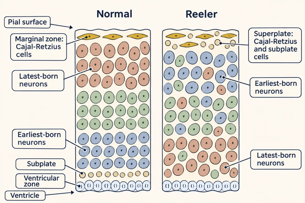
The one idea to carry through this lesson. Neurons are born in order; Reelin signalling is needed to stack that order the right way up. With it, each new wave of neurons tends to finish its climb at the top of the cortical plate, just under the Reelin-rich marginal zone, above the waves born before it: inside-out. Without it, newcomers tend to stop beneath the neurons already there, so the same birth order is laid down roughly upside down.
What the rule covers.
- Which neurons. Radially migrating excitatory neurons of the neocortex. Hippocampal pyramidal cells are also misplaced in reeler, but the hippocampus has its own pattern. In the cerebellum, Purkinje cells fail to spread into a single layer and stay in clumps. Interneurons, which arrive tangentially from the ganglionic eminences, are not covered here.
- Which effect. Placement, in the developing cortex; in the 1973 birthdating comparison, birth timing and the classes of neurons made matched normal mice. The rule describes the outcome, not the message Reelin carries; that question gets its own section.
- A trend, not a law for each neuron. The rule gives the direction, not the scatter seen in real reeler cortex.
- Switching off matters too. Too much active Dab1 also misplaces neurons (the off switch).
Why would a newcomer stop beneath its elders? One leading explanation is that Reelin signalling helps the final step of the climb, and a neuron that cannot take that step tends to halt under the cells already there. That is an explanation, not yet a result; the evidence comes in One neuron’s climb.
Where the signal comes from, and who reads it
Read the transcript of “Matching Mutants, One Relay”
Film transcript: Matching Mutants, One Relay
A signal outside a cell needs a reader inside. Where does Reelin come from, and what reads it? In 1995, D'Arcangelo and Curran cloned the gene broken in reeler. It encodes a very large protein, Reelin. That year, Ogawa, Mikoshiba and colleagues made an antibody, CR-50, that stained Cajal-Retzius cells. In culture, it made normal cortical tissue organize like reeler tissue. It recognizes Reelin, a secreted protein. So Reelin is made at the top of the cortex, where each new wave should stop. Two more mutants, scrambler and yotari, looked like reeler, but had a normal Reelin gene. Both had mutations in Dab1, an adaptor protein. A Dab1 knockout gave the same picture.
Joachim Herz's laboratory removed two lipoprotein receptors, ApoER2 and VLDLR. Without both, the brain was close to reeler. Without one, the defect was milder. Think of a relay race. Remove a runner, and the baton never arrives. But the receptor leg has two runners, with partly overlapping jobs. Lose one, and some signal still gets through. Lose both, and the brain is close to reeler.
Matching mutants suggest one relay. They do not give its order. Predict. Without Reelin, is there more Dab1, or less? And is more of it phosphorylated, or less? There is more Dab1 than normal, but less of it is phosphorylated.
That shows Reelin affects Dab1, but not yet which acts first. So add Reelin to cultured neurons. Dab1 is phosphorylated within minutes. Reelin binds both receptors directly. Then the kinases Fyn and Src add phosphate to Dab1. One explanation for the build-up: phosphorylated Dab1 is destroyed through Cullin 5, and without Reelin less of it is phosphorylated.
Knock down Cullin 5 in migrating neurons, and active Dab1 builds up. The neurons migrate too far. So normal positioning needs both the signal and its shut-off. Takeaway: matching mutants suggest one relay. Adding Reelin and watching Dab1 respond puts Reelin upstream of Dab1; the binding studies place the receptors between them.
From the film
- Reelin is a very large secreted protein made by Cajal-Retzius cells at the top of the cortex, where each new wave should stop.
- Matching reeler-like phenotypes in Dab1 mutants and in mice lacking both ApoER2 and VLDLR suggest one pathway, though they do not give its order.
- Reelin binds the receptors, Fyn and Src phosphorylate Dab1, and phosphorylated Dab1 is destroyed through Cullin 5, so normal positioning needs both the signal and its shut-off.
The source: Cajal-Retzius cells
In 1995 two lines of work began to converge on the same protein. A team led by Gabriella D’Arcangelo and Tom Curran cloned the gene disrupted in reeler, starting from a mouse in which an inserted transgene had broken it. The gene encodes a very large protein that resembles extracellular matrix proteins, and it is made in the developing brain while neurons migrate. They named it Reelin.
The second route used an antibody. Masaharu Ogawa, Katsuhiko Mikoshiba and colleagues made an antibody called CR-50 that stained Cajal-Retzius cells in the marginal zone. Added to cultures in which embryonic cortical cells reassemble, it made normal tissue organize like reeler tissue. In 1997 the two groups together showed that CR-50 recognizes Reelin and that Reelin is a secreted glycoprotein.
That fixes the geometry. Reelin is made by cells at the top of the cortex, the very place where each new wave of neurons should stop. Most Cajal-Retzius cells arrive in the marginal zone from a few sites at the edges of the developing cortex.
Mutants that match
A secreted protein outside the cell needs a receptor and a relay inside. The pieces came from mutants that looked alike.
- Scrambler and yotari. Two more mouse mutants had a reeler-like brain but a normal Reelin gene. In 1997 both turned out to be mutations in Dab1 (Disabled-1), so they are two alleles of one gene. In the same issue of Nature, Jonathan Cooper’s laboratory reported that a targeted knockout of Dab1 (the gene disrupted on purpose) gave the same picture. Dab1 is an adaptor: a protein with no enzyme activity of its own that links one protein to others (see adaptors and scaffolds).
- Two receptors. In 1999 Joachim Herz’s laboratory showed that mice lacking both of two lipoprotein receptors (relatives of the low-density lipoprotein (LDL) receptor, which takes cholesterol-carrying particles into cells), ApoER2 and VLDLR, have a brain close to reeler, while either single knockout is milder.
- Two kinases. Mice lacking both Fyn and Src, two Src-family kinases, have a reeler-like cortex.
Why do matching phenotypes put genes in one pathway? Think of a relay race. Remove a runner and the baton never arrives, and the race fails the same way whichever runner is missing. The receptor leg has two runners whose jobs partly overlap. Lose one and some signal still gets through, so the defects are milder and differ between brain regions; lose both and the brain is close to reeler. When removing Reelin, Dab1 or both receptors gives the same failure, the simplest reading, though only a suggestion, is that they are runners in one race. Nor do matching phenotypes give the order of the runners.
Which comes first?
Predict. In a brain with no Reelin, is there more Dab1 protein than normal, or less? Is more of it phosphorylated on tyrosine, or less? And what would you add to a dish of neurons to find out which acts first?
Check your prediction
Brains lacking Reelin had more Dab1 protein than normal but less of it phosphorylated. That shows Reelin affects Dab1, but a changed steady state alone cannot give the order, because a later step can feed back on an earlier one. The order came from adding the signal: Reelin added to cultured neurons raised Dab1 tyrosine phosphorylation within minutes, and the binding studies below show it acts through the receptors. So Reelin acts upstream of Dab1 activation.
More Dab1 when the signal is missing seems backwards. Phosphorylated Dab1 is marked by a ubiquitin ligase built on Cullin 5 and destroyed (see ubiquitin and proteolysis and turning signals off). This helps explain the surplus: with no Reelin, Dab1 is poorly phosphorylated, so it is probably degraded less efficiently and builds up.
Two papers in one 1999 issue of Neuron, from the Curran and Herz laboratories, showed that Reelin binds ApoER2 and VLDLR directly and that this binding leads to tyrosine phosphorylation of Dab1. Laid out mutant by mutant:
| Mutant | What is broken | Reelin gene | Dab1 protein | Dab1 phosphorylated | Cortex |
|---|---|---|---|---|---|
| reeler | Reelin | broken | more than normal | less than normal | roughly inverted, disorganized |
| scrambler, yotari | Dab1 (two alleles) | normal | little or none (abnormal transcripts) | — | reeler-like |
| Dab1 knockout | Dab1 | normal | none | none | reeler-like |
| ApoER2 and VLDLR both lost | both receptors | normal | built up, as in reeler | expected low, since Reelin acts through these receptors | close to reeler |
| ApoER2 or VLDLR alone | one receptor | normal | — | — | milder, differing between brain regions |
| Fyn and Src both lost | two kinases | normal | — | — | reeler-like |
| Cullin 5 knocked down | the off switch | normal | active Dab1 builds up | the active form builds up | neurons migrate too far |
The pathway reads like this:
Reelin (from Cajal-Retzius cells) → binds ApoER2 and VLDLR → Fyn and Src phosphorylate Dab1 on tyrosine → phospho-Dab1 recruits effectors → phospho-Dab1 is ubiquitinated through Cullin 5 and degraded
Dab1 sits on the receptor tails through its PTB domain, which recognizes an NPxY motif (see protein interaction modules).
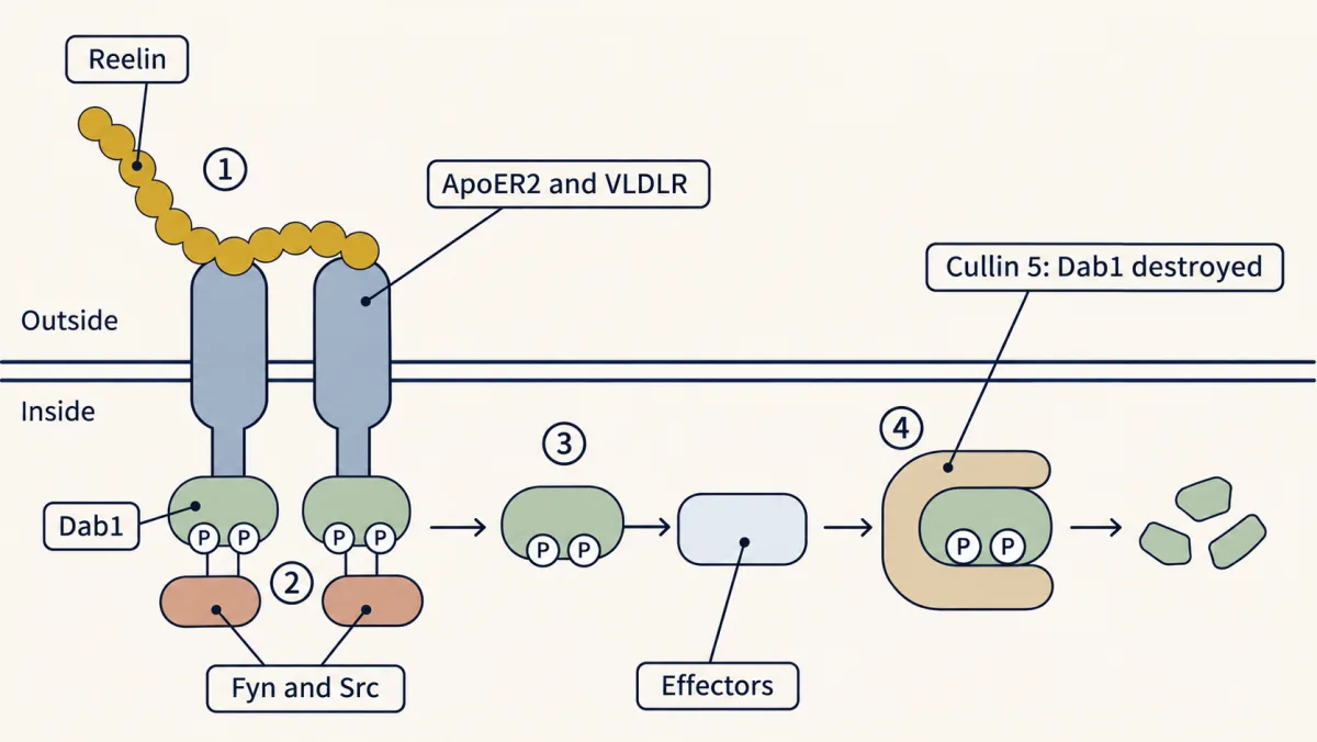
The off switch
The last step of the pathway destroys the signal’s carrier.
Predict. Reduce Cullin 5 in migrating neurons, so Reelin-induced destruction of phosphorylated Dab1 is impaired. Where should those neurons end up?
Check your prediction
In 2007 Cooper’s laboratory knocked down Cullin 5 in migrating mouse neurons. Active Dab1 built up and the neurons migrated too far, piling up at the top of the cortical plate.
Normal positioning needs both the signal and its shut-off.
Needed, but what does it say?
Read the transcript of “Needed, But What Does It Say?”
Film transcript: Needed, But What Does It Say?
Removal shows that a signal is needed. Can it show what the signal says? The fuel in a car is needed for the trip. But it does not choose where the car goes. A permissive signal allows an outcome that something else chooses. An instructive signal chooses the outcome itself. Removing a signal cannot tell the two apart. One test of a spatial message: keep the signal present, but change where it is.
Three simple proposals: stop here, let go of the glial fiber, or come here. Now move the Reelin: below the cortex, into one small spot, or into the medium bathing a slice. Predict. If Reelin means come here, what should happen in each case?
In 2002, Curran's laboratory made Reelin from the ventricular zone of reeler mice. The preplate split, and the ataxia was rescued, though not every defect. Neurons did not migrate down toward the source. In 2010, Nakajima's laboratory made Reelin in one small patch of mouse cortex. Later-born neurons passed earlier-born ones toward the spot, building a small inside-out arrangement around it. In 2004, Jossin, Goffinet and colleagues added Reelin to the medium bathing reeler slices. The developing cortical plate became more organized, without Reelin from its normal source. Whether Reelin stayed even inside the tissue was not shown. No single command fits all three results.
When does a neuron read Reelin? Follow a typical later-born neuron. First it is multipolar, wandering below the cortical plate. Then it takes hold of a radial glial fiber, and climbs. Near the top, it anchors its leading process in the marginal zone and pulls its cell body up: somal translocation. Reelin signalling, through Dab1 and Rap1, has been linked to both the turn and that final step. On one view, Reelin acts less like a command, and more like a switch on adhesion, flipped at particular stages. The essential cue, a gradient, a boundary or an adhesive state, is not settled. Takeaway: Reelin signalling helps each new wave tend to finish its climb at the top.
That alone does not show that neurons steer toward Reelin.
From the film
- Removal shows that a signal is needed, not what it says; changing where the signal is can help tell permissive from instructive.
- Moving Reelin gave results that fit no single command: a source below the cortex rescued some defects, a small spot organized neurons around it, and an even bath organized reeler slices in a short-term assay.
- In mouse cortex, Reelin signalling through Dab1 and Rap1 has been linked to turning multipolar neurons and to the final somal translocation, which fits a switch on adhesion at particular stages.
- Helping later-born neurons settle above earlier ones does not by itself show that neurons steer toward Reelin.
The mutant experiments removed something: Reelin, Dab1, the receptors or the kinases. Removal shows that a signal is needed, not what it says. The fuel in a car is needed for the trip, but it does not choose where the car goes.
Two words make the question exact. A permissive signal allows an outcome that something else chooses; an instructive signal chooses the outcome itself. Removal cannot tell the two apart. One test of a spatial message is to keep the signal present while changing where it is.
There are three simple proposals for what Reelin says.
- Stop here. Reelin in the marginal zone tells an arriving neuron to stop.
- Let go. Reelin tells the neuron to let go of its radial glial fiber, so it can finish its last step and leave room for the next wave to pass.
- Come here. Reelin attracts the neuron’s leading process, the long process it extends ahead of its cell body, and the neuron moves toward the source.
On the first two views, a neuron without Reelin never gets that instruction, so the next wave cannot get past it. The stop-here and come-here proposals treat where Reelin is as information, so they are instructive proposals about position. The let-go proposal can be read either way. The stage-specific view later in this section may be closer to permissive for position, although a signal that flips an adhesion switch at particular stages could still carry information about timing.
Predict. For each proposal, what happens if Reelin is (a) made in the wrong place, below the cortex at the ventricular zone; (b) made in a small spot inside the cortex; (c) added evenly to the medium bathing a slice of reeler cortex, with no source at the top?
Check your prediction
Take the simplest form of each: an attractant that neurons read as a gradient in the tissue, and a stop or let-go response wherever neurons meet enough Reelin.
| If Reelin means | (a) source below | (b) small spot | (c) even bath |
|---|---|---|---|
| come here | neurons turn back down toward the ventricle | neurons gather toward the spot | no help, if the bath leaves no direction to follow |
| stop here | neurons halt near the ventricle, where they first meet Reelin | neurons halt around the spot, without turning toward it | depends on the details: even exposure would halt neurons wherever they first meet enough Reelin, so a rescue needs a threshold, a stage-dependent response or a local context |
| let go | neurons release their fiber where they meet Reelin, and may move on another way | release around the spot, without turning toward it | as for stop here: neurons would let go early unless the response is stage-dependent or local |
Here is what happened.
- (a) Wrong place. In 2002 Curran’s laboratory made Reelin from the ventricular zone of reeler mice, using the nestin promoter. It rescued preplate splitting and the ataxia (the unsteady walk), but not every defect. Neurons did not migrate down toward the source.
- (b) A small spot. In 2010 Kazunori Nakajima’s laboratory made Reelin ectopically in a small patch of developing mouse cortex. The leading processes of migrating neurons gathered in the Reelin-rich spot, cell bodies collected around it, and later-born neurons passed earlier-born ones toward the center. This built a small inside-out arrangement around the source. Location clearly mattered there.
- (c) In the bath. In 2004 Yves Jossin, André Goffinet and colleagues added Reelin, or just its central fragment, to the medium bathing slices of embryonic reeler cortex. The developing cortical plate became more normally organized in this short-term slice assay, which did not measure layering across many waves. So this rescue did not need a gradient from a source at the top. Even supply in the medium is not proof of even supply in the tissue, though: Reelin can bind matrix and cell surfaces and be concentrated locally, so local gradients are not ruled out. The rescue also strains the simplest stop and detachment views, which predict early arrest under even exposure.
No single command fits all three results. Result (a) says Reelin is not simply a positional cue, and a pure attractant is hard to square with it; an attractant fits (c) only if Reelin stayed patchy inside the tissue. A pure stop sign sits badly with (a), where Reelin made below the cortex still let the preplate split, and does not explain why neurons in (b) arrange themselves inside-out around a spot. So the field has moved to asking when a neuron reads Reelin.
One neuron’s climb
Follow a typical later-born neuron. After its birth it becomes multipolar for a while: it has several short processes, wanders in the intermediate zone below the cortical plate, and is not yet oriented toward the surface. Then it turns toward the surface, takes hold of a radial glial fiber and climbs. Near the top it finishes with somal translocation, a step that no longer uses the glial fiber: it anchors its leading process in the marginal zone and pulls its cell body up behind it. (The earliest-born neurons can reach their place by somal translocation alone.) In mouse cortex, Reelin signalling through Dab1 and the small GTPase Rap1 has been linked to two of these stages.
- Turning the multipolar neuron toward the cortical plate, through the adhesion protein N-cadherin (adhesion is how firmly a cell grips its neighbours and surroundings). This happens well below the marginal zone, so Reelin is not read only at the top.
- The final step, somal translocation, again through changes in adhesion.
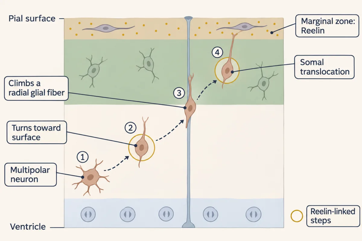
On this view Reelin acts less like a command and more like a switch on adhesion, flipped at particular stages, so what it does depends on the neuron’s stage. Read this way, the three results fit. A neuron needs the switch flipped, but the switch does not need Reelin from its normal source (c), a source below the cortex still rescues some defects (a), and a local source can still organize the neurons that flip their switches around it (b). Whether the essential cue is a gradient, a boundary or a permissive adhesive state is not settled, and the models are not all exclusive. So the guiding idea describes a population outcome, not the message: Reelin signalling helps later-born neurons settle above earlier-born ones. That outcome does not, by itself, show that neurons steer toward Reelin.
Design problem
Design an experiment, in cortical slices or in living mouse embryos, that distinguishes Reelin as a gradient-reading chemoattractant from Reelin as a stop or detachment signal.
Saved in this browser only.
One way to do it
Change where Reelin is, and watch what neurons do.
- Start with reeler mice, so there is no endogenous Reelin to confuse the result. Supply Reelin in one of two patterns: from a focal source placed below the cortical plate (for example, cells engineered to secrete Reelin, or electroporation of a Reelin gene into a patch of the ventricular zone), or uniformly, by bathing slices in Reelin-containing medium.
- Label one cohort of newborn neurons with a fluorescent protein and image their movement by time-lapse in slices, or record their final positions in vivo.
- Controls: the same deliveries into mice lacking Dab1 or both receptors should do nothing, which shows the effect runs through the pathway.
Predict. What would each outcome mean?
- Attractant. Neurons near a focal source turn and move toward it, even downward; seen with the Reelin distribution measured, that would argue for a gradient-reading attractant. Uniform Reelin, if it stays uniform in the tissue, does not restore order.
- Stop signal. Neurons halt where they meet enough Reelin, without turning. Even exposure would then arrest neurons early unless the response is stage-dependent.
- Detachment signal. Neurons leave their glial fiber there, without turning, and may move on another way.
Check. The uniform half has been done in slices: result (c). The same 2004 study found that antibodies that activated the receptors raised Dab1 phosphorylation but did not correct the reeler phenotype. So the bath result does not by itself favor an attractant, although a gradient inside the tissue is not excluded; Dab1 phosphorylation alone is not enough. The focal half, tracking neurons’ paths while measuring where the Reelin is, would be the stronger test of attraction. A mixed result, as published experiments have tended to give, would favor the stage-specific view.
Putting it together
| Experiment | Observed result | What it supports | What it does not establish |
|---|---|---|---|
| Birthdating reeler cortex (1973) | same classes, same schedule; depth roughly inverted, with scatter | a defect of placement, not production | which molecule |
| Preplate in reeler | fails to split; superplate above the cortical plate | the error starts at the first stacking step | why it fails |
| Cloning (1995) and CR-50 (1995, 1997) | a large secreted glycoprotein from Cajal-Retzius cells; the antibody makes normal cultures organize like reeler | the source sits at the top of the cortex | what the signal says |
| Scrambler, yotari, Dab1 knockout (1997) | reeler-like brains | Dab1 is in the pathway | the order |
| ApoER2 and VLDLR knockouts (1999) | both lost: close to reeler; either alone: milder | the receptors are in the pathway, and partly redundant | that Reelin binds them |
| Binding studies (1999) | Reelin binds both receptors and triggers Dab1 tyrosine phosphorylation | the receptors read Reelin directly | — |
| Reeler and Dab1 double mutants (1999) | no worse than either single mutant | suggests one pathway | which gene comes first |
| Dab1 without Reelin, and with Reelin added (1999) | more protein, less phosphorylated; added Reelin raises Dab1 phosphorylation within minutes | Reelin acts upstream of Dab1 activation | that steady-state levels alone give the order |
| Fyn and Src both lost | reeler-like cortex | the kinases are in the relay | — |
| Cullin 5 knocked down (2007) | active Dab1 builds up; neurons migrate too far | the off switch matters as much as the on switch | a general rule linking signal amount to distance |
| Reelin from the ventricular zone (2002) | preplate splitting and ataxia rescued, not every defect; no migration down to the source | a source below the cortex can rescue some defects; not a simple come-here or positional cue | what the cue is |
| Reelin in a small spot (2010) | an inside-out arrangement around the source | a local source can organize neurons | that the normal cue is a gradient |
| Reelin in slice medium (2004) | more normal cortical plate; receptor antibodies raise Dab1 phosphorylation but do not correct it | this rescue does not need a source at the top; Dab1 phosphorylation alone is not enough | that supply in the tissue was even; layering across many waves |
The first rows show a problem of placement; the middle rows name the relay and its order. The last three fit no single simple cue: 2002 argues against a simple come-here or positional cue; 2010 shows that a local source can organize neurons, and is compatible with attraction of leading processes; 2004 shows that a source at the top is not needed for that rescue, without showing even exposure in the tissue. Separate experiments on single neurons fit, without proving, the view that Reelin acts at particular stages, partly through adhesion. What spatial information Reelin carries is still open. You will use the same permissive-versus-instructive test on a survival signal in the neurotrophic hypothesis.
Going deeper
Parallel discovery
A separate team, Hirotsune and colleagues, reported the reeler gene in 1995, the same year the Curran group named Reelin.
An integrin model for the last step
One model ties terminal translocation to inside-out activation of integrin alpha5beta1 and adhesion to fibronectin in the marginal zone, although removing beta1 integrin from mouse neurons does not cause major layering defects, so this step may not be essential.
Reelin beyond the embryo
In humans, rare recessive RELN mutations cause lissencephaly with cerebellar hypoplasia, in which the cortex is thick and smooth or nearly smooth, with few broad folds, and the cerebellum is small (see microcephaly and malformations). In mice, Reelin also modulates synaptic plasticity in the adult hippocampus.
Your notes
Select any text in the lesson to highlight it or add a note to it. Your highlights and notes are saved in this browser and collected in your notebook.
This browser is not letting the site save anything (a private window or strict privacy settings), so highlights and notes will be lost when you leave the page.
How we measure it
Birthdating with tritiated thymidine
A radioactive DNA precursor is given at one stage of development and the tissue is examined later. Neurons that left the cell cycle soon after the pulse stay heavily labeled, so their final position reports where neurons born on that day settle.
Gene knockout and double mutants
A knockout is a mouse in which both copies of a chosen gene have been made non-functional. Crossing two mutants and finding that the double mutant is no worse than either single mutant suggests that the two genes act in one pathway.
Function-blocking antibody in reaggregation culture
Dissociated embryonic cortical cells are allowed to clump and organize again in a dish, with or without an antibody against a candidate protein. If the antibody makes normal cells arrange like mutant cells, the protein it binds is needed for that arrangement.
Phosphotyrosine immunoblotting of a stimulated culture
Neurons are given a candidate signal for a set time, a protein is pulled out with an antibody, and an antibody against phosphorylated tyrosine shows whether and how fast it was phosphorylated. A time course places the response minutes after the signal arrives.
Practice questions on this lesson (3) → MCAT-style, original, each with an explanation
Where this lesson fits
Before thisReview first
- Birth Order and Fate: Temporal Identity and the Inside-Out CortexThis course · Lesson 16
You need the inside-out order of cortical layers from there to see why reeler's roughly inverted order is wrong.
- Radial Migration: Climbing the Glial LadderThis course · Lesson 17
You need the neuron's climb along a radial glial fiber from there to understand where Reelin acts.
- Adaptors, Docking Proteins and ScaffoldsCell Signaling · Graduate school and beyond
Dab1 is an adaptor, and this lesson uses the idea of docking proteins without re-teaching it.
After thisStudy next
- When Building Goes Wrong: Microcephaly, Zika and LissencephalyThis course · Lesson 48
Human RELN mutations give a smooth cortex, taking the Reelin pathway into disease.
- The Neurotrophic Hypothesis TestedThis course · Lesson 29
It reuses the permissive-versus-instructive test on a survival signal, moving the signal rather than removing it.
- Tangential Migration: Interneurons and the Long Way RoundThis course · Lesson 19
Interneurons arrive by another route and settle among the layers that Reelin signalling helps stack.
Elsewhere on the siteFilms, simulations and method films in other courses
- Forward genetic screensMethod film
Scrambler and yotari were found as mutants with a reeler-like walk, the way a screen finds pathway genes.
- Complementation (cis-trans) testMethod film
Matching mutant phenotypes put genes in one pathway, and combining mutations tests whether they hit the same gene.
- Tissue grafting and transplantationMethod film
Moving a Reelin source to a new place tests permissive against instructive signals, as in grafting.
- Cell-free extracts and reconstitutionMethod film
Adding Reelin as an even bath to reeler slices rebuilds the signal in a simple dish assay.
Suggested by an AI model from the lessons themselves, then checked. How we use AI
Finish line
Test yourself
8 questions
Practice and MCAT-style, with explained answers.
Next · Lesson 19
Tangential Migration: Interneurons and the Long Way Round
In a mouse embryo, most of the neurons that will inhibit the cortex are born outside it.
Continue →