Cell Signaling · Unit 7: Space, time and distance
The Shape of a Signal: Size, Duration, Frequency and Place
EGF and NGF both switch on ERK in the same cell. How does the cell tell them apart? It reads the shape of the signal: how much, how long, how often and where.
Pause when the reading reaches a film, and offer to play it. Saved on this device.
Watch the film now, or keep listening
Builds on:Ras to MAPK: The Kinase CascadeCalcium: The Messenger Kept at Arm's LengthCircuits: Amplification, Feedback, Adaptation and SwitchesWhy these?
 ▶ Watch A Late Part Reads Signal Length · 2:20
Read the transcript of “A Late Part Reads Signal Length”
Film transcript: A Late Part Reads Signal Length
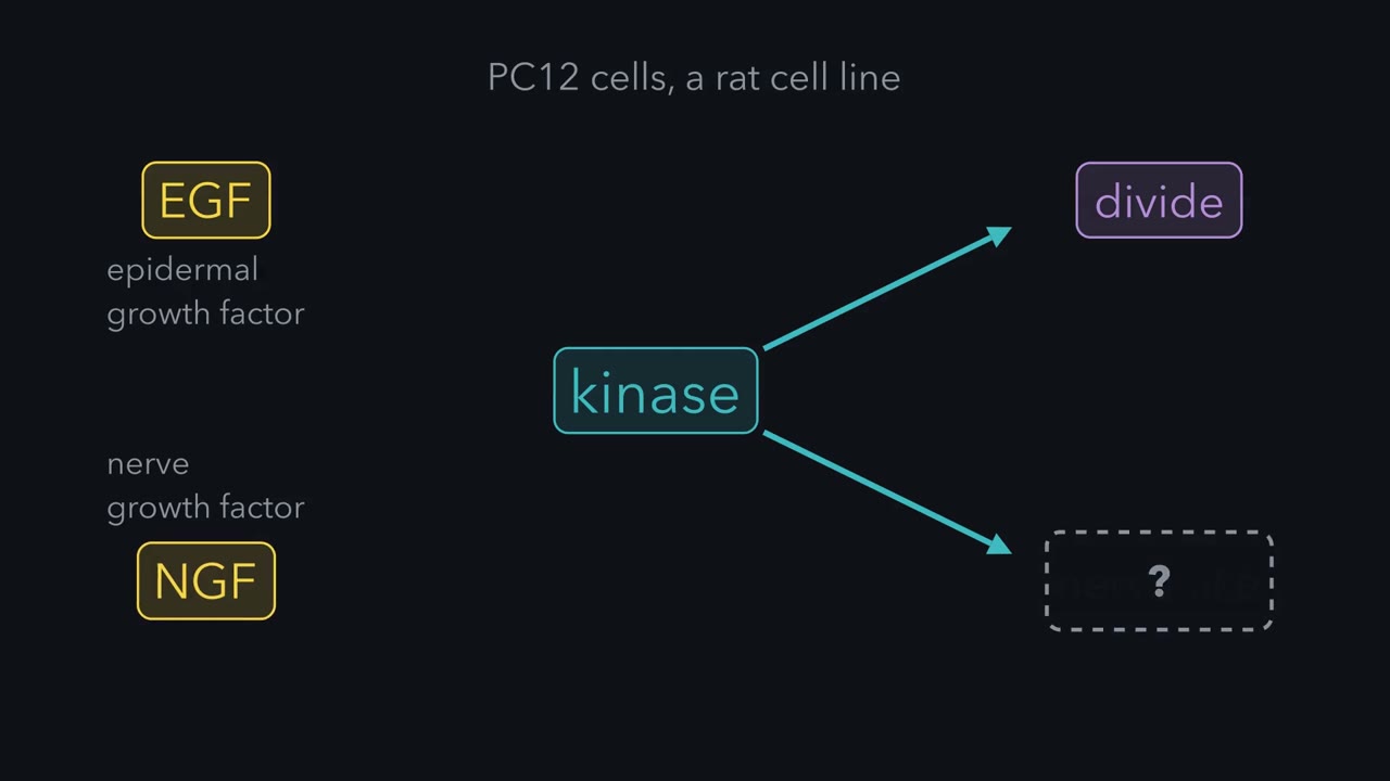
How can one kinase carry two different orders? In a rat cell line called PC12, epidermal growth factor, or EGF, says divide. Nerve growth factor, or NGF, says stop dividing and grow nerve-like processes. Both switch on the same kinase, ERK. The molecule is the same, so the difference must lie in something else about the signal. EGF switches ERK on briefly, and the cell divides. NGF keeps ERK on much longer, and the cell stops dividing and grows processes. Engineered cells carry extra EGF receptors. Given EGF, they keep ERK active for longer, and they differentiate. So length matters, and the pathway's wiring shapes it. A cell has no stopwatch.
What it can do is compare the signal with a delayed product of that same signal. In fibroblasts, ERK switches on a gene called c-fos. The c-Fos protein takes time to make, and once made it is quickly destroyed. So the test is simple: is ERK still on when the new protein appears? Think of a clinic that calls your name a while after you sign in. Only the people still in the waiting room are seen. If ERK is still active when c-Fos appears, phosphates are added near one end of the protein. ERK adds them, and so does a kinase that ERK activates. Those phosphates protect c-Fos. After a brief signal, ERK is already off when c-Fos appears. So c-Fos is made and lost.
After a long signal, ERK is still on, and c-Fos builds up. As a transcription factor, it can then switch on later genes. So in fibroblasts, c-Fos lets the cell tell a brief signal from a long one. The signal reaches c-Fos by a fast route and a slow route, and c-Fos needs both. It needs phosphates from ERK, and it needs the new protein, which takes time to make. This was shown for c-Fos in fibroblasts, and later for several other early gene products. Do not assume every long signal is read this way. Keep this rule: a part that arrives late can time a signal.
A cell reads four things in a signal besides which molecule it is: how much, how long, how often and where. Take a PC12 cell, a line of rat adrenal tumour cells that grow nerve-like processes when given nerve growth factor. One growth factor says “divide” and another says “become neuron-like”, and both say it through the same kinase, extracellular signal-regulated kinase (ERK). If the molecule that carries both orders is the same, the difference must lie in something else about the signal. This lesson is about what that something else can be, and about the parts that do the reading.
The idea: a signal has a shape, and the cell reads it
A cell has a limited set of signalling parts to work with and a great many things to say, so it reuses them. Calcium, cyclic AMP, extracellular signal-regulated kinase (ERK) and NF-κB turn up in pathway after pathway, with a different result each time. A row of arrows cannot explain that. We need a new vocabulary for signaling, and its first four words are how much, how long, how often and where.
Think of a doorbell with one button. A short press, a long press and a run of quick taps are different messages sent down the same wire. A cell reads its signals in a similar way:
- How much. The size of the signal. You know this one from dose-response curves and from the all-or-none switch in signaling circuits.
- How long. Minutes, or hours.
- How often. Many signals arrive as repeated spikes, and the gap between spikes can change.
- Where. At the cell surface, on an internal membrane, or beside one target and far from another.
Reading the last three takes a decoder: a downstream part whose own timing or position makes it answer one pattern and ignore another.
How long: a part that arrives late can time a signal
The lesson on Ras and MAPK gave you the PC12 result. Epidermal growth factor (EGF) switches extracellular signal-regulated kinase (ERK) on briefly, and the cell divides. Nerve growth factor (NGF) keeps ERK on much longer, and the cell stops dividing and grows processes. The length matters, and it comes from wiring: in PC12 cells NGF adds a positive feedback loop to the pathway that EGF does not, and that loop keeps ERK on. PC12 cells engineered to carry extra EGF receptors keep ERK active for longer when they meet EGF, and they differentiate.
A cell has no stopwatch. What it can do is compare the signal with a delayed product of that same signal.
ERK switches on the immediate early genes you met in signaling to the nucleus, and one of them is c-fos. The c-Fos protein takes time to make, and once made it is quickly destroyed. If ERK is still active when c-Fos appears, ERK and a kinase it activates add phosphates near the C-terminal end of c-Fos, and those phosphates protect it. After a brief signal ERK is already off, so c-Fos is made and lost. After a long signal c-Fos builds up, and as a transcription factor it can switch on later genes.
The rule to keep: c-Fos is made late and destroyed fast, and it is kept only if ERK is still active when it appears, so the cell reads a long signal as c-Fos that builds up. This was shown for c-Fos in fibroblasts; do not assume every long signal is read this way. The lesson gives no times, so both axes are relative units and the picture is a sketch of the logic. The dashed line is an arbitrary marker, and the rates are invented. In this sketch, protection lasts only while ERK is on, so c-Fos falls again when ERK goes off; the lesson does not say how fast.
Picture a clinic that calls your name twenty minutes after you sign in. Only the people still in the waiting room are seen. In the cell, the signal reaches c-Fos by a slow route (making the protein) and a fast route (phosphorylating it), and c-Fos builds up only when both arrive.
Leon Murphy, John Blenis and their colleagues showed this for c-Fos in fibroblasts in 2002, and later for several other early gene products. Do not assume every long signal is read this way.
How often: a decoder that holds on between spikes can count
Read the transcript of “Why a Slow Off Switch Can Count”
Film transcript: Why a Slow Off Switch Can Count
How can a cell count? Calcium often rises in brief spikes. In many cell types, a stronger stimulus gives more frequent spikes of about the same height. To read that, a cell needs a part that switches on fast and off slowly. Picture a bucket with a small hole, topped up by one cup of water at each spike. Fill it rarely, and it drains between cups. Fill it often, and the level climbs. Take a decoder that loses its activity with a fixed half-time. The fraction left at the next spike is one half, raised to the gap between spikes divided by the half-time. With a half-time of 20 seconds, spikes a minute apart leave one eighth of the activity. Spikes 10 seconds apart leave about 70 percent to build on.
In 1998, Ricardo Dolmetsch, Keli Xu and Richard Lewis tested human T cells. They imposed calcium spikes of chosen spacing on a line of these cells. Closely spaced spikes activated three transcription factors, NFAT and NF-κB among them. Widely spaced spikes activated only NF-κB. A likely reason is how fast each one switches off. NFAT leaves the nucleus within minutes once calcium falls. In B cells, NF-κB was found to stay in the nucleus. It stayed for more than 16 minutes after a brief calcium spike. So a decoder that holds on between spikes can count.
Calcium signaling showed you that calcium often rises in brief spikes. In many cell types a stronger stimulus gives more frequent spikes of about the same height. One way to read that is with a part that switches on fast and off slowly.
Picture a bucket with a small hole, topped up by one cup of water at each spike. Fill it rarely and it drains between cups. Fill it often and the level climbs. For a decoder that loses its activity with a fixed half-time:
fraction left at the next spike = (1/2) ^ (gap between spikes ÷ half-time of the decoder)
With a half-time of 20 seconds, spikes a minute apart leave one eighth of the activity. Spikes 10 seconds apart leave about 70 percent to build on.
One such part is calcium/calmodulin-dependent kinase II (CaMKII), an assembly of twelve kinase subunits. Calcium-bound calmodulin switches a subunit on. An active subunit can then phosphorylate its neighbour, but only while the neighbour also holds calmodulin, and a phosphorylated subunit stays active after calcium has gone. Fast spikes catch more neighbours loaded at the same moment. In 1998 Paul De Koninck and Howard Schulman gave CaMKII fixed to a surface timed pulses of calcium and calmodulin and found that this calcium-independent activity rose with the pulse rate. That is a test-tube result.
Transcription factors can be sorted by the same rule. In 1998 Ricardo Dolmetsch, Keli Xu and Richard Lewis imposed calcium spikes of chosen spacing on a line of human T cells. Closely spaced spikes activated three transcription factors, nuclear factor of activated T cells (NFAT) and NF-κB among them. Widely spaced spikes activated only NF-κB. A likely reason is how fast each one switches off. NFAT leaves the nucleus within minutes once calcium falls. In B cells, NF-κB stayed in the nucleus long after a brief calcium spike had ended.
Medicine uses this. The brain releases gonadotropin-releasing hormone (GnRH) in pulses. In rhesus monkeys whose hypothalamic lesions had cut off their own GnRH supply, one pulse an hour restored the pituitary’s release of its two reproductive hormones, and the same hormone given continuously shut the release down. Leuprolide is a synthetic relative of GnRH given as a slow-release dose, so that its level stays steady. After a first surge the pituitary goes quiet and testosterone falls, which is the aim in prostate cancer. A drug can change the shape of a signal as well as its size.
Pulses from feedback: a loop with a delay keeps its own time
Where do pulses come from when the stimulus is steady? In signaling circuits you saw negative feedback pull a pathway back toward baseline. Add a delay and the correction arrives late. Think of a shower with a long pipe between tap and head: you turn the tap, nothing changes, you turn it further, and you end up too hot and then too cold. A loop like that can oscillate.
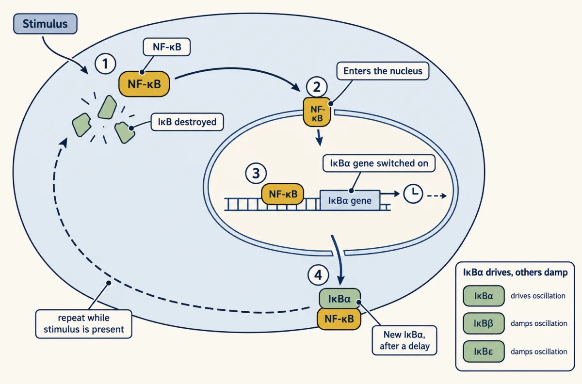
NF-κB sits in such a loop. One of the genes it switches on encodes its own inhibitor, IκBα. Once IκB has been destroyed, as in Toll-like receptors and NF-κB, NF-κB enters the nucleus, new IκBα is made after a delay, and it carries NF-κB back out. If the stimulus is still present, the new IκBα is destroyed and the round can repeat, as films of single cells carrying fluorescent NF-κB have shown in some cell lines (Nelson and colleagues, 2004). A 2002 study that combined a computer model with cells that lacked particular IκB proteins concluded that IκBα drives the oscillation and that the other two IκB proteins damp it.
p53 and MDM2 also form a negative feedback loop, and stress signals covered what Galit Lahav’s films of single cells, made in Uri Alon’s laboratory, showed in 2004: p53 arrives in pulses of about the same size, and a larger dose of radiation gives more pulses on average. The MDM2 loop does not explain them alone: a 2008 study from Lahav’s laboratory found that the pulses are driven by repeated restarts from the upstream kinases ATM and Chk2, and that a second feedback loop, through Wip1, keeps them uniform. Her laboratory later used nutlin-3, a small molecule that blocks MDM2, on a planned schedule to hold p53 steady at the height of one pulse. In the human cancer cells they studied, cells left to pulse after a moderate radiation dose recovered and returned to dividing. Cells held steady often stopped dividing for good.
Is a ring of negative feedback with a delay enough to make a clock? Michael Elowitz and Stanislas Leibler tested that by building one. In the bacterium E. coli they wired three repressors, proteins that switch a gene off, into a ring, each silencing the gene for the next. Each protein takes time to make, so every order to switch off lands late and the three chase one another around the ring. A green fluorescent readout rose and fell with a period of hours in at least 40 percent of the single cells filmed. The clock was noisy, and it ran.
Where: the same kinase means something else in another place
A message can also depend on its address. In cAMP and PKA and adaptors and scaffolds you met A-kinase anchoring proteins, which hold protein kinase A (PKA) beside particular targets. A local rise in cAMP then reaches the PKA parked nearby and may never reach PKA parked elsewhere. A receptor can likewise signal from the cell surface and again from an endosome after it has been taken inside, which is the subject of the next lesson.
A cell can also make a place on demand. In 2016 the laboratories of Michael Rosen and Ronald Vale put twelve purified components from the start of T-cell signalling, the pathway in antigen receptors and checkpoints, on an artificial membrane with no cell present. When a kinase phosphorylated the receptor tail, the downstream proteins gathered into clusters that behaved like liquid droplets. What holds such droplets together is a large number of weak contacts between repeated binding sites of the kind you met in protein interaction modules. The droplets also sorted what entered them. Kinases were concentrated and phosphatases were kept out, which favours a signal that stays on inside.
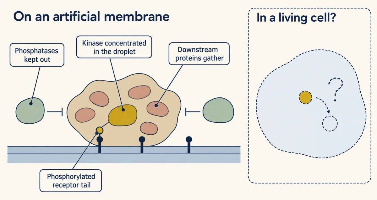
That sorting was shown on a glass slide. In a human T-cell line, LAT mutants that clustered more switched on more extracellular signal-regulated kinase (ERK), but proving that a bright spot in a living cell is a separate liquid is harder. The usual test bleaches a spot with a laser and watches the brightness return, and proteins that bind a fixed scaffold and let go again would look the same as a liquid. For anchored kinases, place is an established part of the signal. For droplets it is established in the test tube and still argued over in the cell.
Design problem
Two hormones raise calcium in the same cell. The first must switch on gene A and not gene B. The second must switch on gene B and not gene A. You may not add a second messenger. Design how the two calcium signals differ in time, and say what property each gene’s activator must have so that it answers only its own pattern.
Saved in this browser only.
One way to do it
With decoders that switch on fast and off slowly, spike rate alone will not do it. In the T-cell experiment, fast spikes switched on everything that slow spikes did, and more. To get two answers that exclude each other, make the signals differ in length and in height together.
Let hormone 1 give one tall, brief spike. Let hormone 2 give a low plateau that lasts.
Gene A’s activator needs a high threshold and a long memory. It ignores the low plateau, switches on at once when a tall spike comes, and stays on long after calcium has fallen. NF-κB, once its inhibitor has been destroyed, behaves this way.
Gene B’s activator needs a low threshold and a short memory. It is sensitive enough to answer the plateau, but it reverses within minutes when calcium falls, so one brief spike is over before enough of it has collected in the nucleus to start transcription. NFAT behaves this way.
Dolmetsch, Lewis and their colleagues saw this split in B cells in 1997. A large, brief rise in calcium activated NF-κB, and a low, sustained rise activated NFAT.
Going deeper
The GnRH experiment
The pulse-versus-continuous experiment described above was done in 1978 by Ernst Knobil’s laboratory.
Who modelled NF-κB oscillations
In 2002 Alexander Hoffmann, Andre Levchenko, Martin Scott and David Baltimore combined a computer model with cells that lacked particular IκB proteins. They concluded that IκBα drives the oscillation and that the other two IκB proteins damp it.
Testing whether a bright spot is a liquid
In 2019 David McSwiggen, Mustafa Mir, Xavier Darzacq and Robert Tjian examined the bleaching test and wrote: “Even proteins that engage in stable, high-affinity binding interactions can show recovery.”
Your notes
Select any text in the lesson to highlight it or add a note to it. Your highlights and notes are saved in this browser and collected in your notebook.
This browser is not letting the site save anything (a private window or strict privacy settings), so highlights and notes will be lost when you leave the page.
How we measure it
Time-lapse imaging of single living cells
A protein is fused to a fluorescent tag and one cell is filmed for hours, so the amount of the protein is read as brightness, frame by frame. An average over millions of cells, as in a Western blot, hides pulses that are out of step with one another. Single-cell films show them.
Calcium clamp
The store-operated calcium channels of a cell are held open and the calcium in the bathing solution is switched on and off. The experimenter then sets the height and spacing of the calcium spikes, and a reporter gene shows which patterns a transcription factor answers.
Reconstitution from purified proteins
Purified proteins are added one at a time to an artificial lipid membrane on a slide. If the behaviour appears, those parts are sufficient for it. Whether a living cell uses them the same way is a separate question that needs a separate experiment.
Fluorescence recovery after photobleaching (FRAP)
A laser bleaches the fluorescent proteins in one small region and the return of brightness is timed as unbleached molecules move in. The method measures exchange. Used alone, it cannot tell a liquid droplet from proteins that bind a fixed scaffold and let go again.
Practice questions on this lesson (3) → MCAT-style, original, each with an explanation
Where this lesson fits
Before thisReview first
- Ras to MAPK: The Kinase CascadeThis course · Lesson 19
It gives the EGF and NGF result in PC12 cells and the ERK cascade whose duration is read here.
- Calcium: The Messenger Kept at Arm's LengthThis course · Lesson 15
It shows calcium rising in spikes, the signal whose frequency is decoded here.
- Circuits: Amplification, Feedback, Adaptation and SwitchesThis course · Lesson 5
It teaches negative feedback and switches, which this lesson extends with a delay to make pulses.
After thisStudy next
- Endocytosis and Signaling EndosomesThis course · Lesson 36
It takes the idea of place further, showing a receptor signaling from an endosome with a different result.
- Long-Distance Signaling in NeuronsThis course · Lesson 37
It applies signal place and timing to a message that must travel from axon tip to nucleus.
Suggested by an AI model from the lessons themselves, then checked. How we use AI
Finish line
Test yourself
8 questions
Practice and MCAT-style, with explained answers.
Next · Lesson 36
Endocytosis and Signaling Endosomes
When a receptor is pulled into the cell, does its signal end, or does it move somewhere new?
Continue →