Cell Signaling · Skim version
The Shape of a Signal: Size, Duration, Frequency and Place
Read the full lesson → Skim the whole course →
The guiding idea
EGF and NGF both switch on ERK in the same cell. How does the cell tell them apart? It reads the shape of the signal: how much, how long, how often and where.
One messenger can carry several messages because a cell reads how much of it there is, how long it lasts, how often it comes and where it is.
-
The idea: a signal has a shape, and the cell reads it
-
How long: a part that arrives late can time a signal
-
How often: a decoder that holds on between spikes can count
-
Pulses from feedback: a loop with a delay keeps its own time
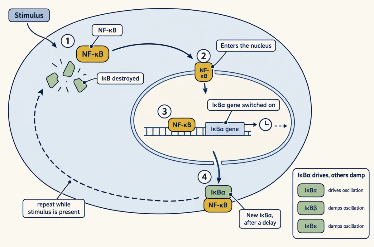
NF-κB switches on the gene for its own inhibitor. After a delay, new IκBα binds nuclear NF-κB and promotes its export to the cytoplasm. If the stimulus remains, IκBα can be destroyed and the cycle can repeat. Hoffmann and colleagues concluded that IκBα drives the oscillation and IκBβ and IκBε damp it. AI-generated illustration -
Where: the same kinase means something else in another place
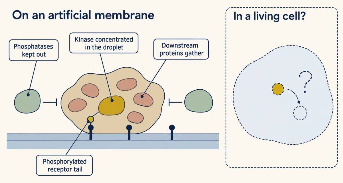
On an artificial membrane supported by glass, with no cell present, membrane-bound LAT and its binding partners formed liquid-like clusters that enriched the kinase ZAP70 and excluded the phosphatase CD45. The drawing is schematic. Whether bright spots in living T cells are a separate liquid phase is still debated. AI-generated illustration -
Going deeper
Where this lesson fits
Before thisReview first
- Ras to MAPK: The Kinase CascadeThis course · Lesson 19
It gives the EGF and NGF result in PC12 cells and the ERK cascade whose duration is read here.
- Calcium: The Messenger Kept at Arm's LengthThis course · Lesson 15
It shows calcium rising in spikes, the signal whose frequency is decoded here.
- Circuits: Amplification, Feedback, Adaptation and SwitchesThis course · Lesson 5
It teaches negative feedback and switches, which this lesson extends with a delay to make pulses.
After thisStudy next
- Endocytosis and Signaling EndosomesThis course · Lesson 36
It takes the idea of place further, showing a receptor signaling from an endosome with a different result.
- Long-Distance Signaling in NeuronsThis course · Lesson 37
It applies signal place and timing to a message that must travel from axon tip to nucleus.
Suggested by an AI model from the lessons themselves, then checked. How we use AI