Cell Signaling · Unit 7: Space, time and distance
Long-Distance Signaling in Neurons
How does a neuron that takes in its survival signal at the tip of its axon get that message to a nucleus centimeters away?
Pause when the reading reaches a film, and offer to play it. Saved on this device.
Watch the film now, or keep listening
Builds on:Finding a Signal: NGF, EGF and the BioassayReceptor Tyrosine KinasesWhy these?
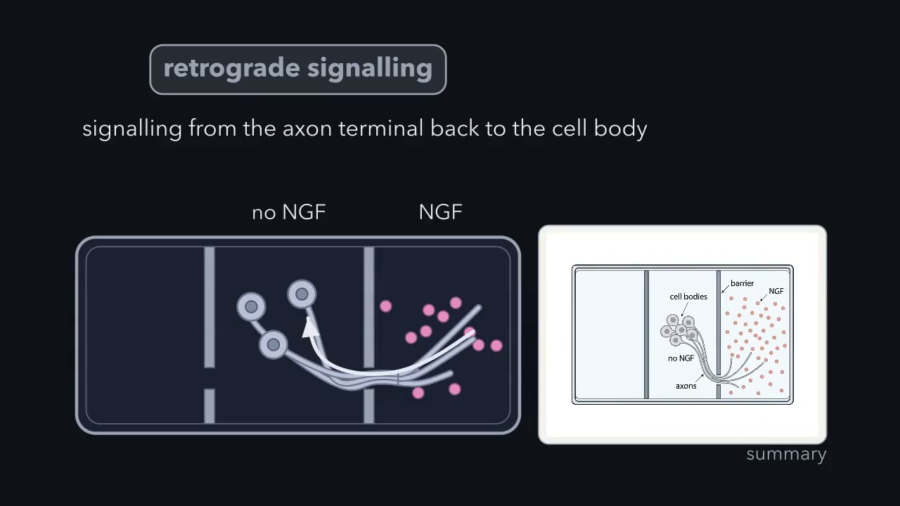 ▶ Watch A neuron can take its survival signal in at the far end of its axon · 2:21
Read the transcript of “A neuron can take its survival signal in at the far end of its axon”
Film transcript: A neuron can take its survival signal in at the far end of its axon
The year is 1977. You grow sympathetic neurons in a special dish. A barrier divides the dish into three chambers. Fluid barely crosses the barrier. Small clusters of cell bodies are in one chamber. Their axons grow under the barrier into the next chamber. So you can keep the fluid around the far ends of the axons separate from the fluid around the cell bodies. After the axons have crossed, you do two runs. 1. You remove NGF from the far chamber only. NGF stays around the cell bodies. 2. You remove NGF from the cell bodies only. NGF stays at the far ends of the axons. Answer three questions. 1. Predict what happens to the axons and to the neurons in the first run.
2. Predict what happens to the axons and to the neurons in the second run. 3. What do the two results together tell you about where a neuron receives its survival signal? Robert Campenot built this dish and published the results in 1977. The dish let Campenot ask where a neuron needs NGF. - First run. When NGF was removed from the far chamber, the axons there stopped growing. Later they appeared to degenerate (break down). This happened even though the cell bodies behind them were in NGF the whole time. - Second run. Removing NGF from the cell bodies had much less effect. Many neurons survived, as long as their axons reached into a chamber that still had NGF.
The axons also did not grow into any chamber that lacked NGF. So the part of the neuron that needs NGF is the part at the target. An axon depends on the NGF in its own surroundings, whatever the fluid around the rest of the neuron contains. A cell body with no NGF of its own can survive on the NGF that its axons collect.
Whatever kept those cell bodies alive had to travel the length of the axon to reach them. NGF could not have reached them through the fluid, because the barrier kept the fluids of the chambers apart. This is the founding result for what the field calls retrograde signalling: signalling from the axon terminal back to the cell body.
A sympathetic neuron collects its survival signal at the tip of an axon that can be a thousand times longer than its cell body is wide, and the genes that must answer are back in the nucleus. The signal gets home inside the axon. In the leading model, nerve growth factor (NGF) and its receptor are taken into a vesicle at the tip, and the motor dynein carries that signaling endosome along microtubules to the cell body.
You met the signal, nerve growth factor (NGF), in the lesson on finding a signal, and its receptor TrkA in the lesson on receptor tyrosine kinases. Here the question is how a message received at one end of a cell reaches the other end, and how you would know which way it went.
A Problem: the signal and the genes are far apart
Sympathetic neurons are the nerve cells that control organs such as the heart and blood vessels. The cell body of one, where the nucleus is, measures a few hundredths of a millimeter across. Its axon, the single long fiber that carries its output, runs out to a target organ, which makes nerve growth factor (NGF), and the axon’s terminals collect it there. Those terminals can be centimeters from the cell body.
What does the cell body need to learn? That its axon has reached a target that makes NGF. It answers with gene expression. The neuron stays alive, and it switches on genes that include the one for TrkA itself and the ones for the tubulin and actin a growing axon is built from.
How does this happen? Rule out the easy answer first. A molecule that spreads by diffusion is slow over long distances, because the time grows with the square of the distance.
time to diffuse a distance x ≈ x² / 2D
Take D as about 10 µm² per second, a typical figure for a protein in cytoplasm. For x = 1 cm, which is 10,000 µm, the time is 5 million seconds, or about 58 days. The survival signal arrives far sooner than that. Something has to be carried, or passed along.
The evidence: NGF at the far end is enough
In an ordinary culture dish one fluid bathes the whole neuron, so you cannot ask which part of the cell needs nerve growth factor (NGF). In 1977 Robert Campenot published a dish that could ask. A barrier sealed to the floor of the dish divides it into chambers. The cell bodies sit in one chamber, their axons grow under the barrier into the next, and the fluid in each chamber can be changed separately.
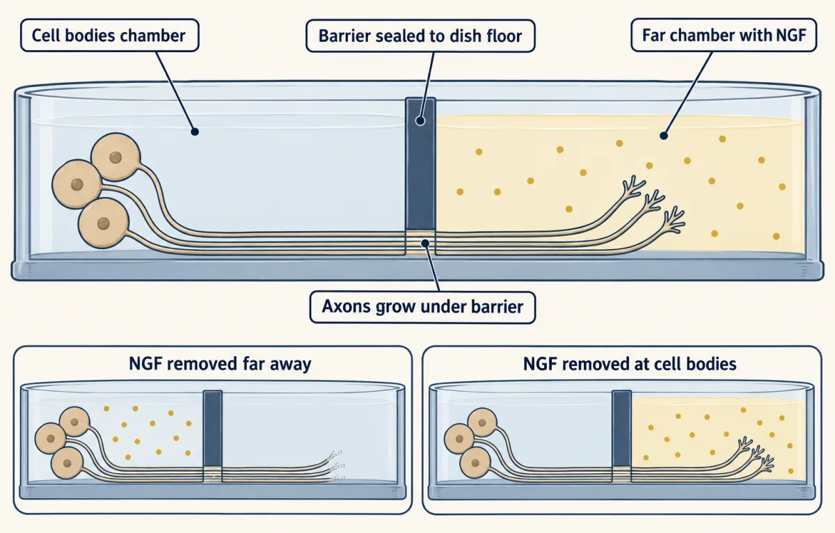
Two runs made the case. When NGF was removed from the far chamber, the axons there stopped growing and later appeared to degenerate, even though the cell bodies behind them were in NGF the whole time. When NGF was removed from the cell bodies instead, many neurons survived, as long as their axons reached a chamber that still had it.
So the part of the neuron that needs NGF is the part at the target. And whatever kept those cell bodies alive had to reach them through the axon, whether carried or relayed, because the barrier kept the fluids apart. This is retrograde signaling: signaling from the axon terminal back to the cell body.
Where does the signal end up? In David Ginty’s laboratory, Antonella Riccio and colleagues applied NGF only to the distal axon, its far end. That activated cAMP response element-binding protein (CREB) in the nucleus, the transcription factor you met in the lesson on signaling to the nucleus.
Be exact about what this shows. It shows where the signal ends. On its own it does not show how the signal traveled. In the same paper Riccio and colleagues reported that taking in NGF and TrkA and carrying them to the cell body were required for the signal.
The model: the endosome as a parcel
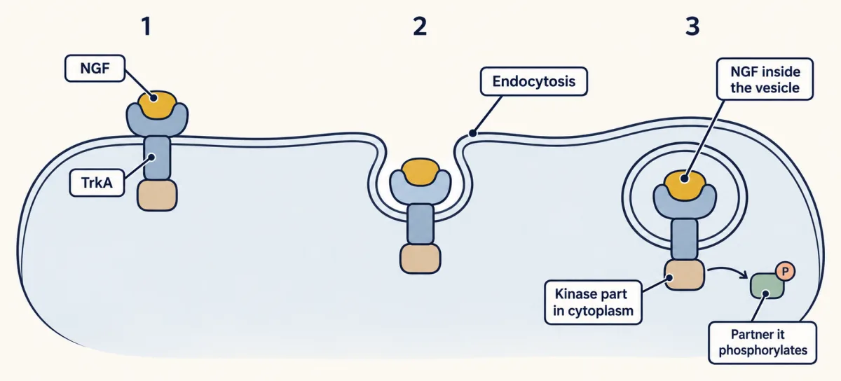
The leading model is the signaling endosome. Nerve growth factor (NGF) binds TrkA at the axon tip, and the pair is taken into the cell by endocytosis, the subject of the previous lesson. Look at how the receptor now sits. NGF is inside the vesicle, still held by the part of TrkA that used to face outward. The kinase part of TrkA is on the other side of the vesicle’s membrane, in the cytoplasm, where it can go on phosphorylating its partners. The vesicle is a patch of active membrane that can be shipped.
Endosomes need motors and roads. The roads are microtubules, long protein filaments whose two ends differ and are called plus and minus. In an axon they all lie the same way round, with their plus ends toward the tip. The kinesins that haul axonal cargo walk toward plus ends, so they carry it out, away from the cell body. Cytoplasmic dynein walks toward minus ends, so it carries cargo back, with the help of a partner complex called dynactin.
A motor-driven vesicle moves at roughly a micrometer per second. At that pace, the centimeter that would take diffusion 58 days takes a motor under three hours.
In this model the parcel reaches the cell body still signaling, and kinases there pass the message on to transcription factors such as cAMP response element-binding protein (CREB).
Not everything is sent home. In Campenot’s dish, axons grew only where NGF touched them, whatever the cell bodies were sitting in. Local mediators? Some of NGF’s work is done where it binds, by signaling proteins beside the receptor at the tip. The survival decision is the part that needs the nucleus.
Carried, or relayed? Why blocking the motor does not settle it
Read the transcript of “Carried, or relayed?”
Film transcript: Carried, or relayed?
A message arrives at the tip of an axon. How does it get home? One answer is a courier. A courier carries one parcel the whole way. The other is a bucket brigade. Each stretch of the axon hands the message to the next, and nothing physical makes the whole journey. The popular test is to block dynein, the motor. In 2004, Heerssen, Pazyra and Segal found that dynein motors transport activated Trk receptors. They found that this transport is needed for the survival of neurons that depend on their target. That is real evidence for a courier. A brigade would fail the same test, though. Suppose each stretch of the axon needs dynein-driven transport for its own local step. Then blocking dynein stops the brigade as completely as it stops the courier.
So the block cannot tell them apart. The relay is a published alternative. In 2002, MacInnis and Campenot reported that retrograde signals can support neuron survival without retrograde transport of NGF itself. In 2003, Ye, Kuruvilla, Zweifel and Ginty titled their paper: Evidence in support of signaling endosome-based retrograde survival of sympathetic neurons. The title claims support. It does not claim proof. Settled: NGF at the terminal is enough, a signal from the terminal reaches the nucleus, and dynein carries activated receptors that survival depends on. Not settled: whether the carried receptor is the only route.
There is a second way to move a message down an axon. Think of a courier against a bucket brigade. The courier carries one parcel the whole way. In the brigade nobody walks far: each stretch of the axon hands the message to the next stretch, and nothing physical makes the whole journey.
The popular test is to block dynein. In 2004 Heerssen, Pazyra and Segal found that dynein motors transport activated Trk receptors, and that this transport is needed for the survival of neurons that depend on their target. That is real evidence for a courier.
A brigade would fail the same test, though. Suppose each stretch of the axon needs dynein-driven transport for its own local step. Then blocking dynein stops the brigade as completely as it stops the courier.
Two alternatives to a carried NGF-TrkA complex had been proposed: a relay along the axon, and a signal that reaches the cell body while nerve growth factor (NGF) itself stays behind. In 2003 Ye, Kuruvilla, Zweifel and Ginty tested these alternatives directly. Survival needed TrkA kinase activity at the axon tip and in the cell body, but blocking TrkA only in the middle stretch of the axon changed neither TrkA signaling in the cell body nor survival, which a relay along the axon would not survive. NGF had to be internalized, and neutralizing the NGF that arrived in the cell bodies took survival away. Together these results argued against a propagated wave and against a signal that leaves NGF behind, and supported a carried NGF-TrkA complex. Later work, which followed single NGF-carrying endosomes along axons and the steps that ready TrkA endosomes for transport, built on it, and the signaling endosome is the leading account. The design problem below asks which experiments could do that.
Here is the score. Settled: NGF at the terminal is enough to support the neuron, a signal from the terminal reaches the nucleus, and dynein carries activated receptors that survival depends on. Not settled: whether the carried receptor is the only route, and whether one kind of parcel does every job NGF asks for.
Two more answers to distance, and a road that fails
See where the endosome ends up
A sympathetic neuron is also a listener. Neurons in the spinal cord make synapses, the contacts where one neuron signals to the next, on the sympathetic neuron’s dendrites, the short branches that collect input. In 2010 Sharma, Deppmann, Harrington and colleagues asked whether nerve growth factor (NGF) from the distant target controls those synapses too. They studied mice that lack NGF and also lack BAX, the death protein from the lesson on apoptosis, so that the neurons live without their survival signal and their synapses can be examined. NGF turned out to be needed for those synapses to form.
How does NGF-TrkA control this? The group grew neurons in a microfluidic chamber, a small relative of Campenot’s dish developed by Noo Li Jeon, using TrkA that carries a tag called Flag. After NGF was given to the axons, tagged TrkA appeared as dots in the dendrites over the next six hours, and the group compared that with the clustering there of postsynaptic proteins, the ones on the receiving side of a synapse. A long-distance relationship? The message that the axon has found its target reaches the side of the cell that receives input.
Make it where you need it
A dendrite has its own distance problem. A synapse can sit a long way from the nucleus, and some changes at a synapse need new protein soon after the signal arrives. Erin Schuman found that the neuron does not ship everything. With Hyejin Kang she applied the growth factor brain-derived neurotrophic factor (BDNF) to slices of hippocampus, a region of the brain, and synapses strengthened. Then they cut the slices so that the synapses were separated from the cell bodies of their own neurons. BDNF still strengthened them, and the effect still depended on protein synthesis. Imaging has since shown a new protein appearing in dendrites, and transcriptome studies have found many different mRNAs in the dendrites and axons of the neuropil, the mesh of fibers where the synapses lie.
When the road fails
A cell built this way depends on its motors. Mutations in DCTN1, the gene for a subunit of dynactin, are found in people whose motor neurons, the neurons that drive muscle, degenerate. Two mouse mutants, Loa and Cra1, carry point mutations in the dynein heavy chain and show nerve loss whose pattern differs with the mutation and the mouse strain. Transport defects have been reported in amyotrophic lateral sclerosis (ALS), a disease of motor neurons, and in Huntington’s, Parkinson’s and Alzheimer’s diseases, although a defect that accompanies a disease is not proof that it causes it. One case names the cargo. In a mouse model of Down’s syndrome that carries an extra copy of App, the gene for the amyloid precursor protein, Salehi and colleagues found that the raised expression of App disrupts the transport of NGF.
Design problem
Two models explain how a survival signal gets from the axon tip to the nucleus. In the first, the receptor itself travels the whole way inside a vesicle. In the second, the receptor stays at the tip and a chain of kinases passes the message along. Design an experiment that could tell them apart. Say first why a drug that stops the dynein motor cannot do it.
Saved in this browser only.
One way to do it
Why the drug fails: both models can need the motor. A courier vesicle needs dynein for the whole trip. A relay could need dynein for each short local step. Stop dynein and both stop, so the result cannot choose.
Ask instead what only one model predicts.
- Mark the receptor at the tip. In a compartmented culture, label TrkA on the axon side only, then look for the label in the cell body. The courier model says it arrives. The relay model says it stays at the tip.
- Compare speeds. See whether a response near the nucleus appears when a motor-driven vesicle would arrive, or sooner. Timing alone is hard to read. It needs the cargo tracked at the same time.
- Break the middle of the chain. Add an inhibitor of the TrkA kinase only to the stretch of axon between the tip and the cell body. A relay that passes TrkA activity along the axon has to go through that stretch, so it should fail. A courier only passes through on its way home. Ye and colleagues (2003) did this, and survival and TrkA signaling in the cell bodies were unaffected. They also found that survival needed TrkA kinase activity at the tip and in the cell bodies, and that neutralizing NGF in the cell-body chamber took survival away.
A caution on step 1: a labeled receptor arriving shows that the trip happens. It does not show that the trip matters. For that you have to block what arrives, as Ye and colleagues did when they neutralized NGF at the cell bodies.
Going deeper
The relay proposal and the timing evidence
In 1997 Senger and Campenot found TrkA phosphorylated in the cell bodies within a minute of nerve growth factor (NGF) reaching the distal axons, long before labeled NGF arrived, and suggested that part of the signal travels by propagation. In 2002 MacInnis and Campenot reported in Science that retrograde signals can support neuron survival without retrograde transport of NGF itself. That result says NGF need not make the trip. It does not say what carried the signal instead. Watson and colleagues (1999) then followed a tagged Trk receptor in living sensory neurons and concluded that the fast signal was the receptor itself, moving along microtubules. The 2003 paper by Ye and colleagues is titled “Evidence in support of signaling endosome-based retrograde survival of sympathetic neurons”, which states its conclusion modestly.
Dates and counts for local synthesis
Kang and Schuman published the slice result in 1996. In 2001 her laboratory watched a fluorescent reporter protein appear in dendrites. In 2012 the laboratory counted 2,550 different mRNAs in the dendrites and axons of the neuropil.
Loa and Cra1
Loa and Cra1 were first reported to lose motor neurons, although later studies found mainly sensory-axon loss, with motor-neuron and neuromuscular-junction findings that differ with the mutation and the mouse strain.
Your notes
Select any text in the lesson to highlight it or add a note to it. Your highlights and notes are saved in this browser and collected in your notebook.
This browser is not letting the site save anything (a private window or strict privacy settings), so highlights and notes will be lost when you leave the page.
How we measure it
Compartmented culture (Campenot chamber)
A barrier sealed to the dish floor separates the fluid around the cell bodies from the fluid around the axons, which grow under the barrier. You can give or withhold a factor at one end of the neuron and measure the response at the other. It shows where a signal acts and whether something must travel inside the axon.
Microfluidic chamber with a tagged receptor
Neurons sit in one compartment and send axons through narrow grooves into a second, and the two fluids stay separate. If the receptor carries a tag, you can follow tagged receptors after stimulating the axons and see where in the cell they turn up. It tests whether the receptor itself makes the trip.
Isolated neuropil slice
A brain slice is cut so that a layer of synapses is separated from the cell bodies of its own neurons. If a change at those synapses still needs protein synthesis, the protein must be made locally. It tests whether translation happens away from the cell body.
Translation reporter in dendrites
The coding sequence for a fluorescent protein is joined to the untranslated region of an mRNA that travels into dendrites. New fluorescence in a dendrite marks protein being made there, provided you can rule out protein made in the cell body and carried out. It lets you watch local synthesis in a living neuron.
Practice questions on this lesson (3) → MCAT-style, original, each with an explanation
Where this lesson fits
Before thisReview first
- Finding a Signal: NGF, EGF and the BioassayThis course · Lesson 2
This is where NGF and its discovery through bioassay are introduced, and this lesson assumes you know the signal.
- Receptor Tyrosine KinasesThis course · Lesson 18
You need TrkA as a receptor tyrosine kinase that stays active after binding NGF to follow the endosome model.
After thisStudy next
- Hacking the Cell: Reporters, Actuators and Genetic ToolsThis course · Lesson 44
The tagged receptors and microfluidic tools used here are extended into reporters and actuators for watching signals in living cells.
Elsewhere on the siteFilms, simulations and method films in other courses
- Still On When It Gets ThereFilm · Developmental Neuroscience
This shows the survival signal from the target traveling home to the cell body.
- Grip, Swing, Let GoFilm · Cell Biology
Dynein walking toward the microtubule minus end, back to the cell body, is the motor that carries the endosome.
- Too Small, or Lost?Film · Developmental Neuroscience
This gives the target-supplied NGF survival evidence that sets up the distance problem.
Suggested by an AI model from the lessons themselves, then checked. How we use AI
Finish line
Test yourself
8 questions
Practice and MCAT-style, with explained answers.
Next · Lesson 38
Exosomes and Extracellular Vesicles: Signals Between Cells
B cells release vesicles that carry their peptide-loaded MHC molecules, and those vesicles switch on only the T cells that recognise that peptide. A scrap of membrane can carry a signal.
Continue →