Developmental Neuroscience · Unit 11: Rebuilding, evolving, and going wrong
What Makes the Human Brain Big
Rewritten on : new text, figures, flashcards and film. Your notes and highlights stay, but the text around them may have moved.
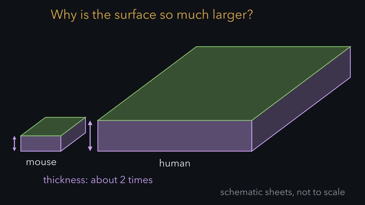
Your cortex is only about twice as thick as a mouse's. Why is its surface roughly a thousand times larger?
Pause when the reading reaches a film, and offer to play it. Saved on this device.
Watch the film now, or keep listening
Builds on:The Ventricular Zone: Radial Glia as Stem CellsBirth Order and Fate: Temporal Identity and the Inside-Out CortexRadial Migration: Climbing the Glial LadderWhy these?
 ▶ Watch Multiply, Don't Add · 3:13
Read the transcript of “Multiply, Don't Add”
Film transcript: Multiply, Don't Add
Your cortex is only about twice as thick as a mouse's. So why is its surface roughly a thousand times larger? Almost all of the gain is sideways. How does a brain build more of the same wall? Compare two kinds of division. In a symmetric proliferative division, one progenitor makes two. If every progenitor does this once, the pool doubles. In an asymmetric neurogenic division, it makes one progenitor and one neuron. The pool stays the same size. The radial glia lining the ventricle, the founders, divide symmetrically first. Later they switch to making neurons. Pasko Rakic showed that young neurons climb radial glial fibres. He argued that they stack into columns.
In 1988, he proposed the radial unit hypothesis. If every unit takes up about the same patch of surface, the number of units sets the area. The neurons per unit set the thickness. Predict. Species B gets three extra rounds of symmetric division, before the switch. Species C gets three extra asymmetric divisions per unit, after it, each adding one neuron. Which species gains more neurons? And which one gains area?
Species B. Doubling three times gives eight times the units, so, with the same patch per unit, eight times the area. Species C gains about three percent more neurons, and no extra units, so no extra area. Doubling multiplies. Adding only adds. Back to the mouse. Ten doublings make about a thousandfold. So, in this model, with every unit the same size, ten extra rounds would close the gap. Our brain is about three times a chimpanzee's. If our cortical area differs by a similar factor, the gap is under two rounds. Rakic made a related argument for primates in 1995. A delay of a few cell cycles before the switch could greatly enlarge the cortex.
In mice, a stabilized form of beta β-catenin kept more progenitors dividing. The cortex grew in area, and buckled into folds. That shows extra founder divisions can enlarge the sheet, not how primates did it. In organoids, human cells took longer than ape cells to become radial glia. The human organoids grew larger. Neither test was done in a developing human brain. Multiply, don't add. Extra rounds of doubling can multiply the neurons a cortex makes. That is the leading explanation for our big cortex, and nearly all of the gain went into area. So for every cell and gene that follows, ask what it changes.
From the film
- Multiply, don't add: a division that makes two progenitors adds a progenitor that keeps dividing, so repeated rounds compound.
- In the radial unit model, extra founder doublings multiply the number of columns, so the cortex gains area.
- Nearly all of the gain in the human cortex went into area, not thickness.
A chimpanzee’s brain weighs roughly a third as much as yours. Yet both are built from the same main classes of cell, arranged in the same six cortical layers, by the same kinds of progenitor.
So what is actually different? You have spent several lessons on how the cortex is made: radial glia lining the ventricle, intermediate progenitors just above them, neurons climbing to an inside-out stack of layers. This lesson asks which steps in that process changed in our lineage, and how anyone has tested it.
A bigger sheet, not a thicker one
Start with the number everyone quotes: about 86 billion neurons in the adult human brain, about 16 billion of them in the cerebral cortex, the mean from four adult men (Azevedo et al., 2009). That is about the number you would predict for a primate brain of its size, so ours is a scaled-up primate brain, not one with unusually many neurons for its size.
What does stand out is the cortex as a sheet. Across mammals, cortical thickness changes little while area changes enormously. The human cortex is only about twice as thick as a mouse’s, but its surface area is roughly a thousand times larger (figures summarized by Pasko Rakic, Nat Rev Neurosci, 2009). Much of that sheet is folded to fit inside the skull.
Predict. Treat the cortex as a sheet whose size is its area times its thickness. By those two figures, the human sheet is about two thousand times the size of a mouse’s. How much of that factor comes from height, and how much from spreading sideways? What kind of growth should you look for in the embryo?
Check your prediction
Height gives a factor of 2 and area a factor of about 1,000, and 2 × 1,000 = 2,000. Almost all of the gain is sideways. Counts suggest that the number of neurons under each square millimetre of surface differs only a few-fold between mouse and human (Rockel et al., 1980; Herculano-Houzel et al., 2008), far less than the roughly thousandfold difference in area. It varies between areas and species, and primate primary visual cortex is the best-known exception. So the extra neurons sit mostly in extra area, not in taller columns. The question for development is not how to build a thicker wall, but how to build a thousand times more of the same wall.
In the lesson on the neurotrophic hypothesis, survival could only trim the neurons that were made. This lesson is about where the number made is set.
The one idea to carry through this lesson. Multiply, don’t add. A division that makes two progenitors adds a progenitor that keeps dividing, so repeated rounds compound; a division that makes one progenitor and one neuron only adds a neuron. The leading explanation for our big cortex is that it has more progenitors working for longer: more rounds of doubling divisions in the early founders, and more amplification by basal progenitors. Nearly all of the gain went into area. A sheet a thousand times larger is about ten doublings’ worth of extra units.
What the rule covers.
- Which question. How many neurons, and how much area. It does not explain folding, the slow maturation of synapses, or which genes changed. Those are tests of the rule or lie outside it.
- Where the doubling happens. In founders at the ventricle, before neurogenesis (the making of neurons) begins, which adds columns; and, as the second section shows, in outer radial glia during neurogenesis. How much of the human gain came from each site is not known.
- A model, not a proof. Extra doubling could produce the numbers, but the numbers alone do not show which divisions happened. A longer neurogenic period, or changes in how neurons survive or pack, also change the total. Extra founder divisions have been tested in mice and in ape and human organoids, and amplification by basal progenitors has been probed by adding a human gene to ferrets and marmosets. None of this was done in a developing human brain.
More columns, not a thicker cortex
The rule compares two outcomes of a progenitor division. In a symmetric proliferative division, the doubling division, one progenitor makes two; when every progenitor does it once, the pool doubles. In an asymmetric neurogenic division, a radial glial cell makes one copy of itself and one neuron, so the pool stays the same size. (Other outcomes exist. A radial glial cell can make itself plus an intermediate progenitor, which goes on to divide into neurons. Making a neuron straight from a radial glial cell is the direct route of neurogenesis, going through an intermediate progenitor the indirect route; outer radial glia add a third. Real lineages mix these, as the next section shows.) Early in development the progenitors lining the ventricle, the founders (first neuroepithelial cells, then radial glia), make doubling divisions. Later they switch to asymmetric division and start making neurons. (The lesson on radial glia as progenitors follows that switch.)
Pasko Rakic, working in the fetal rhesus monkey, had shown that young neurons climb radial glial fibres. He argued that neurons born at one spot in the ventricular zone climb the same fibre and stack up above the patch where they were born, with their birth order setting their layer, the youngest on top. In 1988 (Science) he proposed the radial unit hypothesis. A radial unit is a patch of founders at the ventricle together with the one column of neurons it makes; Rakic called the patch a proliferative unit and the column an ontogenetic column. Columns stand side by side. If every unit takes up about the same patch of surface, with neurons packed the same way, the number of units sets the area of the cortex, and the number of neurons each unit makes sets its thickness.
Predict. Species B is identical to species A, except that its founders go through three extra rounds of doubling division before they switch; each founder starts one unit. Species C instead adds three extra asymmetric divisions to every unit after the switch, each making one neuron directly. Each unit in A makes about a hundred neurons. Which species gains more neurons, and which one gains area?
Check your prediction
Species B. Three rounds of doubling give 2 × 2 × 2 = 8 times as many founders, so eight times as many units: eight times the area and eight times the neurons, with columns as tall as before. Species C gains three neurons per column, 103 instead of 100: about 3 percent more neurons, a slightly thicker cortex and no extra area. In this fixed-column model, if you want area, change the founder phase.
Now return to the opening puzzle. Nearly all of the human gain over the mouse is area, which on this model means units. How many doublings make a thousandfold? Ten, since 2¹⁰ = 1,024. With every unit the same size and nothing else changed, ten extra rounds would turn a mouse-sized sheet into a human-sized one. Our brain is about three times the size of a chimpanzee’s; if our cortical area differs by a similar factor, the gap is fewer than two rounds, since 2¹ = 2 and 2² = 4. Rakic made a related argument for primates in 1995 (Trends Neurosci): a delay of only a few cell cycles before the switch to asymmetric division could account for a large increase in cortical area.
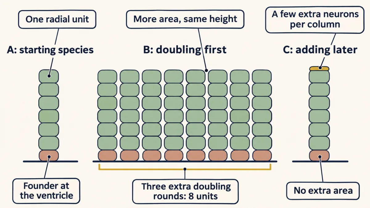
units after k extra symmetric rounds = starting units × 2ᵏ; each extra direct neurogenic division adds one neuron to a unit
Two experiments test whether more founder divisions can do it. Anjen Chenn and Christopher Walsh (Science, 2002) made transgenic mice whose neural progenitors expressed a stabilized form of β-catenin, which kept more of them re-entering the cell cycle. The mice grew enlarged brains with a greater cortical surface area, and the normally smooth cortex buckled into folds. The mice did not live past birth, and their folds came from an enlarged ventricular surface, which is not how primate cortex grows; the experiment shows that more founder divisions can enlarge the sheet, not how it happened in primates. A more direct comparison came from Silvia Benito-Kwiecinski, Madeline Lancaster and colleagues (Cell, 2021), who grew brain organoids from human, chimpanzee and gorilla cells. The human organoids were larger because their neuroepithelial cells took longer to change shape and become radial glia, which gave the founder pool more time to expand.
The model is simplified. Lineage tracing in rodents and ferrets later found that related neurons can spread sideways, and inhibitory interneurons reach the cortex by migrating tangentially from outside it. Its core, the link between the number of units and the area, remains a useful working model. That raises a question. Human neurogenesis lasts months, not the mouse’s week. If that time holds extra divisions, where could they happen, and why would they not simply make our cortex thick?
A second place to double
Read the transcript of “A Second Place to Double”
Film transcript: A Second Place to Double
Human neurogenesis lasts months, not the mouse's week. Where might extra divisions happen? And why wouldn't they simply make our cortex thick? Rakic built his model around progenitors at the ventricle. But in fetal monkey cortex, the zone above them is split in two. What was dividing in the outer part? In 2010, Hansen, Kriegstein and colleagues filmed living slices of human fetal cortex. They found cells with the markers of radial glia, the proteins PAX6 and SOX2. These cells kept a fibre toward the brain surface, but had no process touching the ventricle. Before dividing, a cell moved its cell body up its fibre. Daughters could divide again, or become intermediate progenitors, which soon make neurons.
These are outer radial glia. Later imaging in fetal macaque found them making proliferative divisions too. So, in macaque at least, they can be a second site of doubling. How much can a little doubling matter? Take a toy lineage. One progenitor divides for 60 rounds, and each division adds a neuron. That makes 60 neurons. Predict. Now, in every round, give each progenitor a one in ten chance of making two progenitors instead. Every progenitor keeps dividing. On average, do you get about 60 neurons, a few hundred, or thousands?
Thousands, on average. Each doubling adds a progenitor that keeps dividing, so the pool grows by a tenth each round, like compound interest. Given enough rounds, a little doubling outgrows a lot of adding. So why no thicker cortex?
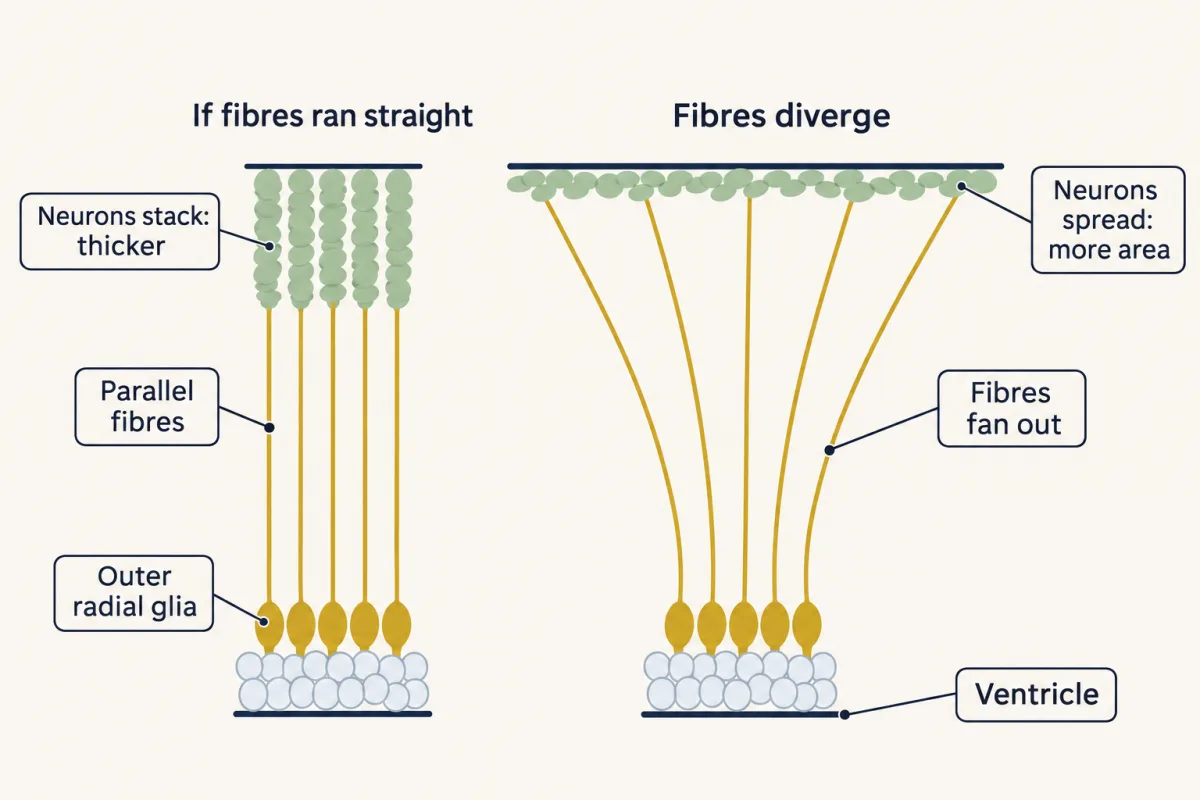
New neurons climb radial glial fibres. If every fibre ran straight up, with packing fixed, what would extra neurons do? In this cartoon, they would stack onto existing columns, and the cortex would thicken. Instead, the fibres diverge as they climb, fanning the scaffold outward. That favours spreading the neurons sideways, into area.
Outer radial glia are not uniquely human. Mice have a few, and ferrets and primates have many. The human outer subventricular zone is especially large. The marmoset, whose cortex is nearly smooth, has them too. So they are not enough for folding on their own. Founders can double at the ventricle. Outer radial glia can double far from it, and their fanned fibres favour area over thickness.
From the film
- Outer radial glia keep a fibre but have no process reaching the ventricle, and can divide far from it.
- In macaque, at least, they can be a second site of doubling.
- Fibres that diverge as they climb favour spreading neurons sideways, into area, rather than thickness.
Rakic built the radial unit model around progenitors at the ventricle. In primates that turned out to be incomplete. Two direction words help here: apical means toward the ventricle, basal toward the brain surface. Progenitors that divide away from the ventricle are basal progenitors, a family with two main members. Intermediate progenitors make neurons after one or a few divisions; this is the indirect route of neurogenesis, and in mouse an intermediate progenitor usually divides once into two neurons, while in primates some divide more than once, so this route can add a few more neurons. Outer radial glia are the subject of this section.
In fetal monkey cortex, Iain Smart, Colette Dehay, Henry Kennedy and colleagues (Cereb Cortex, 2002) described a subventricular zone, the layer just above the ventricular zone, split into an inner and an outer part, the outer subventricular zone. What was dividing there?
In 2010, David Hansen, Jan Lui, Philip Parker and Arnold Kriegstein (Nature) labelled cells in living slices of human fetal cortex and filmed them. In the outer subventricular zone they found cells that expressed PAX6 and SOX2, the transcription factors of radial glia, and kept a long basal fibre toward the brain surface. Unlike classical radial glia, they had no process touching the ventricle. Before dividing, a cell moved its cell body rapidly along its fibre, and then went into mitosis there. Daughters followed in the films could inherit the fibre and divide again, or become intermediate progenitors expressing TBR2. These outer radial glia, in other words, renewed themselves and fed the indirect route of neurogenesis from a site far from the ventricle.
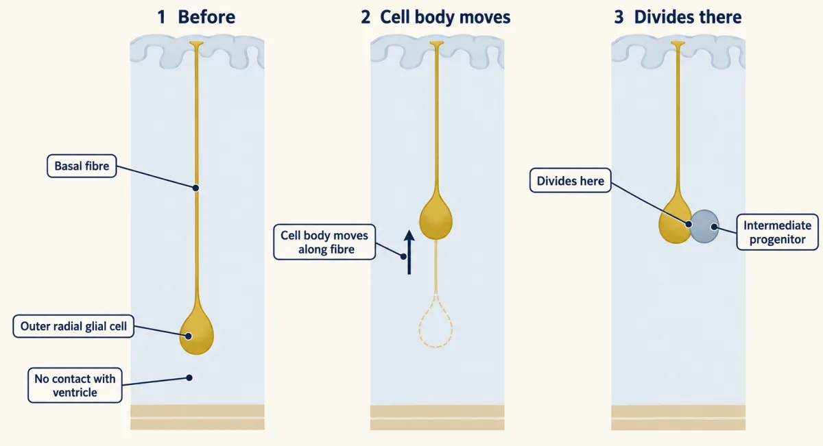
They do more than add. Later live imaging in fetal macaque cortex (Betizeau et al., Neuron, 2013) found basal radial glia of several shapes, with a basal fibre, a process pointing toward the ventricle, or both, and cells that switch between shapes; Hansen’s basal-fibre cell is the classic form, not the only one. These cells also made proliferative divisions, in which both daughters stay progenitors. So, in macaque at least, outer radial glia can be a second site of doubling, one that keeps working during neurogenesis, alongside their asymmetric divisions that make intermediate progenitors and neurons. How much can a little doubling matter?
Predict. Here is a toy lineage, not a measurement. Start with one progenitor and run 60 rounds of division, a number chosen for easy arithmetic, not taken from any species. If every division makes one progenitor and one neuron, the lineage makes 60 neurons. Now give every progenitor, in every round, a one-in-ten chance of making two progenitors instead, and no neuron that round. Every progenitor keeps dividing, and none stops or dies. On average, does the lineage make about 60 neurons, a few hundred, or thousands?
Check your prediction
Thousands: about 2,700 on average. Each doubling adds a progenitor that also keeps dividing, so the pool grows by about a tenth each round, like compound interest: after 60 rounds about 300 progenitors are still cycling (1.1⁶⁰ ≈ 304). Nine tenths of the pool make a neuron in each round, which sums to 9 × (1.1⁶⁰ − 1) ≈ 2,731 neurons. With a one-in-five chance, the same lineage makes about 225,000. Real lineages stop, and real proportions differ, but the shape of the answer holds: over enough rounds, even a small chance of doubling outgrows adding.
Predict. If outer radial glia can add many extra neurons, why don’t they pile up into a thicker cortex? New excitatory neurons climb radial glial fibres. In a cartoon with the surface and the packing of neurons fixed, if every outer radial glial fibre ran straight up, parallel to its neighbours, what would the extra neurons do to the cortex? What would the fibres have to do instead to give area?
Check your prediction
In that cartoon, straight, parallel fibres would stack the extra neurons onto existing columns and make the cortex thicker. Instead, the fibres of outer radial glia diverge as they climb. They fan the radial scaffold outward, so the neurons that climb them can spread sideways. That favours growth in area, and it has been linked to folding in the ferret. How thick and how wide the cortex ends up also depends on how neurons pack and how the tissue grows.
What holds these cells in the radial glial state? Hansen and colleagues also found that many expressed HES1, a Notch target, and that DAPT, a drug blocking γ-secretase, an enzyme Notch needs in order to signal, shifted the balance of cells in cultured slices from the SOX2-high radial glial state toward the TBR2-high intermediate-progenitor state. Because DAPT blocks other γ-secretase targets too, this points to a role for Notch signalling in keeping them in the radial glial state, without proving that Notch alone is responsible.
Outer radial glia are not uniquely human. In the same year Simone Fietz and colleagues in Wieland Huttner’s laboratory described related progenitors in human and in ferret cortex (Nat Neurosci, 2010). A few similar cells exist in the embryonic mouse (Wang et al., Nat Neurosci, 2011; Shitamukai et al., J Neurosci, 2011). The difference is one of degree: these cells are abundant in primates and ferret and rare in mouse, and the human outer subventricular zone is especially large. The marmoset, a monkey with a nearly smooth cortex, also has an outer subventricular zone, so these cells are not sufficient for folding on their own.
Genes that only humans have
Read the transcript of “Testing the Rule in Genes and Time”
Film transcript: Testing the Rule in Genes and Time
A few genes have functional copies in humans, and not in chimpanzees. Do any of them change what progenitors do? No gene has been removed from a developing human brain. So each was tested in another system. First, ARHGAP11B. In embryonic mouse cortex, it increased basal progenitors. In marmoset fetuses, it enlarged the neocortex. In cultured human fetal slices, blocking it reduced basal progenitors. Next, NOTCH2NL, which raises Notch signalling. In mouse cortex, it kept progenitors dividing and delayed neurons. Deleted in human organoids, it made progenitors differentiate early. Both are consistent with more doubling. But the studies described here did not score division modes, so none shows which kind of division changed.
Time matters too. Neurogenesis takes about a week in the mouse, roughly two months in the rhesus monkey, and longer in humans. Extra time allows more rounds, if cell cycles do not lengthen. A steady pool adds in step with time. A growing pool compounds. One more difference lies outside the rule. Human neurons mature slowly. Spine density in prefrontal cortex stays above adult levels into the third decade. SRGAP2C slowed spine maturation in mouse neurons. Is that pace set by the body, or the cells?
Human neurons go into newborn mouse cortex. Whose pace do they keep? They matured over many months, close to a human time course. The mouse neurons around them had long since matured. At least part of the timing travels with the cells.
But maturation shapes connections, not neuron number. It lies outside the rule. And no test shows that one gene made the human cortex. Multiply, don't add: a leading explanation for our large cortex. How much came from founders, outer radial glia, or a longer clock is still open.
From the film
- Human-specific genes such as ARHGAP11B and NOTCH2NL were tested in mouse, marmoset and organoids, not in a developing human brain.
- Neurogenesis lasts longer in humans, and extra time allows more rounds if cell cycles do not lengthen.
- Slow human neuron maturation shapes connections, not neuron number, so it lies outside the rule.
A gene that helped make our cortex big should change what progenitors do: how many there are, how long they keep dividing, how fast they cycle, or what their divisions make. Gene duplication is a common way for a lineage to gain a new regulator. Two of the best-studied duplicated genes or gene families whose functional copies are found in humans and not in chimpanzees have been tested on cortical progenitors; a third, SRGAP2C, acts on neurons and comes up in the next section. They are not the whole list. Note the system each was tested in.
ARHGAP11B. Marta Florio, Huttner and colleagues (Science, 2015) found this partial copy of ARHGAP11A expressed in human cortical progenitors. Electroporated into the embryonic mouse cortex, it increased the number of basal progenitors. Later work from the same laboratory expressed it in ferret embryos and in marmoset fetuses (Heide et al., Science, 2020), where it enlarged the neocortex and increased upper-layer neurons.
NOTCH2NL. Two groups published together in Cell in 2018. Ian Fiddes and colleagues in David Haussler’s group traced these human-specific genes, which derive from part of NOTCH2, and found that deleting them from human stem cells made cortical organoids whose progenitors differentiated early. Ikuo Suzuki and colleagues in Pierre Vanderhaeghen’s laboratory found that NOTCH2NL raises Notch signalling, the signal that holds progenitors back from making neurons. Expressed in embryonic mouse cortex, it kept progenitors dividing and delayed neuron production. The genes sit at 1q21.1, a region where deletions and duplications are associated with smaller and larger heads (the lesson on microcephaly and malformations).
Both sets of results are consistent with more amplification: more basal progenitors, or progenitors held back from making neurons, could follow from extra doubling divisions, but also from more basal progenitors being generated or from slower differentiation. The studies described here did not score division modes, so none shows which kind of division changed; keeping a pool going is not the same as growing it. Most results come from adding or removing a human gene in a mouse, ferret, marmoset or organoid. The closest tests in human tissue used cultured slices of fetal cortex: blocking ARHGAP11B reduced basal progenitors, and knocking down TBC1D3, a gene duplicated repeatedly in ape evolution, reduced outer radial glia. No gene was removed from a developing human brain, and none shows that one gene made the human cortex.
Taking longer: development on a slower clock
The last difference is time. Cortical neurogenesis takes about a week in the mouse and roughly two months in the rhesus monkey (Rakic, Science, 1974), and lasts longer still in humans. A longer neurogenic period gives outer radial glia and other neurogenic progenitors more time to divide. How many neurons that adds also depends on cell-cycle length and on the kind of division. If cycles do not lengthen, a pool that holds steady adds neurons in proportion to the time, and a pool that keeps growing compounds the extra time. The design problem at the end works this out.
Outside the rule: how fast neurons mature
Synapses follow the same slow clock, but this is a different matter: it concerns how connections form, not how many neurons there are. Peter Huttenlocher and Arun Dabholkar (J Comp Neurol, 1997) counted synapses in human cortex and found that synaptic density in prefrontal cortex peaks later, and declines later, than in auditory cortex, with pruning continuing into adolescence. Zdravko Petanjek, Rakic and colleagues (PNAS, 2011) found that spine density on pyramidal neurons of human prefrontal cortex stays above adult levels into the third decade of life. Keeping juvenile features for longer is called neoteny.
SRGAP2C is one candidate for it at the level of single synapses. Cécile Charrier, Franck Polleux and colleagues (Cell, 2012) showed that this truncated copy of SRGAP2 blocks the function of the ancestral protein. Expressed in mouse cortical neurons, it slowed the maturation of dendritic spines and increased their density in the adult. It acts on neurons rather than progenitors.
Predict. Is the slow clock set by the body or by the cells? Human cortical neurons made from stem cells are transplanted into the cortex of newborn mice. If the surrounding tissue sets the pace, how fast should they mature? If the cells carry their own pace, what should you see?
Check your prediction
When the Vanderhaeghen laboratory did this (Linaro et al., Neuron, 2019), the human neurons matured over many months, close to a human time course, while the mouse neurons around them had long since matured. At least part of the timing travels with the human cells.
Putting it together
The table mixes interventions, live observations and comparisons between species. For each, note what was changed or watched and what was inferred, and read the last two columns before the result.
| Experiment | System | Result | What it supports | What it does not establish |
|---|---|---|---|---|
| Stabilized β-catenin in progenitors (2002) | transgenic mouse | more cortical surface area; smooth cortex buckled into folds | more founder doublings can enlarge the sheet | how primates did it |
| Human versus ape organoids (2021) | human, chimpanzee and gorilla organoids | human neuroepithelial cells slower to become radial glia; larger organoids | a longer founder phase in human cells | the same in a developing human brain |
| Live imaging of outer radial glia (2010) | living slices of human fetal cortex | cells with a basal fibre and no ventricular contact divide far from the ventricle; daughters divide again or become intermediate progenitors | a second site of division | how many neurons they add in a fetus |
| Live imaging of basal radial glia (2013) | cultured slices of fetal macaque cortex | some divisions make two progenitors | basal progenitors can amplify during neurogenesis | how much this adds to the human cortex in a fetus |
| γ-secretase blocked | cultured slices of human fetal cortex | lower fraction of SOX2-high cells, higher fraction of TBR2-high cells | γ-secretase activity helps keep outer radial glia in the radial glial state | changes in cell number, no selective cell loss, or that Notch alone is responsible |
| ARHGAP11B added | mouse and ferret embryos; marmoset fetuses | more basal progenitors; in marmoset, an enlarged neocortex with more upper-layer neurons | a human gene can expand cortical output in a primate | that it made the human cortex |
| ARHGAP11B blocked | cultured slices of human fetal cortex | fewer basal progenitors | it acts in human tissue | its effect in a living fetus |
| NOTCH2NL added, or deleted | embryonic mouse cortex; human cortical organoids | progenitors kept dividing, neurons delayed; after deletion, early differentiation | it holds progenitors back from making neurons | how much it contributes in a fetus |
| Neurogenesis timed | mouse, rhesus monkey, human | about a week; roughly two months; longer still | more time for divisions | how many neurons the time adds |
| SRGAP2C expressed | mouse cortical neurons | slower spine maturation, higher adult spine density | outside the rule: maturation | anything about neuron number |
| Human neurons transplanted | newborn mouse cortex | matured over many months, close to a human pace | part of the timing travels with the cells | how the cells encode their pace |
Read down the table and the size experiments show that several routes can lead to more neurons in animals, organoids or cultured tissue: more founder doublings, amplification by basal progenitors, and a later switch to making neurons. None of them scored divisions in a developing human brain, and how much each route contributed in our evolution is still open. None of these, on its own, made us human. The slow maturation of synapses is a separate story.
Design problem: a longer clock and a little doubling
Species P is like species Q, except that its neurogenic period is twice as long, with cell cycles of the same length. Use the toy lineage from the outer radial glia section: one progenitor, 60 rounds in Q and 120 in P. How much more does a P lineage make if every division adds one neuron? How much more, on average, if each progenitor has a one-in-ten chance per round of doubling? And what would you measure in fetal tissue to tell which of the two is happening?
One way to do it
If every division adds, twice the time gives twice the neurons: 120 instead of 60, a gain in proportion to time. With a one-in-ten chance of doubling, the lineage makes about 2,700 neurons on average in 60 rounds and about 830,000 in 120, roughly 300 times more, because the extra time compounds the doubling. To tell the cases apart in tissue, counting progenitors is not enough: new outer radial glia can arrive from the ventricular zone, and doubling can be balanced by cells that stop dividing or die. Film outer radial glia in slices, follow both daughters through their later divisions, tell radial glia from intermediate progenitors, and measure cell-cycle length and cell loss. Then check where the extra neurons went: diverging fibres should favour area over thickness. The numbers are a toy, so the comparison, not the totals, is the point.
Going deeper
How the 86 billion was counted
Frederico Azevedo, Suzana Herculano-Houzel and colleagues (J Comp Neurol, 2009) dissolved each brain region into a suspension of cell nuclei, and counted the nuclei stained for NeuN, a protein found in neuronal nuclei. The method is called the isotropic fractionator. The mean was 86.1 billion neurons, with a standard deviation of about 8 billion, and about 85 billion other cells. So the figure has a real spread, and it says nothing yet about women, children or variation across people.
Where those neurons sit is surprising. About 69 billion are in the cerebellum, more than four times as many as in the cerebral cortex, even though the cortex makes up most of the brain’s mass. The scaled-up-primate description comes from Herculano-Houzel’s comparisons across species: primate brains pack neurons more densely than rodent brains do, so a rodent brain with as many neurons would be far larger.
Other candidate genes and regulatory changes
In mouse, TBC1D3 made cortical progenitors produce cells shaped like outer radial glia and caused local folding (Ju et al., eLife, 2016). Changes outside protein-coding genes matter too. The human version of HARE5, an enhancer of the Wnt receptor gene FZD8, sped up the progenitor cell cycle and enlarged the brain in transgenic mice compared with the chimpanzee version (Boyd et al., Curr Biol, 2015).
Your notes
Select any text in the lesson to highlight it or add a note to it. Your highlights and notes are saved in this browser and collected in your notebook.
This browser is not letting the site save anything (a private window or strict privacy settings), so highlights and notes will be lost when you leave the page.
How we measure it
Isotropic fractionator
A brain region is dissolved into a uniform suspension of cell nuclei, which are counted in a sample; multiplying the total by the fraction that stains for the neuronal nuclear protein NeuN gives the number of neurons. It counts whole structures quickly and does not depend on assumptions about cell density in sections.
In utero electroporation
DNA is injected into the brain ventricle of an embryo in the uterus, and brief electric pulses drive it into the progenitors lining the ventricle. It lets you express a human gene, or knock one down, in one patch of embryonic mouse or ferret cortex and compare it with the untreated side.
Live imaging of cortical slices
Thin slices of fetal cortex are kept alive in culture, a few cells are labelled with a virus or dye, and they are filmed for hours or days. You see where a progenitor divides and what its daughters become, which fixed sections can only infer.
Comparative transcriptomics
The RNAs expressed in progenitor zones, or in single cells, are sequenced in two species and compared. Genes expressed in human progenitors but absent from the chimpanzee or mouse genome become candidates to test by expression in animals or organoids.
Practice questions on this lesson (3) → MCAT-style, original, each with an explanation
Where this lesson fits
Before thisReview first
- The Ventricular Zone: Radial Glia as Stem CellsThis course · Lesson 13
Radial glia as stem cells that switch from doubling to neuron-making divisions is the starting point for the radial unit model here.
- Birth Order and Fate: Temporal Identity and the Inside-Out CortexThis course · Lesson 16
Birth order setting layer position, youngest on top, underlies the idea that one column of neurons stacks above one progenitor patch.
- Radial Migration: Climbing the Glial LadderThis course · Lesson 17
Neurons climbing radial glial fibres is what ties each column of neurons to the patch of progenitors that made it.
After thisStudy next
- Brains in a Dish: Stem Cells and OrganoidsThis course · Lesson 51
Human, chimpanzee and gorilla organoids are a main test system for the founder-phase and gene explanations met here.
Elsewhere on the siteFilms, simulations and method films in other courses
- Copy First, Then ShareFilm · Life Science
Shows the basic idea that new cells come only from existing cells dividing, which the doubling rule depends on.
- Lineage tracingMethod film
The doubling and outer radial glia arguments rest on following all descendants of marked progenitors.
Suggested by an AI model from the lessons themselves, then checked. How we use AI
Finish line
Test yourself
8 questions
Practice and MCAT-style, with explained answers.
Next · Lesson 51
Brains in a Dish: Stem Cells and Organoids
Can a person's skin cells, first reset to stem cells and then grown for weeks in a spinning flask, become a model of their early cortex?
Continue →