Developmental Neuroscience · Skim version
The Ventricular Zone: Radial Glia as Stem Cells
Read the full lesson → Skim the whole course →
The guiding idea
In rodents, radial glia, long seen as mere scaffolding, make most cortical neurons. Why does such a progenitor copy its DNA in one place and divide in another?
Most neurons of the cerebral cortex are made by one kind of cell, the radial glial cell, which for decades was seen mainly as a scaffold for migrating neurons.
From the film: One Cell Across the Wall
- In rodents, a radial glial cell is a stem cell anchored at the ventricle, and each division settles how many of its two daughters stay radial glia.
- Keep two and the radial glial line doubles; keep one and it holds steady while it makes neurons; keep none and it ends.
- A daughter that leaves is spent as a stem cell: it becomes a neuron, or an intermediate progenitor that usually makes two.
From the film: The Lift: A Nucleus on a Timetable
- The nucleus of a radial glial cell rides up in G1, copies its DNA near the top of the ventricular zone in S, comes back down in G2 and divides at the ventricle.
- Pulse labeling showed labeled nuclei out in the ventricular zone first and labeled mitoses at the ventricle later.
- Where a cell divides is a first clue to which kind of progenitor it is; the centrosome fits that, while packing and Notch signals are proposals.
From the film: Scaffold or Source?
- In rodents, sorting, fate mapping, filming and recording all went against the view that radial glia are only a scaffold.
- In the mouse fate map, nearly all cortical projection neurons descended from cells that had used the GFAP promoter, and cortical interneurons did not.
- Each method has a blind spot, such as a dish showing potential and not fate, but the blind spots differ.
- In embryonic rodent cortex, the cell that guides migrating neurons also makes most of them.
From the film: Two Routes to a Neuron
- In direct neurogenesis a radial glial cell divides into a radial glial cell and a neuron; in indirect neurogenesis the second daughter is an intermediate progenitor that, in rodent cortex, usually divides into two neurons.
- Pax6, Tbr2 and Tbr1 mark radial glia, intermediate progenitors and neurons in that order, and removing Tbr2 in mice reduced divisions away from the ventricle and made fewer cortical neurons.
- A daughter that leaves gives up apical contact with the cerebrospinal fluid niche, though losing that contact is not the same as leaving.
From the film: Counting What Stays
- In the counting model, a steady pool at Q of one half still makes neurons; below one half the pool grows and above it the pool shrinks.
- In the model of mouse dorsal cortex, output peaks just after Q passes one half, and moving the crossing one cycle later nearly doubles the total.
- Count radial glia and intermediate progenitors separately: radial glia keep neurogenesis going, and intermediate progenitors multiply what each spent daughter yields.
- The cycle lengthens, mostly in G1, as Q rises, but whether a longer G1 causes the switch is still open.
-
The layout
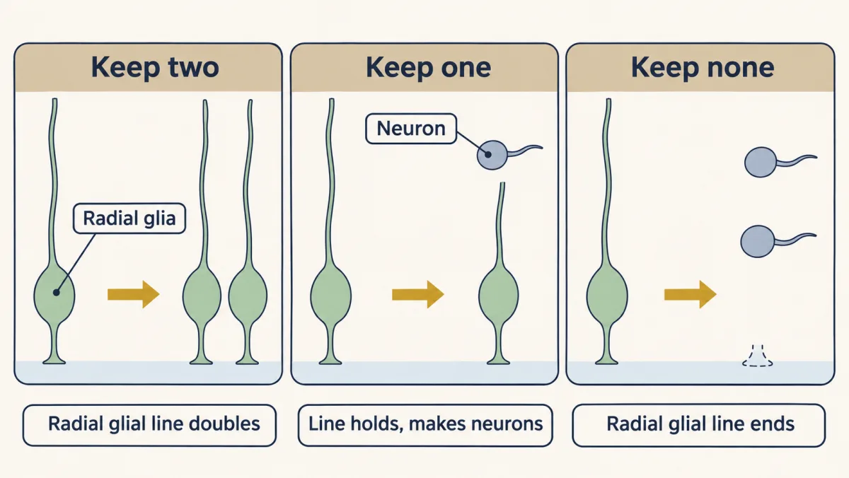
Each radial glial division settles how many daughters stay radial glia. Keep two and the radial glial line doubles; keep one and it holds steady while making neurons; keep none and it ends. Counting those choices round by round sets how many neurons the cortex gets. AI-generated illustration -
The lift: a nucleus on a timetable
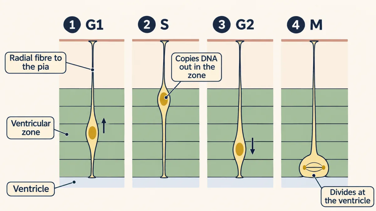
Interkinetic nuclear migration: the radial glial cell keeps its fibre to the pia while its nucleus rides up through the ventricular zone in G1, copies its DNA near the top of the zone in S, comes back down in G2 and divides at the ventricle. AI-generated illustration -
Scaffold or source?
-
Two routes to a neuron
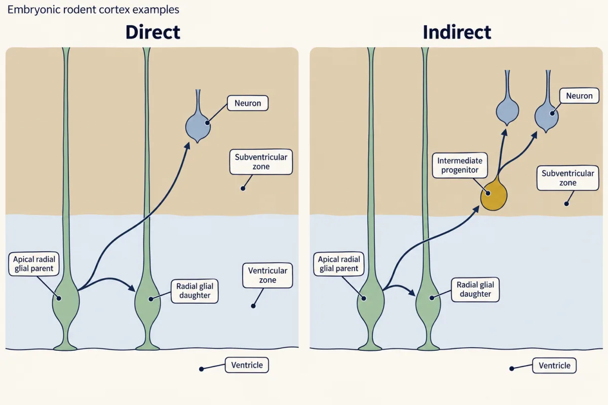
Two routes to a neuron in rodent cortex, shown as example lineages. Direct neurogenesis produces a self-renewing apical radial glial daughter and a neuron. Indirect neurogenesis produces a self-renewing apical radial glial daughter and an intermediate progenitor, which leaves the ventricular surface and divides in the SVZ, usually into two neurons. Intermediate-progenitor yields vary; arrows show ancestry, not migration or relative route frequencies. AI-generated illustration 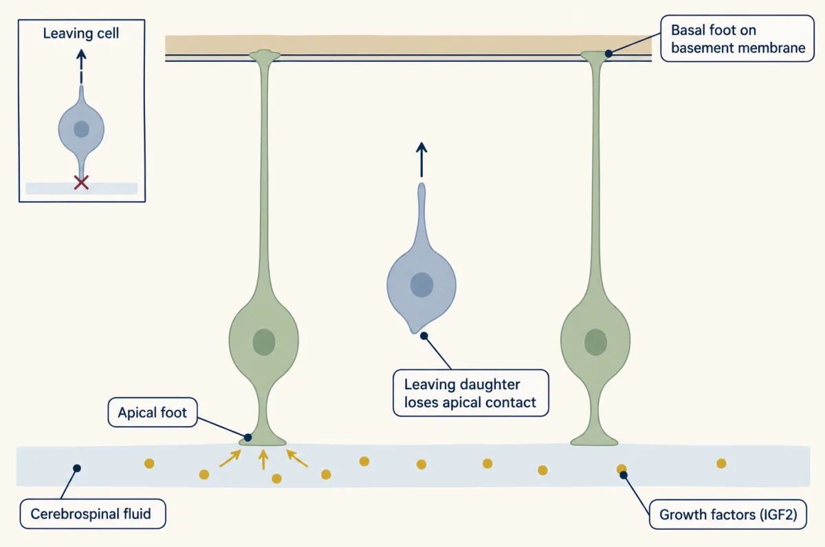
An apical radial glial cell contacts two parts of its niche: cerebrospinal fluid (CSF) at its apical foot and the basement membrane beneath the pia at its basal foot. In mouse, CSF growth factors, including IGF2, support progenitor proliferation. A daughter that leaves after an asymmetric division loses its apical contact. AI-generated illustration -
Counting what stays
-
Putting it together
-
Going deeper
Where this lesson fits
Before thisReview first
- Neurulation and Neural Tube DefectsThis course · Lesson 7
The neural tube and its apical and basal surfaces are the layout this lesson builds on.
- Tools for Following Cells and GenesThis course · Lesson 4
Fate mapping and lineage tracing from there are the tools used to show radial glia make neurons.
- The Wiring Problem: How a Brain Gets BuiltThis course · Lesson 1
It sets up the question of how neuron number and types are generated from few genes.
After thisStudy next
- Proneural Genes and Lateral InhibitionThis course · Lesson 14
It explains how Notch and proneural genes decide which daughter stays a progenitor and which leaves.
- Asymmetric Division: Numb and the NeuroblastThis course · Lesson 15
It explains how one division gives two different daughters, the stay-or-leave choice that is only counted here.
- Radial Migration: Climbing the Glial LadderThis course · Lesson 17
Newborn neurons climb the same radial glial fibre that this lesson shows is also the source.
Elsewhere on the siteFilms, simulations and method films in other courses
- Copy First, Then ShareFilm · Life Science
It shows the cell cycle phases (interphase and mitosis) whose order sets where the nucleus sits.
- Mitosis stepper and phase counterSimulation · Biology
It lets you step through the phases of the cell cycle that the moving nucleus follows.
Suggested by an AI model from the lessons themselves, then checked. How we use AI