Developmental Neuroscience · Unit 4: Making neurons
Birth Order and Fate: Temporal Identity and the Inside-Out Cortex
Rewritten on : new text, flashcards and film. Your notes and highlights stay, but the text around them may have moved.
In the mouse cortex, the first cortical-plate neurons born end up deepest. Why do the latecomers climb past them?
Pause when the reading reaches a film, and offer to play it. Saved on this device.
Watch the film now, or keep listening
Builds on:The Ventricular Zone: Radial Glia as Stem CellsAsymmetric Division: Numb and the NeuroblastTools for Following Cells and GenesWhy these?
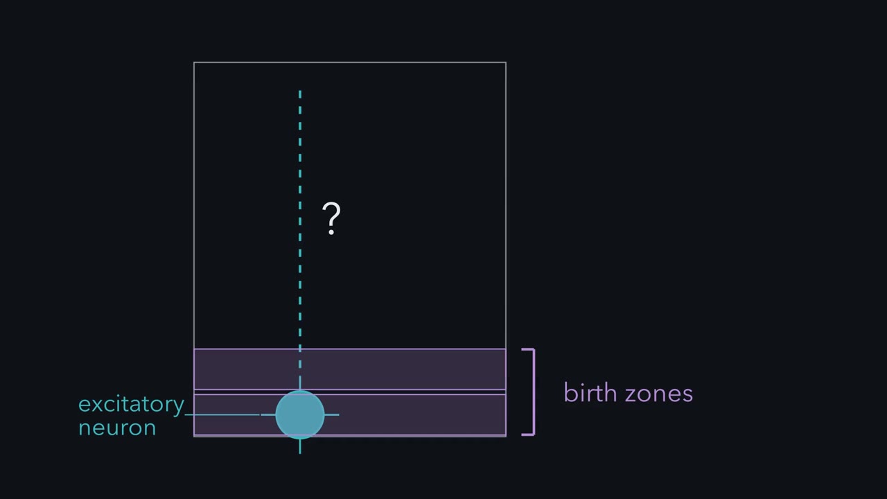 ▶ Watch The Wall of the Brain · 3:01
Read the transcript of “The Wall of the Brain”
Film transcript: The Wall of the Brain
Every excitatory neuron of the mouse cortex is born at the bottom, in the zones beside the ventricle. So how do the layers get in order? Notch and Numb can make two sisters different at a division. But a yes or no alone cannot say which division it was. A stem cell that makes ten different daughters needs more than that. What tells the tenth daughter she is not the first? Picture a slice through the wall of the embryonic mouse forebrain. The ventricle is at the bottom, the pia on top. The ventricular zone lines the ventricle. Many of its progenitors are radial glia. They divide and send a long fibre up to the pia. Just above sits the subventricular zone.
Intermediate progenitors, daughters of radial glia, divide there, usually to make neurons. The first neurons form a thin sheet just under the pia. This is the preplate. The next neurons arrive in the middle of the preplate and wedge it apart. The top half becomes the marginal zone, future layer 1, home of the Cajal-Retzius cells. The bottom half becomes the subplate. Between them, the cortical plate becomes layers 2 to 6. Each new neuron climbs up from below to reach it. Layers are numbered from the surface down, layer 1 on top. In the cortical plate, they are made the other way, from 6 up to 2.
The map raises three puzzles. First, who came first? How can you tell? Second, record or cause? Does the progenitor carry its own schedule, or do its surroundings decide? Third, how many of each? How does one pool make a few neurons, then many, as it first grows and then shrinks?
Here is the one idea to carry. A birthday marks a time. The progenitor's state at that time sets the neuron's temporal identity. The birthday instructs nothing. It is a timestamp. In ferret grafts, laminar fate becomes fixed late in the final cell cycle. Migration and later programs then build the layer. That state has two parts: what the progenitor can still make, its competence, and the cues it reads. In the cortical plate, the earlier the birthday, the deeper the layer. The neuron keeps maturing after it is born. And in normal development, the progenitor's menu gets shorter with time.
The takeaway: cortical-plate neurons are born near the ventricle and climb. Layers 6 to 2 are made in that order.
From the film
- In the cortical plate, the earlier a neuron is born, the deeper its layer.
- A birthday is a timestamp, and the progenitor's state at that time sets the neuron's temporal identity.
- That state has two parts: what the progenitor can still make, and the cues it reads.
- In normal development the progenitor's menu gets shorter with time.
In the mouse cortex, among the excitatory neurons of the cortical plate, the deepest are made first and those nearest the surface are made last. All of them are born below the plate, in the proliferative zones beside the ventricle. So the late neurons must travel past the early ones to reach the top.
That raises two questions, and this lesson answers both. How do you know when a neuron was born? And does its birthday decide what it becomes, or only record when it happened?
You have met one way to make two sisters different: Notch and Numb (see asymmetric division). They can give one daughter one fate and her sister another at a single division. But a yes or no alone does not say which division it was. A stem cell that makes ten different daughters needs more information. Something must tell the tenth daughter that she is not the first.
The layout
Most of the lesson happens on one map, which the first film draws. Picture a thin slice through the wall of the embryonic mouse forebrain, from the fluid-filled ventricle at the bottom to the outer surface, the pia, at the top.
- The ventricular zone lines the ventricle. Many of its progenitors are radial glia, which divide and send a long fibre up to the pia (see the ventricular zone). Some neurons are made there directly.
- The subventricular zone sits just above it. Here intermediate progenitors, daughters of radial glia, divide, usually to make neurons. Many cortical projection neurons arise this way; mouse studies suggest this route may make most of them.
- The cortical plate is where the neurons settle, and it becomes layers 2 to 6. Above it lies the marginal zone (future layer 1, home of the Cajal-Retzius cells) and below it the subplate. Both come from an earlier sheet, the preplate, met again in the next section.
- Each new neuron climbs from the ventricular or subventricular zone up to the cortical plate (that trip is radial migration).
These are the excitatory neurons of the neocortex, the six-layered outer sheet of the mammalian forebrain. Layers are numbered from the surface down: layer 1 on top, layer 6 at the bottom. Keep that in mind, because within the cortical plate layers 6 to 2 are made in the reverse order of their numbers. Layer 1 comes from the early preplate.
The map raises three puzzles.
- Who came first, and how do you know? Every excitatory cortical-plate neuron starts at the bottom, in the ventricular or subventricular zone. How can you tell, weeks later, which layer was made first?
- Record or cause? If a birthday predicts a layer, does the progenitor carry its own schedule, or do the surroundings at the time tell the daughter where to go?
- How many of each? How does one pool of progenitors make a few early neurons, then many later ones, as the pool first grows and then shrinks?
The one idea to carry through this lesson. A birthday marks a time; the progenitor’s state at that time sets the neuron’s temporal identity, and in the cortex that includes its layer. Temporal identity is the part of what a neuron becomes that depends on when it is born. The birthday instructs nothing; it is a timestamp. The state has two parts: what the progenitor can still make (its competence) and the cues it reads. Each system has its own timing: in the ferret cortex, laminar fate becomes fixed late in the progenitor’s final cell cycle; in the fly, a ganglion mother cell inherits the neuroblast’s temporal factors when it buds off, before it divides to make neurons. In the cortical plate, the earlier the birthday, the deeper the layer. The neuron keeps maturing after it is born, so other features are refined later. And in normal development the progenitor’s menu gets shorter with time.
What the rule covers. Excitatory neurons of the mammalian neocortex (mouse, ferret, monkey) and neuroblast lineages of the embryonic fly nerve cord, with interneurons from the medial ganglionic eminence, a ventral birthplace met later, as a partial case. Four limits are flagged where they come up: the preplate sits outside the “earlier, deeper” order; place sets the class of neuron before time acts; cues count as part of the state; and a shrinking menu is the normal course, not a law.
Dating a birthday
Read the transcript of “Dating a Birthday”
Film transcript: Dating a Birthday
Excitatory cortical neurons are born in zones at the bottom of the wall, then climb. So how can you tell, weeks later, when one was born? You date its last S phase, when DNA is copied. That is the final S phase of the cell that divides to make it. Give a pregnant mouse one dose of thymidine carrying tritium, a radioactive form of hydrogen. Only DNA uses thymidine. So only cells copying DNA in the next hour or so are marked. The labeled cell still divides once to make a neuron. Each extra division after that halves the label.
A neuron still heavily labeled weeks later made its last DNA copy while the label was there. The pulse dates its birth. The label marks that S phase. It does not tell the cell anything. Now two models. In model one, each new neuron is added at the bottom and shoves the older ones outward. In model two, each new neuron climbs past the older ones and settles above them. Predict. Pulse on embryonic day 12 then look at postnatal day 21 Where does each model put the heavily labeled cells?
Model one puts them nearest the surface, shoved up by everything added beneath them. Model two puts them deepest. In 1961, Jay Angevine and Richard Sidman gave pulses on different embryonic days. Cells labeled earliest lay deepest, beside the white matter. Cells labeled later lay progressively nearer the surface. Later cells had passed earlier ones. It is a strong tendency, with overlap between days.
Here is the signature evidence, idealized. Heavily labeled cells from an E12 pulse sit mostly in layers 5 and 6. From an E16 pulse, they sit mostly in layers 2 and 3. Lightly labeled cells divided again after the pulse. Born later, at many times, they scatter through many layers. In 1974, Pasko Rakic found the same order in the visual cortex of rhesus monkeys. In the mouse, layers 2 to 6 are made over about six embryonic days. Subplate and layer 6 come first, upper layers last. Then each later wave passes the neurons already in the cortical plate and settles above them.
Notice the exception. The preplate cells are born first, yet they end up both at the top and at the bottom. Earlier means deeper only inside the cortical plate. This is a rule of the neocortex. The retina uses birth order too, but builds its layers another way.
The takeaway: a thymidine pulse dates a birthday, and later-born neurons pass earlier ones to settle above them.
From the film
- A thymidine pulse dates a neuron's birth, because the label marks its last S phase.
- In the neocortex, later-born neurons pass earlier-born ones and settle above them, a strong tendency with overlap between days.
- The preplate is an exception: its cells are born first but end up both at the top and at the bottom.
To date a neuron’s birth, you date its last S phase, the part of the cell cycle when DNA is copied. A neuron never copies DNA again, so its “last S phase” is the final S phase of the cell that divides to make it. Give a pregnant mouse one dose of thymidine carrying tritium, a radioactive form of hydrogen. Only DNA uses thymidine, so only cells copying DNA in the next hour or so are marked. A labeled cell still divides once to make a neuron. A daughter that leaves the cycle at that division keeps the most label, and each extra division halves it.
| Extra divisions before leaving the cycle | Label left in the neuron |
|---|---|
| 0 (left at the first division after the pulse) | full |
| 1 | half |
| 2 | a quarter |
| 3 | an eighth |
The table is idealized: it assumes no label is left for later S phases. A neuron that is heavily labeled weeks later made its last DNA copy while the label was available: the pulse dates its birth and tells the cell nothing. Because the pulse is short, heavy label marks a cohort born around the pulse, not every neuron born that day.
Predict. Two models. (1) Each new neuron is added at the bottom, beneath the ones already made, and shoves them outward. (2) Each new neuron climbs past the ones already made and settles above them. Pulse one litter on embryonic day 12 (E12) and another on E16, and look at postnatal day 21 (P21). Where does each model put the heavily labeled E12 cells?
Check your prediction
Model 1 puts the E12 cells nearest the surface, shoved there by everything added beneath them. Model 2 puts them deepest. In 1961 Jay Angevine and Richard Sidman ran this experiment, pulsing on different embryonic days. Cells labeled earliest lay deepest, beside the white matter, and cells labeled on later days lay progressively nearer the surface. Later cells had passed earlier ones. The rule is a strong tendency, with overlap between days.
This is the signature evidence for “inside-out.” In idealized form, for heavily labeled cells only:
| Layer (surface at top) | Pulse on E12 | Pulse on E16 |
|---|---|---|
| 2 and 3 | few | most |
| 4 | few | few |
| 5 | many | few |
| 6 and subplate | most | few |
Lightly labeled cells, diluted by extra divisions (the lower rows of the first table), are scattered through many layers: they were born later than the pulse, at many times.
The same order was found, in 1974, by Pasko Rakic in the visual cortex of rhesus monkeys. In the mouse, the neurons of layers 2 to 6 are made over about six embryonic days, E11 to E17, and the classes overlap.
Here is what the data look like in the tissue, in three steps.
- Preplate. The first neurons to leave the ventricular zone form a thin sheet under the outer surface.
- Split. The next neurons arrive in the middle of the preplate and wedge it apart. The top half stays under the pia as the marginal zone; the bottom half is pushed down as the subplate. The cortical plate forms between them.
- Inside-out. Each later wave passes the neurons already in the cortical plate and settles above them.
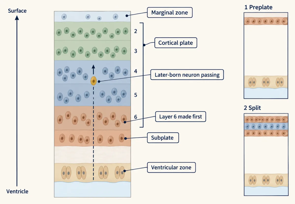
Notice the exception the split creates. The preplate’s cells are born first, yet they end up both at the top, in the marginal zone, and at the bottom, in the subplate. “Earlier, deeper” holds inside the cortical plate.
Inside-out is a rule of the mammalian neocortex, not of the whole nervous system. The retina also uses birth order to bias which cell types its progenitors make, but it does not build its layers by later cells passing earlier ones. How neurons make the climb belongs to radial migration, and what goes wrong in the reeler mouse to Reelin and the reeler mouse. That answers the first puzzle.
Cause or record? Moving a birthday
Read the transcript of “Moving a Birthday”
Film transcript: Moving a Birthday
A birthday predicts a layer. But is the birthday the cause, or only a record? Two explanations are tangled. The progenitor might carry its own schedule. Or the surroundings at the time of birth might tell its daughter where to go. In 1991, Susan McConnell and Christine Kaznowski set the two calendars against each other, in ferrets. Why ferrets? Their visual cortex keeps making neurons after birth. So a newborn kit is a host cortex at the upper-layer stage. They ran the experiment in four steps. First, pulse a donor embryo with tritiated thymidine, at a stage that makes deep-layer neurons. Only cells in S phase take it up. Second, harvest the cells.
Take them out soon after the pulse, or later, in G2, near division. Third, graft them into a newborn kit's cortex, which is making layers 2 and 3. Fourth, find the labeled neurons later. The harvest is the key step. A cell grafted in S phase finishes its cycle in the host. So its daughter is born there. Predict. Where do the daughters settle if the cells were moved in S phase? And if they were moved late in the cycle?
Cells moved in S phase mostly settled in layers 2 and 3, like the host's own neurons. They finished their final cycle in the older host. Its cues could act on them before they divided. Cells moved late in the cycle mostly settled in the deep layers, out of step with the host. Their laminar fate was fixed before the move.
So the grafts bracket commitment. Laminar fate still responds to the host in S phase, and is fixed by a later stage, before division. The exact moment is not known. So the host's cues count as part of the progenitor's state. It is not only a clock sealed inside the cell. Could an old progenitor go back? Run the graft the other way. Predict. Late progenitors go into a young host, which offers deep-layer cues. What do they make?
In 1996, Gretchen Frantz and McConnell did this. The late progenitors still made upper-layer neurons. They had lost deep-layer fates from their menu. So they could not answer the young host's cues. In these ferret experiments, competence narrowed one way. But a 2019 mouse study found that some progenitors moved into a younger host can return to earlier states. The takeaway: the birthday is a record. Schedule or surroundings? Both. Host cues count until laminar fate is fixed late in the cycle. Then the menu usually shortens.
From the film
- A birthday is a record, not a cause.
- In ferret grafts, host cues still act on progenitors moved in S phase, but laminar fate is fixed by a later stage, before division.
- In these ferret experiments late progenitors lost deep-layer fates from their menu, though some mouse progenitors can go back.
Susan McConnell set the second puzzle’s two answers, the progenitor’s own schedule and the surroundings at birth, against each other in ferrets. Their visual cortex keeps making neurons after birth, so a newborn kit is a host cortex at the upper-layer stage, and it can be reached directly.
In 1991 McConnell and Christine Kaznowski ran the experiment in four steps.
| Step | What happens |
|---|---|
| 1. Pulse | Label a donor embryo’s progenitors with tritiated thymidine, at a stage that makes deep-layer neurons. Only cells in S phase take it up. |
| 2. Harvest | Take the cells out either soon after the pulse (most labeled cells still in S phase) or later (labeled cells past S phase, in G2, near division). |
| 3. Graft | Put them into the cortex of an older host, a newborn kit making layers 2 and 3. |
| 4. Read | Later, find the heavily labeled neurons and note their layer. |
The step that matters is the harvest. A cell grafted in S phase finishes its cycle in the host, so its daughter is born there.
Predict. Where do most of the daughters settle if the cells were moved (a) in S phase, and (b) late in the cell cycle?
Check your prediction
(a) Cells moved while in S phase mostly settled in layers 2 and 3, like the host’s own neurons: the host’s cues acted on them before they divided.
(b) Cells moved later in the cycle mostly settled in the deep layers, out of step with the host. Their fate had been fixed before the move.
So the grafts bracket the commitment of laminar fate: not yet made during S phase, made by the later stage, before division. The grafts do not show a single switch, so treat “after S phase, before mitosis” as a working model. A rule that said “the lineage’s own clock decides” would fail result (a).
Could an old progenitor go back? Run the graft the other way.
Predict. Late progenitors, from a stage making upper layers, are moved into a young host making deep layers. A young host offers deep-layer cues. What do the late progenitors make?
Check your prediction
In 1996 Gretchen Frantz and McConnell did this. The late progenitors still made upper-layer neurons. They had lost deep-layer fates from their menu, so they could not answer the young host’s cues. In these ferret experiments competence narrowed one way; some mouse progenitors can go back (Going deeper).
These grafts have limits: they read only the cells that reached the cortex, the cues in the host are only partly known, and a layer position is not a whole identity.
That answers the second puzzle. The birthday is a record, not a cause. And schedule or surroundings? Both: the host’s cues count until the cell commits, and what the progenitor can still make narrows with age.
What a clock is made of: the fly
Read the transcript of “A Clock in the Fly”
Film transcript: A Clock in the Fly
The ferret shows that a progenitor's menu changes with time. But what actually changes inside the cell? The fly asks it in the cleanest form. Each half-segment of its nerve cord has about 30 neuroblasts. Each neuroblast buds off ganglion mother cells, one after another. Each of those divides into neurons or glia. In 2001, Takako Isshiki, Bret Pearson, Scott Holbrook and Chris Doe found a clock of transcription factors. Most neuroblasts switch on four factors in turn. Hunchback, then Krüppel, then Pdm, then Castor. A ganglion mother cell keeps the factor that was on when it budded off. So do its neurons.
The windows overlap, so the sequence is an outline, not a set of sharp steps. One lineage makes this concrete. The neuroblast NB7-1 makes its first five motor neurons, U1 to U5, in that order. In this idealized picture, U1 and U2 come in the Hunchback window, and U3 with Krüppel. U4 comes with Pdm alone. U5 needs Pdm and Castor together. Now read a grid. Rows are place, which neuroblast. Columns are time, which window. The same factor gives a different neuron in a different row. But expression in sequence is only a correlation. To show the factors set identity, take one away, or keep it on too long. Hunchback and Krüppel also pattern the early embryo.
So the tests changed them in the nervous system itself. Without Hunchback in the nervous system, first-born identities were lost in the lineages examined. Predict. Keep Hunchback on long after it would switch off. Do the extra cells look early-born or late-born? And of which lineage?
The lineages made extra cells with first-born features. In NB7-1, extra cells resembled U1 or U2. Each lineage made its own first-born type. Hunchback did not make every first-born cell the same. Prolonged Krüppel made extra second-born cells.
In two thousand six, the same laboratory showed that P D M and Castor specify U four and U five. Birth order had become a genetic program that could be broken and rewired. The takeaway: place picks the class of neuron. Time, read as a cascade of factors, picks the member within it.
From the film
- Most embryonic fly neuroblasts express Hunchback, then Krüppel, then Pdm, then Castor, in overlapping windows.
- Place sets the class of neuron, and time, read as these factors, picks the member.
- Removing or prolonging Hunchback in the nervous system changed first-born identities in the lineages examined.
The ferret shows that a progenitor’s menu changes with time. It does not show what changes. The fly asks the question in its cleanest form. In each half-segment of a Drosophila embryo’s nerve cord there are about 30 neural stem cells, neuroblasts. Each buds off a series of smaller cells, ganglion mother cells, and each of those divides to make neurons or glia. Each neuroblast can be named and found again in every embryo. How do so few make hundreds of neurons and glia of many identities? (Cell number and number of cell types are different counts.)
The answer is a clock of transcription factors. In 2001 Takako Isshiki, Bret Pearson, Scott Holbrook and Chris Doe reported that most neuroblasts express four factors in turn: Hunchback, then Krüppel, then Pdm, then Castor. A ganglion mother cell keeps the factor that was on when it budded off, and so do its neurons. The windows overlap, so the sequence is an outline rather than a set of sharp steps.
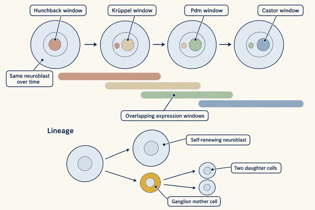
One lineage makes the clock concrete. The neuroblast NB7-1 makes its first five motor neurons, U1 to U5, in that order. Read the row as one place (one neuroblast) and the columns as time (which factors are on); the grid is idealized.
| Neuroblast (place) | Hunchback window | Krüppel window | Pdm, no Castor | Pdm + Castor |
|---|---|---|---|---|
| NB7-1 | U1, U2 | U3 | U4 | U5 |
U5 needs the overlap: Pdm alone goes with U4, Pdm together with Castor with U5. Another neuroblast runs the same windows but makes different neurons in each. Place sets the class; time picks the member.
Expression in sequence is a correlation. To show the factors set identity, take one away or keep it on too long. Hunchback and Krüppel also pattern the early embryo, which complicates tests in the nervous system, so the Doe laboratory changed them in the nervous system itself.
| Hunchback in the nervous system | What the lineages make |
|---|---|
| normal | first-born identities, then later ones, in order |
| removed | first-born identities lost, in the lineages they examined |
| kept on long after it would switch off | ? |
Predict. Fill in the last row. Do the extra cells look like early-born or late-born neurons, and of which lineage?
Check your prediction
With Hunchback kept on, lineages made extra cells with first-born features: in NB7-1, extra cells resembling U1 or U2. Prolonged Krüppel made extra second-born cells. Hunchback did not make every first-born cell the same; each lineage made its own first-born type.
In 2006 the same laboratory showed that Pdm and Castor specify the later U4 and U5 identities in NB7-1. The work turned birth order from a correlation into a genetic program that could be broken and rewired.
How many of each: the Q schedule
Read the transcript of “Grow the Pool, Then Spend It”
Film transcript: Grow the Pool, Then Spend It
One pool of progenitors makes a few early neurons, then many later ones. How? In this simple model, every division gives two daughters. Each daughter either divides again, or leaves the cycle to become a neuron. The fraction that keeps dividing is called P. The fraction that leaves is Q, the leaving fraction. Together they make one. The rules are on screen. The pool after a cycle depends on P. The neurons made depend on Q. Walk 100 progenitors through three cycles, with Q rising each time. Assume no cells die. When Q is 0.3, 100 progenitors make 60 neurons and 140 progenitors. The pool grows. When Q is 0.5, 140 progenitors make 140 of each. The pool holds still.
When Q is 0.7, they make 196 neurons. Only 84 progenitors remain, so the pool shrinks. In 1996, Takao Takahashi, Richard Nowakowski and Verne Caviness measured Q in the mouse cortex. Q was low early. It crossed 0.5 on E14, in the eighth cycle. It rose to about 0.8 by E16. Their model spanned eleven cell cycles. The model covers the ventricular zone only. Intermediate progenitors in the subventricular zone add divisions of their own. Predict. The pool stops growing in the cycle where Q reaches one half. Is that when the most neurons are made?
No. Look back at the table. The next cycle makes more neurons from the same pool. In this rising example, the pool shrinks slowly just past 0.5 while Q climbs, so output rises one more cycle. In general, output rises only if 2 × (1 − this Q) × next Q beats this Q. Later the shrinking pool wins. In the simulation's rising sketch, output peaks in the ninth cycle, the one after Q reached 0.5. About 70 percent of its output comes once Q has reached 0.5. The exact peak depends on how Q rises.
So the cortex first builds its pool, then spends much of it. Neurons spent later go to more superficial layers. The sketch stops at cycle 11 with progenitors left; it does not model how neurogenesis ends. But the model counts how many daughters leave, not which identity they take. Do not read it as layer sizes. Try the simulation below this film. Find the cycle with the most neurons. The takeaway: below 0.5 the pool grows, and above it the pool shrinks. Output can keep rising a cycle after the pool stops growing.
From the film
- In this simple model, the progenitor pool grows when Q is below 0.5, holds at 0.5 and shrinks above 0.5.
- Neuron output can keep rising for a cycle after the pool stops growing.
- The cortex first builds its progenitor pool and then spends much of it, and neurons made later go to more superficial layers.
- The model counts how many daughters leave, not which identity they take, so it does not give layer sizes.
The third puzzle is about numbers. In this simple model, every division gives two daughters. Each daughter either divides again (the proliferative fraction, P) or leaves the cycle to become a neuron (the leaving fraction, Q).
P + Q = 1. Progenitors after one cycle = 2 × P × progenitors before. Neurons made in that cycle = 2 × Q × progenitors before.
Walk 100 progenitors through three cycles, with Q rising each time. Assume no cell death and that every progenitor divides once per cycle.
| Cycle | Q | Progenitors at start | Daughters | Progenitors after | Neurons made |
|---|---|---|---|---|---|
| 1 | 0.3 | 100 | 200 | 140 | 60 |
| 2 | 0.5 | 140 | 280 | 140 | 140 |
| 3 | 0.7 | 140 | 280 | 84 | 196 |
When Q is below 0.5 the pool grows; at 0.5 it holds; above 0.5 it shrinks.
In 1996 Takao Takahashi, Richard Nowakowski and Verne Caviness measured Q in the ventricular epithelium of mouse dorsomedial cortex. Q was low early, crossed 0.5 on E14, in the eighth cycle, and rose to about 0.8 by E16. Their model spanned eleven cell cycles over E11 to E17. It describes the ventricular zone only; intermediate progenitors in the subventricular zone add divisions of their own that the model does not follow.
Predict. The pool stops growing in the cycle where Q reaches 0.5. Is that the cycle in which the most neurons are made?
Check your prediction
No. In the table, the cycle at Q = 0.5 makes 140 neurons, and the next, at Q = 0.7, makes 196 from the same pool of 140. From one cycle to the next, output rises only if 2 × (1 − Q now) × Q next is larger than Q now. Here, just past 0.5 the pool shrinks slowly while Q climbs, so output rises (0.5 to 0.7 gives 1.4 times as many). Later the shrinking pool wins (0.7 to 0.8 gives about 0.69 times as many).
So the cortex first builds its progenitor pool and then spends much of it, and neurons spent later go to more superficial layers. The model counts how many daughters leave, not which identity they take, so do not read its output as the sizes of the layers.
- This cycle
- Progenitor pool after cycle 11
- All neurons made, cycles 1 to 11
P + Q = 1. Below 0.5 the pool grows, above 0.5 it shrinks. The pool is built first and then spent. This is a sketch of the logic: sizes are in units of the starting pool, and only the eleven cycles, the crossing of 0.5 in the eighth and a Q of about 0.8 late come from the lesson's mouse measurements. It assumes no cell death and that every daughter either keeps cycling or leaves.
Try the “Q rising” preset, a sketch with Q of 0.5 in the eighth cycle and 0.8 late, not Takahashi’s fitted curve. Output peaks in cycle 9, and about 70 percent of the eleven cycles’ output comes from cycle 8 on. Now lower only cycle 9’s Q to 0.6: the peak moves to cycle 10, because the peak, like that share, depends on the shape of the curve. Then set every cycle to 0.5 and watch the pool hold still. The sketch ends at cycle 11 with progenitors still left; it does not model how neurogenesis ends. In this two-outcome model the pool would empty only in a cycle with Q = 1, and real progenitors also switch to making glia.
What a birthday leaves behind
Read the transcript of “What a Birthday Leaves Behind”
Film transcript: What a Birthday Leaves Behind
A neuron's layer is one record of its birthday. What else does a birthday leave behind? Each layer's neurons carry identity genes that serve as markers. Markers read identity without knowing position. In mouse, Tbr1 is high in subplate and layer 6 neurons. Ctip2 and Fezf2 are high in layer 5 neurons that project below the cortex. Satb2 marks callosal neurons, which send axons to the other hemisphere. Most sit in the upper layers. Expression overlaps, and position alone does not define a class. The cortical schedule so far describes excitatory neurons made by dorsal progenitors. They release glutamate. Cortical interneurons release GABA. In rodents, most are born in the ganglionic eminences of the ventral forebrain.
They travel up into the cortex. For most cortical neurons in rodents, a dorsal birthplace means excitatory, a ventral one inhibitory. Interneurons from the medial ganglionic eminence follow a similar birthdate rule, settling inside out. Not every interneuron does. One question is still open. Does a mammalian progenitor run a clock like the fly's? Predict. Grow one early progenitor alone in a dish, with no cortex around it. Does it still make neurons in order?
Sally Temple's laboratory did this in 2006. The clones made deep-layer markers before later markers. That points to a schedule within the lineage. Relatives of the fly factors do have timing jobs in mouse. But a full cascade like the fly's has not been shown in cortex. It could be a cascade. Or it could be a gradual change in competence, shaped by feedback from neurons already made.
Now return to the one idea. The progenitor's state at a birthday sets the neuron's temporal identity, its layer included. The pulse dated birthdays. The ferret grafts showed laminar fate is set late, cues included, and that the menu shortens. The fly showed what a temporal state can be made of. And Q counted how one pool is built and then spent.
The takeaway: a birthday is a timestamp. What the progenitor can make then, and the cues it reads, set the neuron's temporal identity.
From the film
- A neuron's layer is one record of its birthday, and identity-gene markers such as Tbr1, Ctip2 and Satb2 are another, though expression overlaps.
- In rodents, for most cortical neurons a dorsal birthplace goes with excitatory neurons and a ventral one with inhibitory interneurons.
- Whether mammalian progenitors run a transcription factor clock like the fly's is still open.
Each layer’s neurons carry identity genes that serve as markers, so identity can be read without knowing position, as the dish experiment below does. In mouse, Tbr1 is high in subplate and layer 6 neurons; Ctip2 and Fezf2 are high in layer 5 neurons that project below the cortex; Satb2 marks callosal neurons, which send axons to the other hemisphere and are mostly in the upper layers. Expression overlaps, and position alone does not define a class.
The schedule in this lesson describes the excitatory, glutamate-releasing neurons made by dorsal progenitors. Most cortical interneurons, which release GABA, are born in rodents in the ganglionic eminences, bulges in the floor of the forebrain (the ventral telencephalon), and travel into the cortex (see tangential migration). This is the other half of “place sets the class”: for most cortical neurons in rodents, a dorsal birthplace goes with excitatory neurons, a ventral one with inhibitory interneurons. Interneurons from the medial ganglionic eminence follow a similar birthdate rule, settling inside-out. Not every interneuron does (Going deeper).
Does a mammal run a clock like the fly’s?
When the laboratory of Sally Temple grew single mouse cortical progenitors alone in culture (Shen and colleagues, 2006), clones made neurons with early, deep-layer markers before neurons with later markers. That points to a schedule within the lineage, which fits the ferret result. Homologs of the fly factors do have temporal jobs in mouse. A full cascade like the fly’s has not been shown in cortex. Whether vertebrate progenitors run a transcription factor cascade, or a gradual change in competence shaped by feedback from the neurons they have already made, is open.
Putting it together
The three puzzles share one answer: the birthday is a timestamp, and the progenitor’s state then sets temporal identity. The thymidine pulse dated the birthdays and showed that later cells pass earlier ones. The ferret grafts showed that laminar fate is set late in the final cycle, cues included, and that the menu shortens. The fly showed what a temporal state can be made of: a cascade of transcription factors you can break or prolong. And Q counted how one pool is built and then spent.
How each new neuron makes its climb past the older ones is the subject of radial migration.
Going deeper
Why only DNA takes up the label
Cells take up thymidine by the salvage pathway, in which thymidine kinase adds the first phosphate (see nucleotide metabolism). Its deoxyribose sugar is why it goes into DNA and not RNA. BrdU and EdU are thymidine stand-ins read by an antibody or by click chemistry; the exact labeling window depends on the label and the dose.
Seven-up times the switch
Another factor, Seven-up, is needed to switch Hunchback off on time (Kanai and colleagues, 2005).
Intermediate-stage grafts
In 2000 A. R. Desai and McConnell found that ferret progenitors at an intermediate stage, making layer 4, could be pushed forward to upper layers but not back to layer 6. Together with Frantz and McConnell’s result, this is the ferret evidence that competence narrows one way.
How some mouse progenitors go back
In mouse, late apical progenitors moved into younger hosts can return to earlier states by sensing Wnt, while intermediate progenitors cannot (Oberst and colleagues, 2019). That is why this lesson calls the shrinking menu the normal course, not a law.
Fly-factor relatives in the mouse
Ikaros, a relative of Hunchback, has a temporal job in the mouse retina. The COUP-TF genes, relatives of the fly’s Seven-up, act in the switch from making neurons to making glia.
Timing the Q schedule
The cell cycle lengthens over the same days, from about 8 hours early to about 18 hours late. That lengthening is how eleven cycles fit the six days from E11 to E17, with the eighth near E14.
Interneurons in transplants, and those that do not follow the rule
In transplants, interneurons from the medial ganglionic eminence take their layer from cues met in their last division, as projection neurons do (Valcanis and Tan, 2003). Interneurons from the caudal ganglionic eminence go mainly to superficial layers, and their layer follows birth date less closely (Miyoshi and colleagues, 2010).
Your notes
Select any text in the lesson to highlight it or add a note to it. Your highlights and notes are saved in this browser and collected in your notebook.
This browser is not letting the site save anything (a private window or strict privacy settings), so highlights and notes will be lost when you leave the page.
How we measure it
Birth-dating with labeled thymidine, BrdU or EdU
Give one pulse of a labeled DNA precursor, wait until the animal is older, and find the heavily labeled cells. Each later division halves the label, so a heavily labeled neuron is one whose last S phase overlapped the pulse. The exact labeling window depends on the label and the dose.
Cumulative and paired labeling to measure the leaving fraction
Label cells in S phase with one marker, then add a second marker shortly afterward and keep it present. Cells with only the first marker are a cohort that has just left S phase. Daughters that re-enter S phase pick up the second marker, and those that do not have left the cycle, which gives the fraction Q.
Heterochronic transplantation
Label progenitors in a donor of one age and place them in a host of another age. Where their daughters settle tells you whether the donor's schedule or the host's surroundings set fate.
Identified lineages with loss and misexpression
In the fly, a single neuroblast and its progeny can be recognized by position and markers. Removing a factor, or keeping it on too long, shows whether it specifies the neurons born in its window.
Practice questions on this lesson (3) → MCAT-style, original, each with an explanation
Where this lesson fits
Before thisReview first
- The Ventricular Zone: Radial Glia as Stem CellsThis course · Lesson 13
The progenitors whose output and state this lesson times are the radial glia and intermediate progenitors introduced there.
- Asymmetric Division: Numb and the NeuroblastThis course · Lesson 15
You need to know how one division gives two sisters different fates before asking what tells later daughters apart.
- Tools for Following Cells and GenesThis course · Lesson 4
Labeling and following cells to learn when and where they were born is assumed when reading the birth-dating experiments.
After thisStudy next
- Radial Migration: Climbing the Glial LadderThis course · Lesson 17
It explains how later-born neurons climb past earlier ones, the movement that builds the inside-out order.
- Reelin and the Reeler MouseThis course · Lesson 18
The reeler mouse shows what happens when the inside-out order fails because migrating neurons cannot stop in the right place.
- Tangential Migration: Interneurons and the Long Way RoundThis course · Lesson 19
It follows the ventrally born interneurons that reach the cortex from the ganglionic eminences.
Elsewhere on the siteFilms, simulations and method films in other courses
- How One Set Becomes TwoFilm · Biology
The thymidine label marks S phase, so knowing the cell cycle's DNA-copying phase makes the dating logic clear.
Suggested by an AI model from the lessons themselves, then checked. How we use AI
Finish line
Test yourself
8 questions
Practice and MCAT-style, with explained answers.
Next · Lesson 17
Radial Migration: Climbing the Glial Ladder
How does a newborn neuron cross a thick, crowded wall to reach its layer, and what brings its nucleus along?
Continue →