Sensory Systems · Unit 4: Proprioception and interoception
Proprioception: Sensing the Body's Own Position
Rewritten on : new text, flashcards and film. Your notes and highlights stay, but the text around them may have moved.
Vibrate a tendon at 100 Hz and a person with their eyes closed misjudges the position of the elbow, by as much as 40 degrees when the muscle is contracting.
Pause when the reading reaches a film, and offer to play it. Saved on this device.
Watch the film now, or keep listening
Builds on:Piezo Channels and the Physics of ForceThe Low-Threshold Mechanoreceptors: One Touch, Several SignalsSensory Neurons by Their Genes: the Atlas EraWhy these?
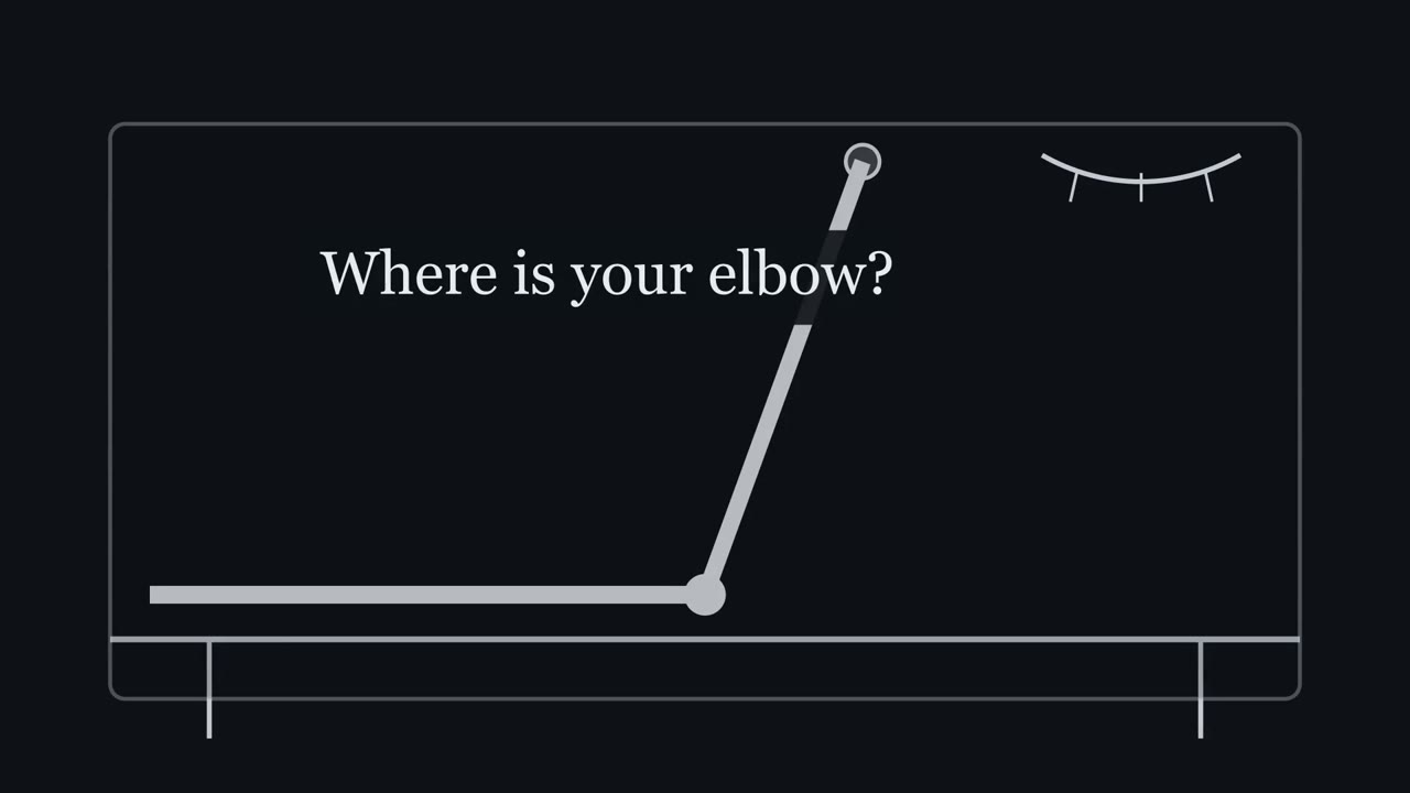 ▶ Watch Read as Stretch · 2:51
Read the transcript of “Read as Stretch”
Film transcript: Read as Stretch
Close your eyes. Where is your elbow? You know without looking. But how? Inside limb muscles lie stretch sensors called muscle spindles. They sit alongside the ordinary fibres. Stretch the muscle, and the spindle stretches too. Its sensory endings fire faster. The primary ending, called type Ia, is especially sensitive to small, rapid stretches. A vibrator on a tendon delivers exactly that. Many tiny stretches, every second. Goodwin, McCloskey and Matthews held a vibrator on the biceps tendon, at 100 Hz. The elbow did not move. The biceps runs in front of the elbow. It gets longer as the arm straightens. Predict. If the brain reads the biceps spindles' firing as biceps stretch, which way should the elbow seem to move?
Toward a straighter arm. Subjects judged the elbow to be where it would be if the vibrated muscle had been stretched. Vibrate the triceps, behind the elbow, and the arm seemed more bent. During contraction, the error could reach 40 degrees in that experiment. It is not a standard size.
A spindle's firing carries no label of its cause. With the arm still and unseen, the brain read it as stretch. The one idea: limb position is an estimate, built mainly from spindle signals of muscle length. Skin, joint and motor-command signals are added to that estimate. So in a still, unseen arm, vibrating one tendon usually biases the estimate toward that muscle being stretched. A typical direction, not a guaranteed response. And when proprioceptive input is severely impaired, accurate movement depends heavily on vision. In touch and pain, capsaicin opened the heat channel TRPV1 on pain fibres. It felt hot, with no heat. Here, a vibrator drives spindle endings while the joint stays still.
One caveat. Primary endings are the likely carriers. In a small leg-muscle sample, they tended to respond more to low-amplitude vibration than secondary endings. But in contracting muscle, secondary endings and tendon organs respond too. So primary endings are not shown to act alone. So vibrating one tendon can make a still limb feel moved. The brain reads the extra spindle firing as stretch.
From the film
- Limb position is an estimate built mainly from muscle spindle signals of muscle length.
- A spindle's firing carries no label of its cause, so vibrating one tendon of a still, unseen limb usually biases the estimate toward that muscle being stretched.
- When proprioceptive input is severely impaired, accurate movement depends heavily on vision.
Close your eyes, and a vibrator pressed on your biceps tendon at 100 Hz can make you misjudge where your elbow is, by as much as 40 degrees when the muscle is contracting, though the joint has not moved.
You know where your elbow is without looking. That knowledge is not a direct reading. It is an estimate, built from sensors you cannot feel, and the right stimulus can fool it.
Motor units and reflexes already covered spindle anatomy, the Ia afferent, the tendon organ, the stretch reflex and gamma drive, and touch and pain mentioned that two people lacking PIEZO2 are poor at sensing limb position. We start at what is left: transduction, the lines that feed position sense, and what losing them costs. For how adaptation is measured, see sensory coding.
The illusion, and the rule it reveals
Start with the experiment in the opening line, and predict before you read the result.
A muscle spindle is a stretch sensor. It lies alongside the ordinary fibres of a muscle, so when the muscle is lengthened the spindle is lengthened too, and its sensory endings fire faster. Its primary (Ia) ending is especially sensitive to small, rapid stretches. A vibrator pressed on a tendon delivers exactly that: many tiny stretches every second.
Goodwin, McCloskey and Matthews (1972) held a vibrator at 100 Hz on the biceps or triceps tendon of human subjects and asked them to judge the angle of the elbow. The elbow did not move.
Predict. The biceps runs in front of the elbow and gets longer as the arm straightens. If the brain takes the biceps spindles’ firing to mean “the biceps is being stretched”, which way should the elbow seem to move?
Check your prediction
Toward a straighter arm. Subjects judged the elbow to be where it would have been if the vibrated muscle had been stretched: more extended with the biceps vibrated, more flexed with the triceps vibrated. During contraction the error could reach 40 degrees in that experiment.
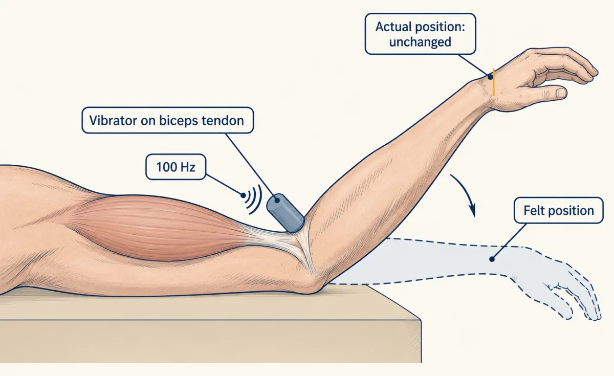
With the arm still and the eyes shut, the brain read the extra biceps firing the way it usually reads it: as stretch.
The one idea to carry through this lesson. Your sense of limb position is an estimate, built mainly from muscle spindle signals of muscle length, with skin, joint and motor-command signals added. A spindle’s firing carries no label saying what caused it. So when one tendon of a still, unseen limb is vibrated, the estimate is usually biased toward that muscle being stretched: a typical direction, not a percept that every rise in spindle firing must produce, since motor state, stimulation history and other inputs matter. And when proprioceptive input is lost, accurate movement comes to depend heavily on vision.
This is the rule from touch and pain, “follow the line”, applied to muscle. There, capsaicin opened the heat-sensitive channel transient receptor potential vanilloid 1 (TRPV1) on pain fibres and felt hot with no heat. Here, a vibrator drives spindle endings while the joint stays still.
Run the rule on three cases, the expected responses in single-tendon trials. In each the subject cannot see the arm; in the first, someone else moves it, and in the other two it stays still. The triceps runs behind the elbow, so it gets longer as the arm bends.
| What happens at the muscle | Which stretch line fires faster | Typical percept in these conditions |
|---|---|---|
| The elbow is straightened by hand | Biceps spindles, because the biceps lengthens | The elbow is extending. Correct. |
| The biceps tendon is vibrated; the elbow stays still | Biceps spindles, driven by the vibrator | The elbow is extending. An illusion. |
| The triceps tendon is vibrated; the elbow stays still | Triceps spindles, driven by the vibrator | The elbow is flexing. An illusion. |
These rows give the usual direction of the bias; an illusion can be absent, or change during prolonged stimulation (Seizova-Cajic and colleagues, 2007). The end of the lesson lists the rule’s other limits.
One caveat on which endings carry the illusion. Primary endings are the likely carriers. With microneurography, a fine electrode recording single nerve fibres in awake people, Roll and Vedel (1982) recorded 15 primary and five secondary endings from tibialis anterior and extensor digitorum longus, and found that primary endings tended to be more sensitive to low-amplitude tendon vibration. That is a tendency, not a clean test. But in contracting muscle the vibration responses of primary, secondary and tendon organ afferents overlap. So the experiment shows that muscle afferent signals bias the percept, not that Ia endings act alone.
The rest of the lesson asks what turns stretch into current on the line, what happens when the line goes silent, and whether anything besides perception depends on it. First, a short account of the lines themselves.
Which lines feed the estimate
A review of the human literature concludes that limb-position information is generated by populations of afferents (incoming sensory fibres) in muscle, skin and joints, not by one receptor, with muscle spindles the main contributors, and that signals from the brain’s motor areas also contribute (Proske and Gandevia, 2012).
Muscle spindles: length, and its rate of change. Inside the spindle’s capsule lie intrafusal fibres, small specialised muscle fibres separate from the ordinary force-producing ones. The sensory endings sit on their central region. A spindle has one primary (Ia) ending and, usually, one or more secondary (II) endings. Both signal static muscle length, and Ia endings add sensitivity to the rate of change of length. So “Ia is velocity and II is position” is wrong: Ia carries both, and II endings respond to the rate of change too, though less. The contractile ends of the intrafusal fibres, the poles, are driven by gamma motor neurons. Picture it: gamma drive contracts the poles, the poles stretch the middle, and the endings there fire more. So gamma drive sets the gain, how strongly the endings respond to a given stretch. Static and dynamic gamma neurons also shift, in different ways, how much the endings respond to held versus changing length.
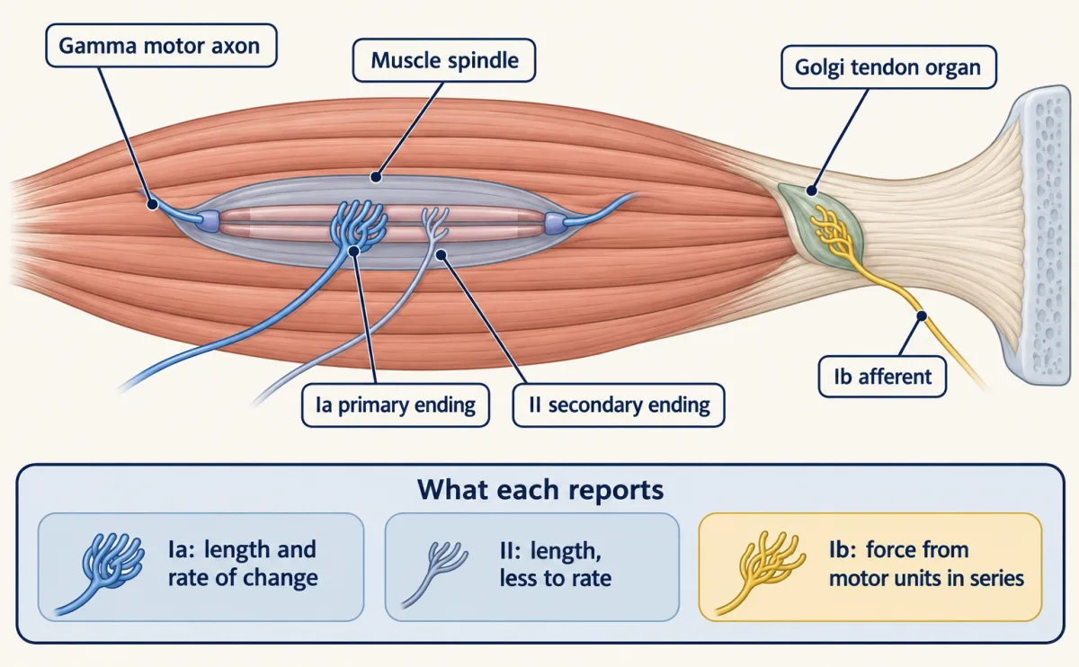
Golgi tendon organs: force. A tendon organ sits at the myotendinous junction and has one Ib afferent. It is highly sensitive to contraction of the motor units in series with it, so it reports force from those units. It is not an injury alarm that waits for dangerous tension.
Joint afferents: mostly the limits. Joint receptors were once credited with signalling joint angle. Burke, Gandevia and Macefield (1988) recorded from the median and ulnar nerves at the wrist of six conscious people while their finger and thumb joints were rotated passively. Most joint afferents fired near the limits of rotation, not across the range. There were exceptions: three tonically active afferents responded throughout the physiological range, and only one, from the thumb interphalangeal joint, responded in a single direction throughout it. Most of the afferents they sampled came from skin, and many skin afferents also fired with joint movement. The authors concluded that joint afferents matter mainly when spindle afferents cannot contribute.
That joint afferents fire mostly near the limits does not confine skin input to the limits. The tendon organ’s force line returns at the end of the lesson.
The transducer: Piezo2 on the stretch line
Read the transcript of “The Channel on the Line”
Film transcript: The Channel on the Line
If the brain trusts the stretch line, what turns stretch into a signal on it? A candidate is Piezo2, a mechanically activated channel. Force opens it. Woo and colleagues tested it in mice, in three ways. First, where it is. A reporter showed Piezo2 in the sensory endings on spindles and tendon organs. Second, what current it carries. Poked cell bodies of labelled neurons gave rapidly adapting currents. Third, does the ending need it? They used Cre driver lines to remove Piezo two in the cells those lines reach, including proprioceptors. Neither driver is specific to proprioceptors. Then they stretched an isolated muscle and recorded from its sensory nerve.
Predict. With Piezo2 gone from these neurons, what happens to stretch-evoked firing? It was sharply reduced, with either Cre line. And the mice moved with severely uncoordinated limbs. Not quite all of it went. In one of eight muscles per line, some stretch response remained. That could be Piezo2 left behind by incomplete deletion. So it is called the principal transducer. The leftover does not prove a second channel. Nor does it rule one out. So Piezo2 is the principal channel on the stretch line. It starts the signal the brain reads as stretch. Now a puzzle. In cultured cell bodies, the current fades with a time constant of 5.6 milliseconds.
Assume one exponential. By 20 milliseconds, only about 3% % is left. Zoom out to one second. The curve lies flat on the floor. Yet a spindle must report a muscle held at a new length for seconds. And Piezo2 is needed for that held response. So how does a channel this fast do it? A cell body in a dish may not behave like an ending in muscle. Other channels may carry part of the held response. And the ending has machinery of its own. Vesicle-released glutamate helps sustain firing in a held stretch. The full pathway is not settled. Piezo2 is the principal transducer on the stretch line. How it supports a held signal is still open.
From the film
- Deleting Piezo2 with two Cre lines, neither specific to proprioceptors, sharply reduced stretch-evoked firing in mouse muscle, so Piezo2 is the principal transducer on the stretch line.
- A small stretch response remained, which may be Piezo2 left by incomplete deletion; it neither proves nor rules out a second channel.
- How a channel whose current fades within milliseconds in cultured cell bodies supports a held stretch is still open.
If the brain trusts the stretch line, what turns stretch into a signal on it? A candidate is Piezo2, a mechanically activated channel, covered in Piezo channels and the physics of force. Woo and colleagues (2015) tested it in mouse.
The key tool is conditional deletion. A Cre driver is a mouse line that makes the enzyme Cre recombinase where a chosen gene is active. Crossed to a mouse whose Piezo2 gene is flanked by Cre target sites (floxed), it removes the gene in the cells the driver reaches, including their descendants. Pvalb-Cre is driven by the parvalbumin gene, which proprioceptors express, along with some other neurons. HoxB8-Cre reaches sensory neurons in the caudal body generally, and also spinal cord neurons and glia, not only proprioceptors. Two drivers with different off-target cells make a shared result more convincing.
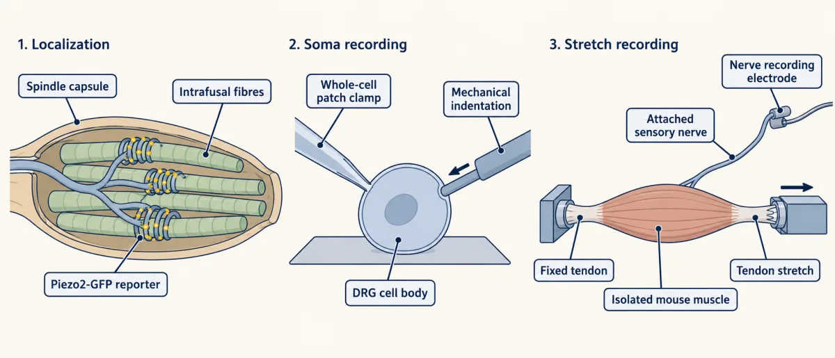
Woo and colleagues supplied three kinds of evidence.
- Where it is. A Piezo2-GFP reporter showed the protein in proprioceptor endings that innervate spindles and tendon organs of postnatal hindlimb muscle.
- What current it carries. Whole-cell patch clamp with mechanical indentation of dissociated adult dorsal root ganglion (DRG) cell bodies, labelled with the parvalbumin-driven Cre line, recorded rapidly adapting currents.
- Whether the ending needs it. They deleted Piezo2 with Pvalb-Cre or with HoxB8-Cre, then stretched an isolated extensor digitorum longus muscle, with its deep peroneal nerve, from adult male mice and recorded its sensory fibres.
Predict. If Piezo2 is the channel that turns stretch into current at the ending, what should its deletion do to stretch-evoked firing? And what should the mice look like?
Check your prediction
Stretch-evoked firing was sharply reduced, with either Cre line. Both lines of mice also moved with severely uncoordinated limbs and abnormal limb positions from about one week after birth.
A small stretch response remained (one of eight muscles for each Cre line still increased its firing on stretch, mostly in atypical ways), which the authors noted could reflect Piezo2 left behind by incomplete deletion. So the careful word is principal transducer: the residue does not prove a second channel, and it does not rule one out.
Worked problem: the held-stretch puzzle
In the cell-body recordings, 23 of 25 labelled neurons had rapidly adapting mechanically activated currents, and the inactivation time constant of those rapidly adapting currents was 5.6 ± 0.33 ms, as reported.
Step 1, a fraction. 23/25 = 0.92, so 92% of this sample of 25. Parvalbumin labels proprioceptors and some other neurons, so this is not 92% of proprioceptors.
Step 2, a decay. Assume the current decays as a single exponential from its peak with tau = 5.6 ms. That is our assumption, not the paper’s. The fraction left at time t is exp(-t/tau).
fraction remaining = exp(-t / tau)
At 10 ms, exp(-10/5.6) = exp(-1.786) = 0.168, about 17%. At 20 ms, exp(-20/5.6) = exp(-3.571) = 0.028, about 3%.
- Fraction of peak remaining
- 0.168
- As a percent of the peak
- about 17%
- Arithmetic
- exp(−10.0/5.6) = exp(−1.786) = 0.168
In this sketch: one exponential from the peak, τ from Pvalb-labelled adult mouse DRG cell bodies in culture (Woo and colleagues, 2015), no rise phase, no spikes, no ending.
Rule to keep: fraction remaining = exp(−t/τ), and it is a current in cultured cell bodies under indentation, not a spindle afferent firing spikes. The 25 dots are a count in one sample, not part of the curve.
Predict. A spindle has to report a muscle held at a new length for seconds. Push the same equation to one second. What is left of the current, and what does that tell you?
Check your prediction
At 1 s, exp(-1000/5.6) is about 10−78: effectively nothing. A current like this one, on its own, could not carry a signal through a held stretch.
Step 3, the puzzle. Yet Piezo2 is needed for exactly that kind of signal. In Woo’s muscle recordings, the stretch response was the firing rate while the stretch was held, minus the baseline, and deletion sharply reduced it. So a channel whose current is almost gone 20 ms after a poke in a cultured cell body is needed by an ending that reports held length. How?
Part of the answer is in what the measurement is. This is a current in a cell body in culture, not a current at a spindle ending. It says how fast the channel population inactivates under indentation, not how a spindle afferent adapts when it fires spikes. Beyond that, other channels may carry part of the held response, and the ending has machinery of its own that tunes its excitability. Some of that machinery has been tested directly. In adult mouse muscle-nerve preparations, added glutamate raised firing during held stretch, while reducing vesicular glutamate packaging, by drug or with one copy of the VGLUT1 gene, impaired it (Than and colleagues, 2021). Voltage-gated sodium channels in the afferent also support firing during the hold (Espino and colleagues, 2022). So vesicle-released glutamate has experimental support as a contributor to the held response, but the full pathway from Piezo2 activation to sustained firing, and how much each piece matters, is not settled.
Take the line away: people
Read the transcript of “When the Line Goes Quiet”
Film transcript: When the Line Goes Quiet
What happens to a person when proprioceptive input is lost? Picture reaching for a cup in a lit room, with your stretch lines silent. Predict. What would you have to rely on to know where your arm is? Mainly your eyes. Vision does much of the work. Take it away too, and reaching becomes far less accurate.
Chesler and colleagues reported two unrelated young people with inactivating variants in both copies of PIEZO2. Reaching was tested with and without sight. Without vision, their uncoordinated movements and misjudged reaches became markedly worse. Yet they could still walk, talk and write, though one walked only with support. An older case shows the daily cost. A man known as I.W. had lost large-fibre sensation below the neck. He lacked light touch and position sense. But he kept pain, temperature and the sense of muscle fatigue. In testing, he could make no new movement without watching it. Every new movement had to be watched. Both cases show the rule's second half.
With proprioceptive input lost, accurate movement depends heavily on vision. Neither case silences spindles alone; touch was affected too. In mice, the line turned out to matter for the skeleton too. Deleting Piezo2 with a parvalbumin driver curved the spine in some mice by postnatal day 60. Deleting it in bone and cartilage lineages did not curve the spine, at the ages tested. The authors read this as non-autonomous. The curve shows in the skeleton, but the gene was lost in parvalbumin-labelled neurons. How the signal reaches the skeleton is not known. One limit to remember. Near a joint's limits, and for the feeling of effort, the stretch rule is not the whole story.
So limb position is an estimate built mainly from the stretch line. Lose that input, and vision does much of its work.
From the film
- When proprioceptive input is lost, vision does much of the work of knowing where the arm is, and without sight reaching becomes far less accurate.
- Two people with inactivating PIEZO2 variants and a man without large-fibre sensation show this, though neither case silences spindles alone.
- In mice, deleting Piezo2 with a parvalbumin driver curved the spine in some animals by postnatal day sixty, and how the signal reaches the skeleton is not known.
Mice can show which gene the line needs. Only people can show what losing it is like.
Predict. Picture reaching for a cup in a lit room, with your stretch lines silent. What would you have to rely on to know where your arm is? And what happens when that source is taken away too?
Check your prediction
Mainly the eyes. With the stretch lines silent, vision has to do much of the work of position sense. Take vision away as well, and reaching becomes far less accurate; for one man, new movements became impossible. Two kinds of patient show this.
People without PIEZO2. Chesler and colleagues (2016) reported two unrelated females with compound inactivating PIEZO2 variants, who presented at 18 and 8 years and were last examined at 19 and 10. Both had congenital hip dysplasia (a hip joint malformed from birth) and progressive scoliosis, and tendon reflexes were absent (except one brachioradialis reflex in the younger patient). The investigators tested the sense of direction with 10-degree angular joint movements in a forced-choice task, and tested reaching with and without sight. Proprioceptive ataxia, uncoordinated movement, and dysmetria, misjudging the distance a reach should cover, became markedly worse without visual cues. Their report says they could still perform tasks such as walking, talking and writing, though one walked only with support. Two people are a clinical series, not a prevalence. The human data show that loss of the gene goes with the deficit, and they do not show which endings account for each part.
A man without large-fibre sensation. An older case shows what such loss means day to day. Cole and Sedgwick (1992) studied an adult man, known as I.W., with a complete loss of large-fibre sensation below the neck that had lasted more than 16 years. He lacked light touch and proprioception below the neck but kept pain, heat, cold and the sense of muscular fatigue. In their testing no novel movement was possible without visual feedback, though he could hold a posture or continue a simple repetitive movement. That separates sensory loss from motor loss.
Their main test was of force. With vision and movement allowed he told weights apart almost as well as controls, 20 g in 200 g, but with eyes shut he could tell only 200 g from 400 g. The authors concluded that a crude sense of effort remained.
Both kinds of patient show the rule’s second half in the clinic. Neither case is a selective silencing of spindles, since PIEZO2 loss also affects touch and I.W. lost all large-fibre input.
What else the line is for: the skeleton
The rule is about perception. Losing proprioceptor Piezo2 turned out to matter for something else as well.
First, a term. An effect is non-autonomous when the defect appears in one tissue (here, the skeleton) but comes from losing the gene in another (here, parvalbumin-labelled sensory neurons, which include proprioceptors). An autonomous effect would come from losing the gene inside the bone or cartilage cells themselves.
If a muscle sensor is removed, would the spine notice? The same lab had already shown (Blecher and colleagues, 2017) that mice lacking proprioceptive neurons, Runx3 mutants, develop scoliosis, and that mice lacking spindles, Egr3 mutants, have a milder form. Assaraf and colleagues (2020) then asked about the channel. They used Pvalb-Cre to delete Piezo2 in mice. Serial in vivo CT at postnatal days 40, 60 and 90 (P40, P60, P90) showed scoliosis by P60 in some of these animals, and hip histology showed signs of dysplasia by P14. Spine histology at P10 showed no apparent differences from controls in bone, cartilage or surrounding soft tissue.
Predict. To ask whether the curve comes from the neurons or from the skeleton itself, the authors also deleted Piezo2 with Col1a1-Cre or Col2a1-Cre, which target the bone-forming (osteogenic) and cartilage-forming (chondrogenic) lineages. If the effect is non-autonomous, what should those deletions do?
Check your prediction
They should not reproduce the spinal curve, and they did not, at the ages tested. The authors interpreted the results as a non-autonomous role: proprioceptive Piezo2 acts on the skeleton through a route that does not need Piezo2 inside the bone cells tested.
In the same paper, Runx3 and Egr3 mutants also had hip abnormalities, and their spines curved in an S shape, while the Piezo2 deletion gave a single C-shaped curve.
What the paper does not show is the intervening pathway. The human finding came first: Chesler’s two patients had both, and those authors proposed that prenatal loss of proprioception caused them. Human cases show association, not the causal tissue. Parvalbumin-driven Cre also labels cells that expressed parvalbumin at some time, and not only proprioceptors.
Design problem
Given the Assaraf results above, design experiments that decide whether the skeletal phenotype needs proprioceptive feedback in a postnatal window or continuously in adults.
Saved in this browser only.
One way to do it
Timing. Use an inducible deletion of Piezo2 in proprioceptors and open it at two times: early, before alignment is established, and later, after it. Image the same animals with serial CT at matched ages. If only the early deletion gives scoliosis, and the late deletion is shown to be as complete and is followed as long, a postnatal window is needed. If the late deletion also curves the spine, feedback is still needed after alignment is set; deletions at more ages, or reversible silencing, would show whether the need is continuous.
Channel versus cell. Piezo2 deletion, Runx3 loss of proprioceptive neurons and Egr3 loss of spindles give differently shaped curves. To ask why, compare Piezo2 deletion with a generic reversible silencing of the same neurons and with loss of the proprioceptor population, scored by the same CT measures. First verify that silencing abolishes firing, and match the timing and targeted population across manipulations. A difference between deletion and verified complete silencing would then point to transduction through other channels, or to incomplete deletion. If cell loss has effects beyond verified complete silencing, test roles of the cells beyond firing, such as structural or trophic ones. If silencing proves incomplete, resolve that before interpreting the difference.
Controls and scope. Use littermate controls and a second, independent driver, since one Cre line is not a pure type. Keep the ages tested in the claim.
Limit. Even a clean answer shows the requirement in the mouse, not the causal tissue in human families.
Where the idea stops
- It covers position and movement, not force and heaviness. Tendon organs report force, and a sense of effort that comes with the motor command contributes too, as I.W.’s weight test showed.
- Near the limits of a joint’s range, it is not the whole story. Proske (2023) proposed that near elbow extension, joint signals combine with spindle signals to carry joint-limit information. Treat that as a hypothesis.
- It predicts the usual direction of an illusion, not its size. The 40 degrees was the error during contraction in one experiment, not a standard size.
- It is stated for one vibrated muscle. Vibrating opposing muscles together can largely cancel the illusion (Gilhodes and colleagues, 1986), which suggests the estimate weighs opposing muscles against each other.
- It does not explain the skeleton. The spinal phenotype is a separate consequence of losing proprioceptor Piezo2.
Going deeper
How vibration frequency shapes the illusion
In Roll and Vedel’s (1982) elbow illusions, the perceived velocity of movement rose up to about 70 to 80 Hz in a 10 to 120 Hz series before falling. Those illusions and their leg-muscle recordings come from different limbs and were not recorded together.
Spindle endings are not passive
Bewick, Reid, Richardson and Banks (2005) worked in adult rat hindpaw lumbrical nerve-muscle preparations ex vivo. The dye FM1-43 was taken up and released at primary endings in a calcium-sensitive way, which indicated recycling of synaptic-like vesicles about 50 nm across. Added glutamate raised the excitability of Ia endings to stretch, and the compound PCCG-13 lowered it even with no glutamate added. The authors interpreted this as an endogenous peripheral glutamatergic system. The pharmacology did not establish the receptor (the authors suggested a phospholipase D-linked metabotropic one), and it was done in rat. Its role in the held response was tested later in mouse (see the worked problem).
Candidates for the residual current
Later reviews name DEG/epithelial sodium channel (ENaC) channels, including ASIC subunits found in spindle endings, as candidates for any current beyond Piezo2’s, but their role is not settled.
More families with PIEZO2 loss
Delle Vedove and colleagues (2016) described ten affected people in four consanguineous families, with homozygous variants in each family (three frameshifts and one two-exon deletion), and a picture that included arthrogryposis, muscular atrophy, perinatal respiratory distress and scoliosis.
Eight groups, or five: proprioceptor atlases and their disagreement
The classical picture has three classes: Ia, II and Ib. In 2021 two papers asked what single-cell RNA sequencing finds (for the method, see sequencing and genomes).
Wu and colleagues (Nature Communications 12:1026) sorted proprioceptors from the C5 to T1 dorsal root ganglia of adult mice (postnatal day 54, PV-Cre crossed to Ai14), sequenced them by Smart-seq2, kept 1,109 neurons after quality control and reported eight molecular subgroups. The analysis first found seven clusters, and one heterogeneous cluster was subdivided to give eight. Oliver and colleagues (Nature Communications, Molecular correlates of muscle spindle and Golgi tendon organ afferents) sampled neurons labelled by an intersection of PV-Cre and Runx3-FlpO, at all segmental levels in adults of at least P56, and reported five clusters. They validated Ia (calretinin, calbindin 1, vGlut2) and Ib (Pcdh8, Pou4f3) by markers and tissue, and assigned the rest to group II only provisionally, proposing Nxph1 as a possible group II marker that needs more validation.
Do the counts conflict? Not necessarily. They differ in labelling, age range, sampling depth and how clusters were cut. Neither is a human atlas. A cluster is a molecular class, not a proven function, and the number of groups depends on the data set and the method. The practical lesson is to cite the paper with the count. Related atlases are in the sensory neuron atlas; for how proprioceptors depend on neurotrophin signalling in development, see Trk and p75.
Wu and colleagues also tested what experience does. Adult C57BL/6J males started four weeks of free-wheel or locked-wheel housing at eight weeks of age. Running changed the marker-defined composition of Ia subtypes, but the total number of brachial Ia proprioceptors was unchanged. That could be marker regulation, conversion of subtype, or something else, and lineage tracing would be needed to choose among them.
Your notes
Select any text in the lesson to highlight it or add a note to it. Your highlights and notes are saved in this browser and collected in your notebook.
This browser is not letting the site save anything (a private window or strict privacy settings), so highlights and notes will be lost when you leave the page.
How we measure it
Microneurography
A fine electrode is inserted through the skin into a peripheral nerve of an awake person and records the spikes of single afferents while the limb is moved or the muscle is vibrated. It gives the firing of identified endings in humans but samples few units, from accessible nerves.
Tendon-vibration psychophysics
A vibrator is held against a tendon and the person reports or matches where the limb seems to be. A systematic error in the direction expected from stretching the muscle shows that muscle afferent signals bias the percept, but it does not by itself show which ending class did it.
Conditional gene deletion with ex vivo muscle-nerve recording
A Cre driver removes a floxed gene in the cells it reaches, and an isolated muscle with its nerve is stretched while sensory firing is recorded. It tests whether the gene is needed for the ending's response, only in the cells the driver reached and at the ages tested.
Single-cell RNA sequencing of labelled proprioceptors
Genetically labelled neurons are dissociated, sorted and their transcripts read cell by cell. Clustering then groups neurons by expression. A cluster is a molecular class, and it becomes a functional type only when anatomy, physiology or behaviour is tied to it.
Practice questions on this lesson (3) → MCAT-style, original, each with an explanation
Where this lesson fits
Before thisReview first
- Piezo Channels and the Physics of ForceThis course · Lesson 2
You need how Piezo channels turn force into current to follow the Piezo2 transducer argument here.
- The Low-Threshold Mechanoreceptors: One Touch, Several SignalsThis course · Lesson 4
This lesson builds on reading what a sensory line signals, as touch neuron classes do for skin.
- Sensory Neurons by Their Genes: the Atlas EraThis course · Lesson 3
The proprioceptor atlas disagreement here assumes you know how single-cell clusters define sensory neuron groups.
After thisStudy next
- Vagal Sensory Neurons: The Body Reports to the BrainThis course · Lesson 16
It extends the study of Piezo-dependent body sensing from limb position to internal organs.
- Balance: Two Codes for Every Head MovementThis course · Lesson 25
It takes the same body-position estimate further, with balance signals combined with other senses.
- Combining Senses and Predicting ThemThis course · Lesson 29
It builds on the vibration illusion and vision taking over when proprioception is lost, as senses are weighted.
Elsewhere on the siteFilms, simulations and method films in other courses
- Where Every Command EndsFilm · Neuroscience
It shows how the stretch signal from the spindle drives reflex action on motor neurons.
- A Predictor That Learns from ErrorFilm · Neuroscience
It shows how movement prediction uses sensory feedback, and what clumsiness follows when that estimate fails.
- Every Response Has a Running TimeFilm · Organismal Biology
It follows the stretch-reflex route from receptor through sensory neuron to effector that carries the spindle signal.
Suggested by an AI model from the lessons themselves, then checked. How we use AI
Finish line
Test yourself
8 questions
Practice and MCAT-style, with explained answers.
Next · Lesson 16
Vagal Sensory Neurons: The Body Reports to the Brain
Light on one set of vagal sensory axons stops an anaesthetized mouse breathing, and light on another set makes it breathe fast and shallow. What does each set report, and how would you know?
Continue →