Sensory Systems · Unit 6: Hearing and balance
Balance: Two Codes for Every Head Movement
Rewritten on : new text, flashcards and film. Your notes and highlights stay, but the text around them may have moved.
Under review Parts of this lesson are still being checked.
- The direction of the plasticity switch reported by Rowan and colleagues (2018) is being checked; Going deeper states the in-vivo result as a decrease in VOR gain.
The text stays up while the check is done. If you spot a problem, report it. All lessons under review.
People with severe loss in both balance organs can see the world bounce as they walk. Yet in monkeys, head-motion neurons go largely quiet for a turn the animal makes itself, which may keep the head-steadying reflex from fighting it. How does one nerve report motion fast and precisely, yet let the brain set your own turns aside?
Pause when the reading reaches a film, and offer to play it. Saved on this device.
Watch the film now, or keep listening
Builds on:The Hair Cell's Transduction ChannelThe Cochlear Amplifier and Otoacoustic EmissionsFive Ways to Turn a Stimulus into a CurrentWhy these?
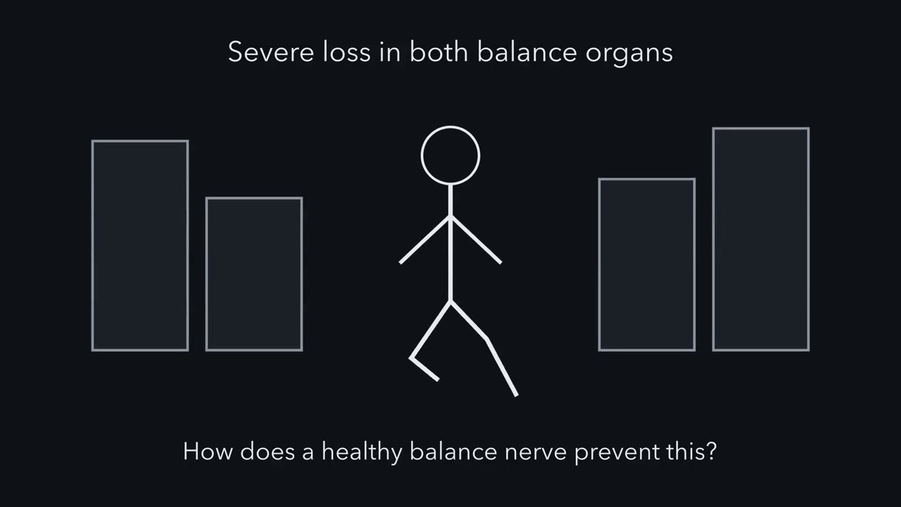 ▶ Watch Report It Twice · 2:57
Read the transcript of “Report It Twice”
Film transcript: Report It Twice
People with severe loss in both balance organs can see the world bounce as they walk. How does a healthy balance nerve prevent that? Canals sense rotation; otoliths sense linear acceleration and tilt. Each organ sends both regular and irregular fibres. From here on, we follow canal fibres and head rotation. With the head still, both fire fast. A regular fibre ticks like a clock. An irregular one fires with uneven gaps. Now a slow head turn. The regular fibre's rate rises and falls with head velocity, how fast the head is moving. Now a sudden change. The irregular fibre, with its higher gain, responds more strongly, and its spikes lock to the event.
Yet regular fibres detect small motions well. How can the lower-gain fibre win? Pause and predict. One made-up fibre has three times the gain, but ten times the CV. Which detects a small, slow turn first? A change in rate counts only if it stands out from the noise. For our made-up counting decoder, threshold is noise divided by gain. The quiet, low-gain fibre wins. Its threshold comes out lower. If our counting decoder must report within a tenth of a second, both thresholds rise, and the regular fibre's lead shrinks. So what is the irregular fibre good for? Replay the same jittery head motion many times. Pause and predict. If irregular spikes were just noise, could their timing tell one motion from another?
Their precisely timed spikes told different motions apart. Irregular at rest, precise when it matters. Neither channel keeps reporting a steady spin. In a long turn at constant speed, the canal signal fades in both. So here is the one idea. The nerve reports head motion on two overlapping channels, whoever caused it. In the canals, regular fibres follow how fast the head is turning, in a steady, precise rate. Irregular fibres add higher gain, more response to how quickly the turn changes, and well-timed spikes. In the head-posture neurons tested, your own turn is largely cancelled, when neck feedback matches the prediction. Fast, precise reports are thought to help hold the world still.
Muting matched, self-made turns in the tested neurons may help the head reflex spare the turns you intend.
From the film
- The vestibular nerve reports each head movement on two overlapping channels, whoever caused it.
- In the canals, regular fibres follow how fast the head is turning in a steady rate, while irregular fibres add higher gain, more response to how quickly the motion changes, and well-timed spikes.
- In the head-posture neurons tested, your own turn is largely cancelled when neck feedback matches the prediction, which may help the head reflex spare the turns you intend.
People with severe loss in both balance organs, for example from an antibiotic that damages hair cells, can find that the world blurs or jumps as they walk, and in the dark they often become unsteady and can fall. Their vestibular input to the vestibulo-ocular reflex (VOR) is greatly reduced; when gaze is held on a target, this reflex turns the eyes against head rotation to hold the image still. Healthy balance organs report each head movement fast enough for the eyes and neck to counter it. Yet a different reflex, the one that steadies the head on the neck, would fight every turn you make yourself if it took the turn for a disturbance. How does one nerve report motion both precisely and quickly, and how does the brain set your own turns aside?
This lesson answers the two puzzles in turn. The first is about speed and precision, and the nerve answers it with two overlapping kinds of signal. The second is about authorship: the nerve reports a movement, but something must decide whether you made it.
Hearing and balance gave you the canals, which sense rotation and over ordinary movements report roughly the head’s rotational velocity, the otoliths, which sense linear acceleration and tilt, and the three-neuron reflex arc. This lesson starts one step later, at the nerve fibres (afferents) that leave those organs, and one step further in, at the first central neurons that read them.
Two channels for one head movement
The afferents carry head motion to the brain, where it drives the reflexes that keep vision and posture steady and feeds the sense of heading (Eatock, 2025). They can be split two ways. The split by organ tells you what a fibre reports. The split by regularity, which this lesson is about, tells you how it reports it, and every organ sends both kinds.
In mammals both kinds fire at a high rate with the head still, so each can signal motion in either direction by speeding up or slowing down. Regularity at rest is measured by the coefficient of variation (CV) of the intervals between spikes: roughly a few per cent for the most regular fibres, tens of per cent for the most irregular. Regularity is a continuum, and “regular” and “irregular” name its two ends. One more term: a fibre’s gain is how much its rate changes for a given motion.
| What the organ senses | Regular afferents | Irregular afferents | |
|---|---|---|---|
| Canals | rotation of the head | rate follows angular velocity, closely over everyday turns | higher gain; respond more to how quickly the rotation changes; precisely timed spikes |
| Otoliths | linear acceleration and tilt | rate follows linear acceleration, including gravity’s pull in a held tilt | higher gain; respond more to rapid changes; adapt faster to a held tilt |
These are overlapping tendencies of two populations, not two exact channels.
The signature: two spike trains
Picture one canal fibre of each kind side by side through four conditions.
| Regular afferent | Irregular afferent | |
|---|---|---|
| Head still | fires like a clock | a similar high rate, with uneven gaps |
| Slow head turn | rate rises and falls smoothly with head velocity | rate changes more for the same turn (higher gain) |
| Fast change in motion | keeps following velocity | responds more strongly, with spikes that lock to the fast event |
| Long turn at steady speed | signal fades, with the canal | signal fades, and it adapts faster |
Read down the columns. Most of a regular canal fibre’s information is in its rate. An irregular fibre favours higher frequencies: as movements get faster it responds more and more to how quickly the velocity is changing, roughly the head’s acceleration, and it carries more information in the precise timing of its spikes (Eatock, 2018; Lacquaniti and colleagues, 2023).
The fading in a long turn belongs to the canals, as Hearing and balance showed. A held tilt is different: it changes how gravity pulls along an otolith fibre’s preferred direction, so regular otolith fibres keep reporting it for as long as it lasts.
A paradox: lower gain, better detection
Here is something that should puzzle you. In the canals, regular fibres have lower gain, yet recordings show they detect small rotations better.
Predict. Take two made-up fibres, each at rest near 100 spikes per second. The regular one has a gain of 0.5 spikes per second per degree per second, and a CV of 0.05. The irregular one has three times the gain, 1.5, but ten times the CV, 0.5. Which detects a slow, small head turn first? One hint: a rate measured over a stretch of time wobbles, and the wobble is bigger for a fibre with a bigger CV.
Check your prediction
A change in rate is useful only if it stands out from the rate’s own wobble. For a simple made-up decoder that counts spikes in a window, call the detection threshold the turn that shifts the mean rate by one standard deviation of that wobble: threshold = noise / gain. A rough rule for long windows puts the rate’s standard deviation at √(CV² × rate / time).
- Regular, over 1 second, by the rough rule: √(0.05² × 100 / 1) = 0.5 spikes per second of noise, so the threshold is 0.5 / 0.5 = 1 degree per second.
- Irregular, over 1 second: √(0.5² × 100 / 1) = 5 spikes per second of noise, so the threshold is 5 / 1.5 = about 3.3 degrees per second.
The quiet, low-gain fibre wins, because its noise is far smaller and its gain only three times smaller. For so regular a fibre the rough rule is already off at 1 second: counted exactly, for model spike trains and a window placed at a random moment, the noise is about 0.65 against 5.0 spikes per second, and the thresholds are about 1.3 against 3.3 degrees per second. The ranking holds, with the regular fibre ahead about 2.6-fold.
Now suppose our counting decoder must report a brief change within 0.1 second, so its window shrinks to 0.1 second and holds only about ten spikes. Counted exactly, the thresholds rise to about 7.1 and 10.9 degrees per second, and the regular fibre’s lead shrinks to about 1.5-fold. In this toy, a fast local rate estimate is much less reliable, which leaves room for the irregular fibre’s other strength; other decoding strategies, and real afferents’ frequency-dependent gains, can shift the trade-off.
Irregular at rest, precise when it matters
“Irregular” describes a fibre with nothing to report. Give it something fast and the picture changes. Researchers played the same noisy head motion, roughly like natural movement, again and again, and compared the spike trains from each repeat. A fibre whose spikes are locked to the motion fires at nearly the same moments on every repeat; one whose spikes are pure noise does not. Here information means how well a spike train tells one motion from another.
Predict. If an irregular fibre’s spikes were just noise, could their timing tell one motion waveform from another?
Check your prediction
It could not: their timing would carry nothing about the motion. Instead, an irregular afferent’s precisely timed spikes told different waveforms apart, while a regular afferent carried more of its information in its rate (Lacquaniti and colleagues, 2023).
The one idea to carry through this lesson. The vestibular nerve reports head motion on two overlapping channels, whoever caused it. In the canals, regular afferents follow how fast the head is turning in a steady, precise rate, and irregular afferents add higher gain, more response to how quickly the motion is changing, and precisely timed spikes; otolith afferents show the same contrast for linear acceleration and tilt. Centrally, in the head-posture neurons tested in monkeys, the response to a turn you make yourself is largely cancelled when neck feedback matches what the motor command predicts. They pass on the surprise.
The idea covers mammalian afferents and the vestibular-only neurons recorded in monkeys later in this lesson; not every central vestibular neuron cancels self-generated motion. The next sections take it apart: where each channel comes from, how the fast one may be made fast, and what the brain subtracts.
Where each channel comes from
Each channel has a home in the organ, as a population pattern. Mammalian vestibular organs contain two hair-cell types. A type I cell is flask-shaped, and its afferent ending, a cup-shaped calyx, almost completely encloses the basolateral membrane. A type II cell is cylindrical and receives several small bouton endings. Both types occur across each organ, in proportions that vary by organ and species. Each organ also has a central zone, the striola of the otolith maculae and the centre of each canal crista, and a peripheral zone around it.
One afferent can carry more than one kind of ending, which gives three kinds of fibre. The table maps populations, not single cells: regular discharge can come from fibres with calyx endings, irregular discharge from fibres with only boutons, and there is no compelling evidence that the calyx alone sets a fibre’s regularity (Contini and colleagues, 2022).
| Fibre | Its endings | Mostly found in | Usual discharge |
|---|---|---|---|
| Calyx-only | calyces on type I cells | central zone | irregular |
| Dimorphic | calyces on type I cells and boutons on type II cells | both zones | irregular in the central zone, regular in the peripheral zone |
| Bouton-only | boutons on type II cells | peripheral zone | regular |
How was this map made? For decades, researchers recorded single afferents, filled them with dye, reconstructed their endings, and matched each fibre’s firing to the zone and endings it came from, in chinchillas, turtles and other species (reviewed by Contini and colleagues, 2022).
What is each channel for? In the canals, the rate channel tracks angular velocity and detects small rotations with lower thresholds; regular otolith afferents also give a sustained signal of orientation relative to gravity. Reviews propose that the timing channel gives the reflexes that steady gaze and posture fast, precisely timed input during rapid movement. But silencing irregular fibres in monkeys left the vestibulo-ocular reflex (VOR) largely unchanged for the stimuli tested, so how much each reflex needs the fast channel is still open (Minor and Goldberg, 1991).
The fast channel can also be read in the clinic. Sound and bone-conducted vibration mostly activate irregular otolith afferents from the striola. That is why the vestibular-evoked myogenic potential (VEMP), a reflex response of neck or eye muscles to sound or vibration, reports on this pathway (Pastras and colleagues, 2023).
A synapse that need not wait for vesicles
Read the transcript of “A Synapse That Does Not Wait”
Film transcript: A Synapse That Does Not Wait
Many irregular fibres start at type I hair cells, each wrapped in a cup-shaped ending called a calyx. Could this synapse help make them fast? A type I cell is flask-shaped, almost enclosed by its calyx. A type II cell is cylindrical, with small boutons. Calyx-only fibres, and many irregular fibres, come from each organ's central zone. Type I cells occur in both zones. That is a population pattern. A tempting shortcut says type I cells release no vesicles. Wrong. Both types release glutamate from vesicles. In sustained release, type I cells in mature mouse tissue released about ten times less than type II cells. Now a second route. Non-quantal transmission is a change in calyx voltage with no vesicle fusion and no gap junctions.
At a chemical synapse, a vesicle must fuse, glutamate must cross, and receptors must open. Each step adds delay. Pause and predict. Remove otoferlin, or a calcium channel that release needs. Can a type I cell still make its calyx fire?
Yes. Without either protein, type I cells still made the calyx fire, in mouse tissue in a dish. The test was whether spikes survive, not whether the signal is normal. How could that work? The cleft between the cell and its calyx is very narrow. When the hair cell depolarizes, more potassium current flows out into that thin space. Potassium slowly builds up there. Raise potassium in the cleft, and potassium's reversal potential at the calyx moves up. With the other conductances held fixed, this shift tends to depolarize the calyx. The current also makes the cleft itself positive, quickly. Less potassium leaves the calyx, or some enters, so it depolarizes.
We met potassium first, but in a model, the cleft potential acts first and potassium later. That order is a prediction. Keep the evidence apart. Mouse knockouts and young rat calyces are measurements. So are turtle recordings of both cells together, which found a slow coupling and a much faster one.
Two routes leave a type I cell. One waits for vesicles. One does not.
From the film
- Type I hair cells release glutamate from vesicles, as type II cells do, though sustained release in mature mouse tissue was about tenfold smaller.
- In mouse tissue in a dish, type I cells lacking otoferlin or a needed calcium channel could still make the calyx fire.
- Cleft potassium and cleft potential can move the calyx without vesicles; the animal recordings are measurements, while the model's order of events is a prediction.
Many irregular fibres start at central-zone type I cells and their calyces, so the synapse there is one place to look for their speed, though not the only one. Begin with what the two cell types share. Both hair-cell types release glutamate by calcium-dependent quantal release, in packets of one vesicle each, at ribbon synapses, release sites where a protein ribbon tethers vesicles (Spaiardi and colleagues, 2022). The tempting shortcut, type I as the non-vesicular cell and type II as the vesicular one, is wrong.
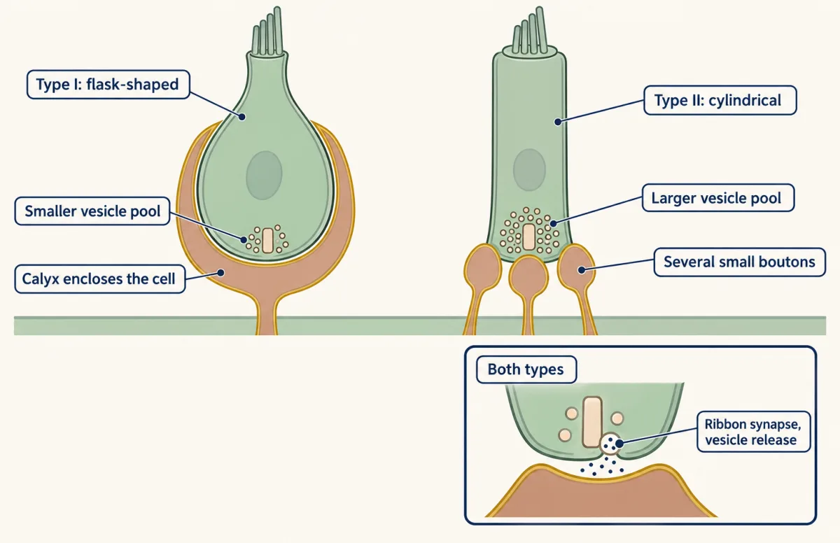
What differs is how much each cell releases, and whether it has another route. Release can be counted from membrane capacitance, because each fusing vesicle adds membrane. In mature mouse utricles, sustained calcium-evoked exocytosis measured this way was about tenfold smaller in type I than in type II cells. For type II cells, the large vesicle pools would favour sustained, tonic or low-frequency transmission. Type I cells release fewer vesicles, and they may have a second route that needs none.
In non-quantal transmission the hair cell changes its calyx’s voltage directly, through what it does to the cleft they share. Formally, it is a change in calyx potential without vesicular exocytosis and without gap junctions (Govindaraju and colleagues, 2023). Its appeal is speed. At a chemical synapse a vesicle has to fuse, glutamate has to cross the cleft, and receptors have to open, and each step adds delay. The fast electrical part of a non-quantal route skips those steps, while its potassium part builds more slowly, and reviews propose that it evolved to give the reflex pathways fast, reliable input (Eatock, 2018).
Predict. Otoferlin and CaV1.3, a calcium channel, are two proteins that vesicle release in hair cells needs. Remove either one in mature mice. If vesicle release were a type I cell’s only way to drive its calyx, what would the calyx do?
Check your prediction
It would stop responding to the hair cell: the spikes the hair cell evokes would disappear, whatever firing the calyx makes on its own. Instead, in the mature mouse utricle, type I cells lacking otoferlin or CaV1.3 could still drive calyx action potentials (Spaiardi and colleagues, 2022). The authors took that to support a functional role for non-quantal transmission. The test was whether spikes survive, in vitro, not whether the signal is normal.
How the cleft can move the calyx
The calyx wraps the type I cell so closely that the cleft between them is very narrow, and ions leave it only slowly. When the hair cell depolarizes, more potassium current flows out of it into that thin space. Potassium accumulates there, and the current, crossing the cleft’s resistance on its way out, makes the cleft electrically positive. Two consequences follow. The action potential lesson’s balance-point rule explains the first: for a compartment with fixed conductances, one common outside voltage and no other net current, the steady voltage is the average of its reversal potentials, weighted by each conductance. The calyx breaks the common-outside condition, because its inner face sees the cleft and its outer face the bath, so here the rule gives the direction of a change, not the absolute voltage. The second consequence needs only a subtraction.
Predict. The calyx’s inner membrane, the one facing the cleft, has potassium channels, and potassium’s reversal potential there (EK, the voltage at which potassium stops flowing) depends on the potassium concentration in the cleft. With illustrative values of 140 mM potassium inside the calyx and 37 °C, a cleft at 7 mM gives an EK of about −80 mV. Raise the cleft to 20 mM and EK is about −52 mV. Which way does the calyx’s voltage move?
Check your prediction
Up. Raising cleft potassium moves EK toward positive, so, with the other conductances fixed, the weighted average moves up and the calyx tends to depolarize. The 7 mM is the model’s resting cleft value; the 20 mM is made up to show the direction.
The second consequence is faster. Follow it in three steps.
- Current leaving the hair cell makes the cleft itself positive.
- The potassium current across the calyx’s inner face is gK × (Vcalyx − Vcleft − EK), so a more positive cleft, like a higher EK, shrinks the driving force: less potassium leaves the calyx there, or some enters.
- The calyx depolarizes as the cleft potential changes, with no vesicle to fuse.
In the model of Govindaraju and colleagues, the electrical potential in the cleft changes first and gives the initial change in driving force on the calyx; potassium accumulation contributes later. That order is a model output, not a measurement in the cleft.
Three kinds of evidence, three animals
The evidence comes from different animals and methods.
| Preparation | What was done | What it showed | Measured or modelled |
|---|---|---|---|
| Mature mouse utricle, in vitro | capacitance; otoferlin and CaV1.3 knockouts | type I exocytosis about tenfold smaller; calyx spikes survive the knockouts | measured |
| Neonatal rat saccule, in vitro | hair cell and calyx recorded separately | maturing channels shorten the receptor potential’s rise; some immature calyces show non-quantal transmission with little delay | measured |
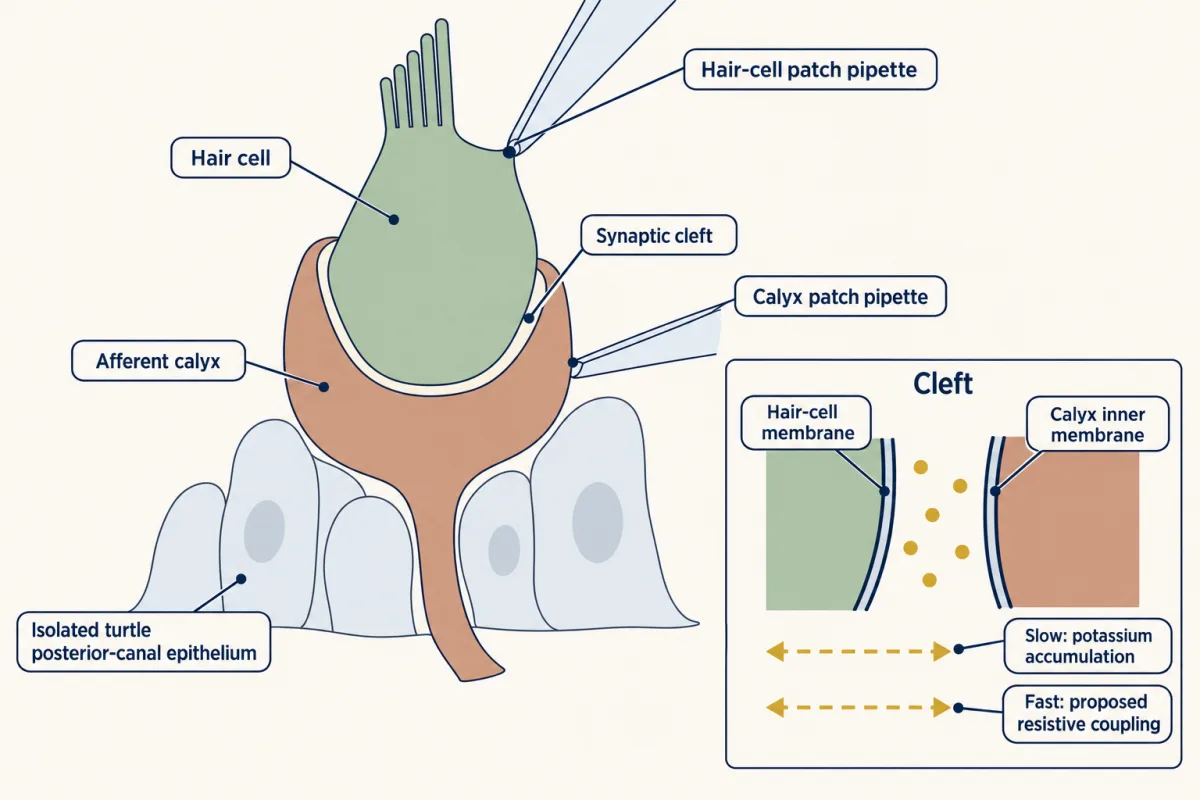
| Isolated turtle posterior-canal epithelium | dual voltage clamp of hair cell and calyx together | a slow component and a much faster one, each working in both directions | measured, in turtle |
| Finite-element model with adult rat geometry | electrodiffusion computed in the cleft | cleft potential first, potassium accumulation later | predicted |
In the turtle, the slow component fits potassium building up in the cleft. For the fast one, which accumulation cannot explain, Contini, Holstein and Art proposed potassium-sensitive resistive coupling: the two cells behave as if joined through a resistance, so current in one changes the other’s voltage almost at once. In both directions means current or potassium from either cell changed the other. These are turtle recordings, not mammalian latencies.
The fast channel is built during development. In mice, as low-voltage-activated potassium channels of the KV1 and KV7 families are acquired over the first postnatal weeks, hair-cell receptor potentials become faster, transmission from type I cells gains a non-quantal component, and afferent firing becomes more irregular (Eatock, 2025).
Passing on the surprise
Read the transcript of “Pass On the Surprise”
Film transcript: Pass On the Surprise
When you turn your own head, why does the reflex that steadies your head not fight you? That reflex treats head motion as a disturbance. Your own turn would look like one. Does the ear tell the two apart? In alert monkeys, canal afferents responded to both. So any cancelling must happen in the brain. Roy and Cullen recorded vestibular-only neurons, thought to drive the neck reflex that steadies the head. The brain has two clues to a self-made turn. One is the motor command, which predicts the neck's feedback. The other is the neck's actual sense of stretch. Pause and predict. A passive turn. An active turn with matching neck feedback. An active turn with mismatched feedback.
Which ones drive the neuron strongly? The passive turn did. So did the active turn with mismatched feedback. Only the matched active turn was cancelled. In this made-up example, nearly all of the response is cancelled. This is not the ear switching off. The afferents kept firing. This muting may help keep the head-steadying reflex from opposing a turn you intend. It does not mean you stop sensing your own head movements. A prediction must stay right. Brooks and colleagues recorded neurons in a cerebellar output nucleus, the rostral fastigial nucleus. Their monkeys turned their heads against a resistive load. As the animals adapted, these neurons' vestibular sensitivity fell.
When the load was suddenly removed, it rose again. These are recordings. They show what the neurons did, not that they caused the learning. Finally, a vestibular implant stimulates the canal branches of the nerve when both ears have lost their signal. It varies its pulses to represent head rotation. It does not recreate the nerve's two natural channels, and it sends no otolith signal. In eight adults, some balance measures improved. Hearing in the implanted ear fell in all but one. A cohort this small shows feasibility and a trade-off, not that the device works for everyone. The nerve reports motion on two channels. Where neck feedback matches the prediction, these head-posture neurons pass on mainly the surprise.
From the film
- In the neurons tested in monkeys, the response to a head turn is cancelled only when the turn is self-made and neck feedback matches the motor command's prediction.
- This is not the ear switching off: canal afferents kept firing, so any cancelling happens in the brain.
- A vestibular implant supplies a motion-modulated rotation signal without the nerve's natural two channels or an otolith signal, and in a small cohort hearing in the implanted ear fell in all but one.
Now the second puzzle: how can the head reflex spare your own turns? Does the ear itself tell a self-made turn from an imposed one? It does not. Semicircular-canal afferents in alert monkeys respond to head motion whether the animal causes it or something else does (Cullen and Minor, 2002). So any cancellation of your own movement must happen centrally.
Roy and Cullen (2004) recorded vestibular-only neurons, neurons of the vestibular nuclei that respond to head motion but not to eye movements, in alert macaques. These neurons are thought to drive the vestibulocollic reflex, which turns the neck against an imposed head movement to steady the head. For a voluntary turn the brain has two signals to compare. It has an internal copy of the motor command sent to the neck muscles, called an efference copy, from which it can predict the neck’s feedback. And it has the neck’s actual sense of stretch and position (neck proprioception).
Predict. Compare one head turn in three conditions: the monkey is turned passively; the monkey turns its own head and the neck feedback matches the motor command; the monkey turns its own head but the neck feedback is made not to match. In which conditions does a vestibular-only neuron respond strongly?
Check your prediction
It responded strongly to the passive turn and to the active turn with mismatched feedback, and weakly to the active turn with matched feedback. In the neurons tested, cancellation of the response to active motion required neck proprioceptive activation that matched the feedback expected from the motor command. Neither the motor command nor neck feedback alone was enough.
Illustration with made-up numbers. Suppose a passive turn peaking at 20 degrees per second evokes a 40 spikes per second modulation, a sensitivity of 2.0 spikes per second per degree per second. An active turn of the same size evokes 4 spikes per second, so sensitivity is 0.2 and the response is 1 - 0.2/2.0 = 90 percent cancelled. If neck feedback is made to mismatch the prediction, the sensitivity is expected to climb back toward 2.0.
- Canal afferent modulation
- drawn the same in all conditions
- Central sensitivity (spikes/s per deg/s)
- 2.0
- Central modulation (spikes/s)
- 40
- Percent cancelled = 1 − Sactive/Spassive
- 0
In this sketch: linear sensitivity (modulation = sensitivity × peak head velocity), relative timing, other velocities scaled by this sketch, and the afferent drawn in relative units because the lesson gives it no value. The numbers are the lesson’s made-up illustration, not data. The cancellation rule is from Roy and Cullen (2004), vestibular-only neurons in four alert macaques.
Rule to keep: in the neurons tested, cancellation needed neck feedback that matched the motor-command prediction. It is not the periphery switching off, it does not locate the comparator, it is not shown for every vestibular neuron, and it is not unconditional.
Two points keep this accurate. The periphery did not switch off; the afferents kept firing. And the muting may help the head-steadying reflex spare a voluntary turn; it does not mean you stop sensing your own head movements. Multisensory integration and prediction takes up this kind of prediction across the other senses.
Keeping the prediction calibrated
A prediction is only useful if it stays right, so it must be updated when the head or the world changes. Brooks, Carriot and Cullen (2015) recorded rostral fastigial neurons, neurons of a cerebellar output nucleus that also carry head-motion signals, in rhesus macaques turning their heads voluntarily against a resistive load. As the animals adapted, neuronal vestibular sensitivity fell. On catch trials, when the load was unexpectedly removed, sensitivity rose again.
That is what a prediction-error signal should do: shrink as the new head becomes predictable, and return the moment the prediction is wrong, so the pattern is consistent with updating of an internal model. Neural and behavioural learning ran at statistically similar rates, and recordings alone cannot show that these neurons cause the learning.
Reflexes need calibration too. The VOR’s gain, the size of the eye movement made for a given vestibular input, must be well calibrated to keep images stable on the retina, and visual error can drive it up or down through the cerebellum (Kimpo and colleagues, 2014). That is a different circuit with a different learning problem. Purkinje-cell circuitry belongs to the cerebellum, and the reflex arcs to motor units and reflexes.
Rebuilding the signal: a vestibular implant
A vestibular implant tries to restore the motion signal when both ears have lost it. Chow and colleagues (2021, New England Journal of Medicine) implanted one prosthesis in each of eight adults with adult-onset bilateral vestibular hypofunction, meaning weak or absent function in both balance organs. The device electrically stimulated the three semicircular-canal branches of the vestibular nerve, varying its pulse rate and amplitude to represent rotational head motion. Measured against the one idea, it supplies a motion-modulated signal for rotation that does not reproduce the nerve’s natural regular and irregular code, and it encodes no otolith signal for linear acceleration or tilt.
At six months, two of five posture and gait measures had within-participant change intervals that excluded zero, though the intervals were not adjusted for multiple comparisons. Self-reported dizziness handicap fell. Hearing in the implanted ear was reduced in all but one of the eight, severely in three.
A cohort this small, in one age band and without an independent implantation control, establishes feasibility and a trade-off, not population efficacy.
Design problem
Reviews propose that the type I calyx’s non-quantal route helps the fast, irregular channel stay fast. Design an experiment in mature mice that tests this idea, and say which result would count against it.
Saved in this browser only.
One way to do it
Record single afferents, classify each as regular or irregular from its resting intervals, and fill it with dye so you know whether it ends in a calyx. Measure what the fast channel is for: latency and spike-timing precision to rapid stimuli such as vibration, with responses to slow rotation as a comparison. Then remove the non-quantal route while leaving quantal release intact. That is the hard part. The low-voltage-activated potassium channels that come with non-quantal transmission also speed the hair cell’s own receptor potential, so a knockout could slow the signal before it reaches the synapse. Check quantal release by capacitance and record the hair cell’s receptor potential, so that any change can be pinned on the synapse.
Choose the control with care. Regular afferents are not a clean control, because some carry calyx endings, and removing the calyx’s route would reach them too. Use dye-identified bouton-only fibres, which have no calyx, as the control that should change little, and compare calyx-bearing irregular fibres with calyx-bearing regular ones. If irregular, calyx-bearing afferents keep their latency and timing precision after the non-quantal route is gone, the idea is in trouble. Judge the result on latency and timing, not on regularity, which the calyx does not set alone. A reflex readout, such as a vibration-evoked VEMP, would show whether any change matters for behaviour.
Going deeper
This section keeps the numbers, preparations and methods behind the main line.
Thresholds and the averaging window
The rough rule in the paradox treats a spike train as a renewal process, with independent, identically distributed intervals, in which the count variance approaches CV² times the mean count as the window grows. It needs a window long enough for edge effects to be negligible, and a large mean count is not enough for a very regular train. For made-up gamma-process spike trains with the lesson’s rates and CVs, and a window placed at a random moment, the exact count variance gives the 1 s noise quoted in the paradox and, over 0.1 s, about 3.6 and 16 spikes per second. For the regular fibre at 1 s, with 100 spikes in the window, the count variance is about 0.42 against the rule’s 0.25, because the long-window expansion carries an extra edge term of about (1 − CV⁴)/6, about 0.17 here. It comes from where the window edges fall between evenly spaced spikes, and at 0.1 s it supplies most of the regular fibre’s noise. A simulation of 200,000 such windows agrees with these values to within about 1 percent. These are toy numbers: real thresholds also depend on the stimulus, on correlations between spikes and on how downstream neurons read the train. With real afferents, Sadeghi, Chacron, Taylor and Cullen (2007) reported lower angular-velocity detection thresholds for regular than for irregular canal afferents, despite the regular fibres’ significantly lower gains.
Does the vestibulo-ocular reflex need the fast channel?
In a classic functional-ablation study, Minor and Goldberg (1991) applied large inhibitory galvanic currents to the inner ear of alert squirrel monkeys, which silenced irregular afferents selectively. The eye movements evoked by the head rotations tested were largely unchanged, so the regular channel can carry the VOR, at least for those stimuli.
Counting vesicles from capacitance
Spaiardi and colleagues worked in acutely dissected mouse utricles from postnatal days 18 to 28, in vitro, and measured exocytosis as a rise in membrane capacitance. They converted capacitance to vesicle number with 37 aF per vesicle. A later study from the same group showed that a large transient capacitance change in type I cells comes from the gating of a potassium conductance, not from vesicle fusion (Spaiardi and colleagues, 2025), so only the sustained component counts as release.
Worked problem: from capacitance to vesicles. Suppose, in a made-up recording, a depolarizing step raises a type II cell’s capacitance by 74 fF.
Step 1: units. 74 fF is 74,000 aF.
Step 2: divide. 74,000 aF / 37 aF per vesicle = 2,000 vesicles.
Step 3: scale. A cell with about tenfold smaller exocytosis would show about 7.4 fF, or about 200 vesicles. The conversion is an assumption borrowed from the paper, so the count is an estimate, not a head count.
The three preparations, in numbers
Songer and Eatock used excised rat saccules at postnatal days 1 to 9, deflected single bundles, and recorded hair cells and, separately, calyces at room and at body temperature. The shortening of the receptor potential’s rise time by low-voltage-activated channels was seen by days 7 to 9.
In the turtle recordings, a representative fast time constant, at a holding potential of minus 70 millivolts, was 32 microseconds; that is one illustrated response, not a population latency. The mean resistive coupling across 17 pairs was 522 plus or minus 577 pS, a spread larger than the mean.
Worked problem: what does 522 pS buy? Take the mean coupling and an assumed 10 mV change in hair-cell voltage while the calyx voltage is held fixed. Treating the coupling as a simple transfer conductance, the calyx current changes by 522 pS x 10 mV = 5.2 pA. The 10 mV is a teaching assumption, not a measurement, and the real spread across pairs means individual pairs would differ widely.
The Govindaraju model used adult rat morphology and channel parameters from mouse and rat recordings at room temperature, and its output was compared with turtle recordings. In it, the cleft-base potential rose from 1.9 to 6 mV in 0.43 ms after excitatory stimulation, and taller modelled calyces reduced spike latency. None of these model numbers was measured in the cleft.
The field is unsettled in an instructive way. A direct measurement of cleft potential or potassium in a mature mammalian calyx would test the model, and so would a manipulation that changes calyx height or potassium-channel density and then reads latency. Neither experiment has been reported in the work described here.
The cancellation experiment
Roy and Cullen’s study appeared in the Journal of Neuroscience in 2004. They recorded in four alert macaques, with magnetic search coils tracking head and gaze, and varied conditions such as head restraint and neck torque. The comparator circuit was not located.
Learning rates in the load experiment
The study (Nature Neuroscience, 2015) used two adult male rhesus macaques. The learning constants were 34 plus or minus 7 trials for peak head velocity and 24 plus or minus 8 trials for neuronal sensitivity. For an exponential course, a constant is the number of trials to cover about 63 percent of the change. The two did not differ significantly (Wilcoxon test, p > 0.05). Time constants measure rate, not onset, so they cannot say whether neurons or behaviour changed first.
A different circuit: how the VOR’s gain is taught
This is the floccular circuit, not the rostral fastigial circuit of the load experiment.
What teaches the cerebellum? Masao Ito proposed in 1972 that climbing-fibre signals carry visual error to the flocculus (as described in Lisberger’s 2021 review). The modern picture is conditional. Kimpo and colleagues (2014, eLife) recorded floccular Purkinje cells in two rhesus monkeys, where complex spikes are a one-to-one readout of climbing-fibre spikes. During training for increased gain, climbing-fibre activity predicted a lower Purkinje output on the next trial. During training for decreased gain, the same climbing fibres responded just as strongly, yet their activity had no trial-by-trial effect on Purkinje output. The authors read these correlations as gating: a climbing-fibre error signal does not always trigger plasticity. In adult male mice, the same group paired climbing-fibre photostimulation with 1 Hz, plus or minus 10 degrees per second rotation in darkness. Appropriately timed pairing induced a gain increase, but the protocols tested did not produce associative gain decrease beyond vestibular-only habituation. So stimulation sufficed for one direction under those conditions; it did not prove climbing fibres are required in natural learning, nor that they are irrelevant to a decrease.
Rowan and colleagues (2018, Neuron) added that inhibition shapes the teaching signal. In cerebellar slices from mice aged 6 to 15 weeks, coincident activation of molecular-layer interneurons reduced climbing-fibre-evoked dendritic calcium, to 61.3 plus or minus 3.2 percent of control, and strong activation could reverse the sign of the plasticity that climbing-fibre activity induces at parallel-fibre synapses. In awake mice, co-activating climbing fibres with those interneurons, timed to the end of ipsiversive head turns, produced a decrease in VOR gain instead of an increase.
Reading the implant cohort
The eight participants were aged 50 to 66. Beyond the two posture and gait measures whose intervals excluded zero, a third had a lower bound of exactly zero; with five endpoints and two positive, the unadjusted intervals invite a correction for multiple comparisons.
Pure-tone thresholds in the implanted ear rose by 3 to 16 dB in five participants and by 74 to 104 dB in three, a severe loss in that ear. That is a rise in all eight, though the authors counted hearing as reduced in all but one; read the threshold changes rather than the count.
Worked problem: group medians are not paired improvements. The Dizziness Handicap Inventory median fell from 79 before implantation to 19 at six months, and the paper reports a median within-participant improvement of 50 points.
Step 1: difference of group medians. 79 - 19 = 60 points.
Step 2: the paired statistic. Pair each participant first, subtract, then take the median of the eight differences. The paper gives 50.
Step 3: why they differ. A median is not a linear summary, so the median of paired changes can differ from the difference of group medians, even when one person supplies both group medians. The 60 is not a correction of the paper’s 50; it answers a different question.
The missing control. The design is single-arm and within-participant, so in the baseline-to-six-month comparison surgery, device use and time are confounded. The blinded, randomised same-day comparison with a constant-stimulation mode helps: it holds the surgery and the elapsed time fixed and addresses expectancy and spontaneous recovery, so it isolates an immediate effect of motion-modulated stimulation. It cannot estimate the long-term effect of the operation or of months of adaptation, so a control that separates those from the stimulation is still missing.
Your notes
Select any text in the lesson to highlight it or add a note to it. Your highlights and notes are saved in this browser and collected in your notebook.
This browser is not letting the site save anything (a private window or strict privacy settings), so highlights and notes will be lost when you leave the page.
How we measure it
Dual patch clamp of hair cell and calyx
One pipette records the hair cell and another records the calyx, so presynaptic current and postsynaptic response are measured at once. It lets an experimenter separate a slow potassium accumulation from a fast resistive coupling.
Membrane-capacitance assay of exocytosis
When vesicles fuse, the cell membrane gains area and its capacitance rises. Dividing a capacitance jump by a per-vesicle value converts it to a vesicle count, with the assumption that the conversion holds for that cell. In type I hair cells a fast transient capacitance change comes from potassium-channel gating, not vesicle fusion, so only the sustained component counts as release.
Single-unit recording in alert macaques
A fine electrode records one neuron while the animal makes or receives head movements, with search coils tracking head and gaze. It shows what a neuron did in that task, and it is correlational unless paired with a manipulation.
Finite-element electrodiffusion modelling
The cleft is divided into small elements and the electrical potential and ion concentrations are computed in each. It predicts variables that cannot be measured in a space this narrow, and its outputs are predictions until a measurement tests them.
Practice questions on this lesson (3) → MCAT-style, original, each with an explanation
Where this lesson fits
Before thisReview first
- The Hair Cell's Transduction ChannelThis course · Lesson 22
You need how hair cells turn bending into current to follow how type I and II cells drive their afferents.
- The Cochlear Amplifier and Otoacoustic EmissionsThis course · Lesson 23
It covers the hair cell's active machinery and ear physiology that the balance organs share.
- Five Ways to Turn a Stimulus into a CurrentThis course · Lesson 1
It gives the fast, hair-cell transduction timing that this lesson's fast channel builds on.
After thisStudy next
- Combining Senses and Predicting ThemThis course · Lesson 29
It takes the idea of cancelling self-generated motion and applies prediction across the other senses.
- Restoring Senses: Implants, Prostheses and Optogenetic VisionThis course · Lesson 30
It takes the vestibular implant trade-off further, alongside other implants that replace a damaged sense organ.
Elsewhere on the siteFilms, simulations and method films in other courses
- One Cell, One SwitchFilm · Neuroscience
It shows how a hair cell's bundle deflection toward the tallest stereocilia opens channels, which underlies head-motion sensing.
- Every Response Has a Running TimeFilm · Organismal Biology
It follows the receptor-to-effector route of a reflex, the same route the head and eye reflexes use.
Suggested by an AI model from the lessons themselves, then checked. How we use AI
Finish line
Test yourself
8 questions
Practice and MCAT-style, with explained answers.
Next · Lesson 26
One Receptor per Neuron: How Olfactory Neurons Choose
A mouse olfactory neuron holds about a thousand receptor genes and uses one copy of one. How does it choose, and what keeps it from changing its mind?
Continue →← Lesson 24 · Hidden Hearing Loss, Gene Therapy and Regeneration