Sensory Systems · Unit 6: Hearing and balance
The Hair Cell's Transduction Channel
Rewritten on : new text, flashcards and film. Your notes and highlights stay, but the text around them may have moved.
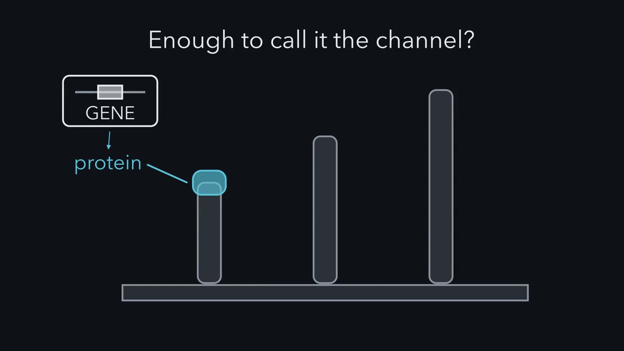
A gene is required for hearing, and its protein sits at the right place. What else must be shown before it counts as the channel?
Pause when the reading reaches a film, and offer to play it. Saved on this device.
Watch the film now, or keep listening
Builds on:Piezo Channels and the Physics of ForceFive Ways to Turn a Stimulus into a CurrentWhy these?
 ▶ Watch One Current, Three Factors · 2:35
Read the transcript of “One Current, Three Factors”
Film transcript: One Current, Three Factors
A gene is needed for hearing, and its protein sits in the right place. Is that enough to call it the channel? Start with the current. A hair bundle is a staircase of stereocilia, in rows that rise in height. Tip links connect the tip of each shorter stereocilium to the side of its taller neighbour. Deflect the bundle toward its tallest row. The tip links are thought to pull channels open near their lower ends. With the cell held at a negative voltage, cations flow in, and the current is inward. This is the conventional transduction current. The current rises within a fraction of a millisecond. That is too fast for a chain of second messengers.
So force is thought to open the channel directly. Plot the size of the peak current against bundle displacement. The S-shaped curve saturates when nearly all channels are open. That current comes from many channels. Three things set it. First, how many working channels sit at the tip links. Call that N. Second, how much current one open channel passes. That is the unitary current, small i. Third, the chance that each channel is open. That is the open probability, P_open. For channels that are alike, multiply the three. That gives the average current from the whole bundle. Predict. A mutation halves that average current. Which of the three factors did it change?
From the average alone, you cannot tell. Halve the channels, the unitary current, or the open probability. Each gives the same average current. A trafficking factor, an anchor or a lipid-modifying enzyme could all be needed. Deleting any one would silence the cell. So a lost current cannot tell you which factor you removed. Evidence about permeation speaks more directly to the pore. Which ions pass, and how much current does one open channel carry? So do a residue reached in a working channel, and activity of the protein on its own. But partners can change permeation too. So the case rests on several rungs together. Carry one question through every experiment.
Which factor did it test, and what did it rule out?
From the film
- The mean current from a hair bundle is the product of the number of working channels, the current through one open channel and the open probability.
- A lost current cannot tell you which of those three factors was removed.
- Evidence about permeation, a residue reached in a working channel and activity of the protein on its own speaks more directly to the pore, and the case rests on several rungs together.
A hair cell fails to hear when a gene is missing, and the gene’s protein sits in the right place. Is that enough to call the protein the channel?
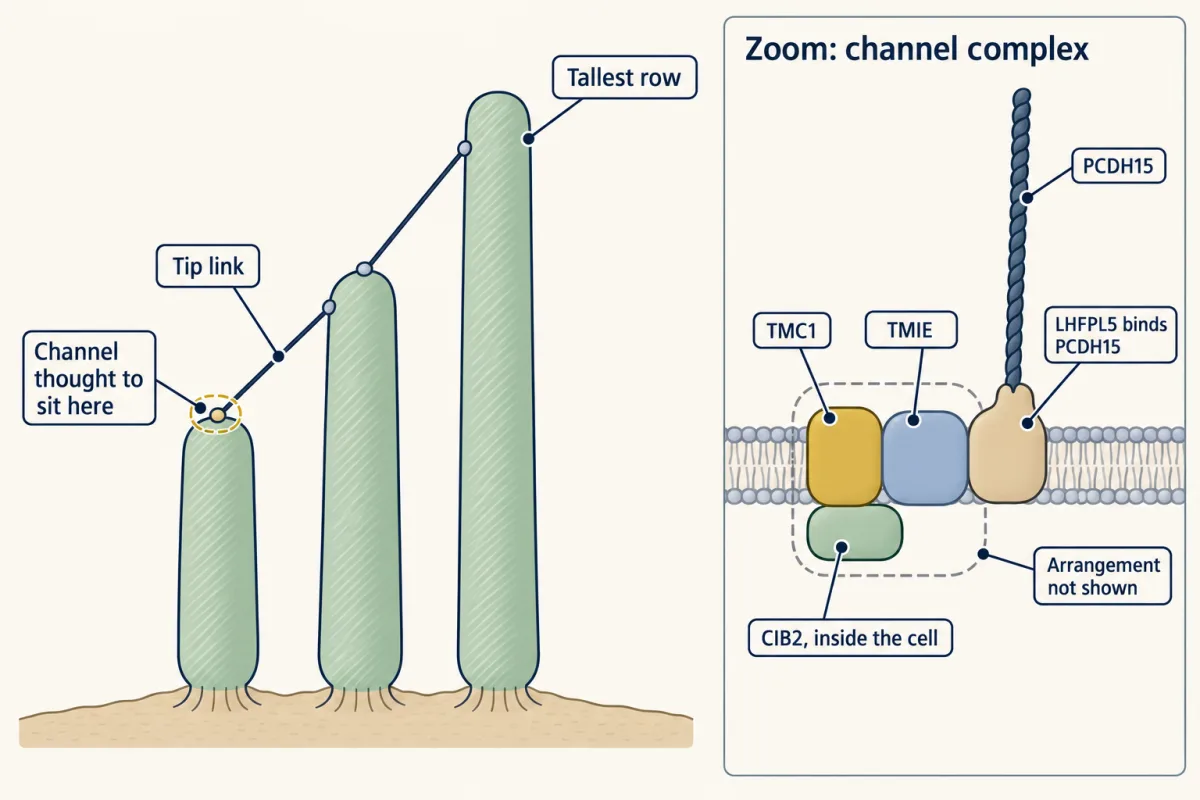
It is not, and the history of TMC1 shows how much more is asked. The best-supported answer so far is that TMC1, a membrane protein found through the genetics of inherited deafness, and its close relative TMC2 form or line the pore of the transduction channel, which is thought to sit at the lower end of the tip link, and that they work inside a complex with partner proteins. Of the two, TMC1 is the one needed for hearing in the mature mouse cochlea.
If tip links are new to you, read Hearing and balance first: it covers tip links, the gating spring and the driving force, and gives TMC1 one sentence as a pore component. Here we start where that sentence stops. The gating logic of channels is in ion channels as receptors, and earlier in this course mechanotransduction channels compare candidates across senses.
First comes the current and one equation for reading any experiment on it; then the experiments rung by rung, the partner proteins and adaptation.
What a transduction current looks like
A hair bundle is a staircase of stereocilia, in rows that rise in height. Tip links connect the tip of each shorter stereocilium to the side of its taller neighbour. Deflect the bundle toward its tallest row and the tip links are thought to pull channels open near their lower ends. Cations flow through the open channels; the recordings here hold the cell at a negative voltage, so the current is inward.
The current opened by that deflection is the conventional transduction current. It is inward at negative voltages and would be outward above its reversal potential, near 0 mV. Picture three features of it.
| Feature | What you see | Why it matters |
|---|---|---|
| Direction | Deflection toward the tallest row opens channels | This direction defines the conventional current; some other mechanically activated hair-cell currents have the reverse polarity (Going deeper) |
| Speed | The current rises within a fraction of a millisecond | That is too fast for a chain of enzymes and second messengers (signal molecules made inside the cell), so force is thought to open the channel directly; how the force reaches it is still open |
| Grading | Peak current plotted against bundle displacement gives an S-shaped curve | More displacement opens a larger share of the channels, until nearly all are open and the current saturates |
The recordings use a pipette on the voltage-held cell while a stiff probe or a fluid jet moves the bundle. Each experiment asks the same thing: what happened to this current, and why?
One current, three factors
The current from one bundle is the sum of many channels. Three things set it:
- N, how many working channels sit at the tip links.
- i, the unitary current: the current through one open channel. In a recording it appears as a single step-like event, so it is also called the single-channel current.
- Popen, the open probability: the chance that a given channel is open, or the fraction of time it spends open.
For channels that are alike, at a fixed voltage and ionic conditions, the mean current from the bundle is their product, I = N × i × Popen; if two kinds of channel are present, add their currents. The instantaneous current flickers around this mean as channels open and close. To compare channels you also need the unitary conductance, g, which removes the effect of the voltage: g = i / (V − Erev), where V is the holding potential and Erev is the reversal potential, the voltage at which no net current flows through the open channel. V − Erev is the driving force. This g is the same at every voltage only if the current-voltage relation is a straight line, which the problems here assume; real transduction currents are not quite linear, so a g is quoted with its voltage and ions.
Worked problem: a conductance from a real recording. Beurg and colleagues (2006) recorded from rat inner hair cells (the cochlea’s main sensory cells; outer hair cells mostly amplify) in 1.5 mM external calcium. The mean inward unitary current was 15.0 pA at -84 mV. Take the reversal potential as +3 mV and assume a linear current-voltage relation.
- (a) The driving force is -84 - (+3) = -87 mV. Conductance g = i / (V - Erev) = (-15.0 pA) / (-87 mV) = 0.172 nS, or about 172 pS.
- (b) For a made-up ensemble of 100 identical, independent channels at open probability 0.20, the mean current is I = N × i × Popen = 100 × (-15.0 pA) × 0.20 = -300 pA, an inward current of 300 pA. The count and probability are teaching assumptions, not measurements.
- (c) Why quote the calcium? The same paper estimated about 260 pS for inner hair cells at 0.02 mM calcium, about one and a half times the 172 pS you just computed at 1.5 mM. Calcium is thought to impede the passage of monovalent cations (singly charged ions such as sodium and potassium), so a conductance is incomplete without its ionic conditions.
g = i / (V - Erev), and the mean ensemble current is I = N × i × Popen
- Driving force V − Erev (Erev = +3 mV)
- Conductance g = i / (V − Erev), from −15.0 pA at −84 mV
- Unitary current i at this V (linear assumption)
- Open channels: expected N × Popen | drawn
- Expected ensemble current I = N × i × Popen
- Simulated current = drawn open count × i
In this sketch: a linear current-voltage relation (assumed, and extended in both directions from the one measured point at −84 mV, so everything away from that point is extrapolated, not measured), Erev taken as +3 mV (the lesson’s assumption), identical independent channels, rat inner hair cells P6 to P11, 1.5 mM external calcium (Beurg and colleagues, 2006), room temperature (19 to 22 °C), no adaptation. The −15.0 pA is the lesson’s mean unitary current (15.0 ± 1.6 pA, SD, n = 7, taken as inward) at −84 mV; every other number follows from it by the formula.
Not shown here: that there is one hair-cell conductance (the same paper estimated about 260 pS at 0.02 mM calcium, and in the same paper’s low-calcium estimates, rat outer hair cells rose from 145 to 210 pS between sampled low- and mid-frequency positions); that N and Popen are measurements; or that a conductance identifies which protein forms the pore.
Rule to keep: g = i / (V − Erev), and the whole ensemble current is I = N × i × Popen. A conductance is quoted with its ionic conditions, and a negative current is inward.
Predict. A mutation halves the ensemble current in (b), from -300 pA to -150 pA. Which of the three factors did it change?
Check your prediction
From the mean current alone, you cannot tell. Halve N, halve i or halve Popen, and each gives exactly -150 pA. A knockout is the extreme case: setting any one factor to zero gives zero current, so a trafficking factor, an anchor or a lipid-modifying enzyme could each be needed for the current. To learn which factor changed you need more information, such as resolved single-channel events or the size of the current’s fluctuations, which differ between the three cases.
The one idea to carry through this lesson. The mean current you record from a hair bundle is a product, I = N × i × Popen. Removing a protein that any one of the three factors needs silences the cell, so a lost current cannot tell you which one you removed. What speaks more directly to the pore is evidence about permeation (which ions pass, and how much current one open channel carries), a residue that a reagent can reach in a working channel, and force-gated activity of the protein outside the hair cell. Partner proteins can change permeation too, so the case for a pore rests on several rungs together, never on one.
The idea covers the conventional, tip-link-gated current in mammalian hair cells. It says nothing about how force reaches the channel (the gating spring and the lipid), and it is a way to read experiments, not a claim that any single experiment is decisive.
The ladder: what each rung rules out
A protein can be required for a current without forming the pore, so the field asks for a ladder of evidence. Two sets of terms first. A knockout inactivates a gene, and a null allele makes no working product (a double null lacks working copies of both Tmc1 and Tmc2); a rescue supplies a working copy in that background. Permeation is how ions pass through the open channel, and selectivity is which ions it prefers. Each rung rules out a different alternative; only some of them bear on a single factor of the equation.
| Rung | The test | What it speaks to | What it rules out | What it cannot show |
|---|---|---|---|---|
| Localization | The protein is detected at the lower end of the tip link, where the channel is thought to be | Presence at the proposed site | Weighs against a protein found only elsewhere, within the assay’s resolution | That it conducts, or that this pool causes the current |
| Requirement | The conventional current disappears when the gene is removed | The product | A protein the current does not need | Which factor was lost |
| Rescue | Re-expressing the gene restores the current in the mutant background | The product | Supports the missing gene as the cause, given expression and vector controls | Which factor, a pore, or compensation by an overexpressed substitute |
| Permeation | A change in the protein’s sequence changes which ions pass, not only how much current flows | Selectivity of the open channel, and perhaps i | Weighs against a role only in delivering or anchoring unchanged channels | Pore lining, because a partner can change permeation too |
| Acute accessibility | A reagent applied to a cell that already carries current alters permeation through an engineered residue | Whether a residue reachable from outside affects permeation in a working channel | Altered synthesis or delivery of the protein | That the residue lines the pore rather than sits beside it; the protein alone, because native partners are still present |
| Purified activity | The isolated protein, in a membrane with no hair cell around it, gives force-gated current in a reduced system | Whether the protein alone can conduct and be gated by force | Weighs against a need for the rest of the hair cell for gating | The native complex or its force coupling; species and lipid matter |
No single paper climbs every rung, and the rungs differ in species and preparation. The first rung is taken here as a working assumption, not reviewed, which is why the channel is “thought to sit” at the lower end of the tip link.
Requirement and rescue
Read the transcript of “Change It While It Works”
Film transcript: Change It While It Works
If deleting a protein cannot reveal the pore, what experiment can? In 2011, Kawashima and colleagues removed both TMC1 and TMC2. Hair cells lacking both had no conventional current. Putting back either gene restored it. That puts the TMC proteins in the pathway. But loss and rescue act on the whole product, so they cannot separate a pore from its helpers. The sharper test keeps the channel working, and changes one amino acid. The Beethoven mouse carries such a change. Methionine 412 of mouse TMC1 becomes lysine. In 2013, Pan and colleagues recorded from inner hair cells lacking Tmc2. So every channel had to use TMC1. Predict. If TMC1 only delivered or anchored the channel, what would a point mutation change?
If only the delivery of normal channels changed, there would be fewer channels. A coupling defect would mean fewer open. Each open channel would be unchanged. But the Beethoven channels let through less calcium. That calcium result has been replicated. Whether the single-channel current also shrinks is disputed.
One residue in TMC1 changed which ions pass. A deletion can never show that. But a partner beside the pore can also change what passes. So this rung narrows the candidates. Next, change the channel while it works. In 2018, Pan and colleagues made TMC1 with single cysteine substitutions. Delivered into hair cells lacking both genes, they restored a current.
Then they added a reagent that cannot cross the membrane. It reacts with cysteine in water. Predict. The current is already flowing when the reagent arrives. If permeation changes, what does that rule out? It rules out altered synthesis or delivery. The current existed before the reagent was added. A residue reached from outside, whose change alters which ions pass, points to a water-filled route. That is what a pore would provide. The cells still had their native partners, and a reachable residue may sit beside the pore. No purified mammalian TMC1 was tested alone. Purified, truncated turtle TMC1 and budgerigar TMC2, in liposomes, responded to pressure.
That supports force-gated activity in a reduced system. The proteins were not mammalian, and this was not a native complex. Each rung ruled out a different alternative. None alone proves the pore.
From the film
- Loss and rescue of TMC1 and TMC2 put them in the pathway but cannot separate a pore from its helpers.
- A point mutation that changes which ions pass, as in the Beethoven mouse, narrows the candidates, though a partner beside the pore can also change what passes.
- A reagent that alters permeation in a channel already carrying current rules out altered synthesis or delivery, but the native partners were still present.
Kurima and colleagues (2002) linked human TMC1 mutations to dominant deafness (DFNA36) and to recessive deafness (DFNB7/B11). The same paper found a Tmc1 deletion in the deaf dn mouse and Tmc1 mRNA in mouse hair cells. That was genetic evidence: the gene matters for hearing and for cochlear hair cells. It did not say what the protein does.
The first functional rung came from mice, and it held a puzzle. Early postnatal hair cells in Tmc1 mutant mice can still show transduction currents. Kawashima and colleagues (2011) recorded from neonatal mouse hair cells in excised tissue, and removed Tmc1’s relative Tmc2 as well.
Predict. If TMC1 and TMC2 can each do the same job, what should cells lacking both show? And why might the single Tmc1 mutant have hidden the effect?
Check your prediction
Cells lacking both Tmc1 and Tmc2 had no conventional transduction current. In double-null cultures, expressing either TMC1 or TMC2 restored it. So the two genes can substitute for each other, and Tmc2 is expressed only transiently in the early postnatal cochlea, which is why a Tmc1 single mutant can still show early currents.
Read through the equation, requirement and replaceability in neonatal mouse hair cells both act on the whole product: they put the TMC proteins in the pathway without separating a pore from a factor the pore needs. The double-null cell also gave later groups a clean background for testing altered versions of the protein.
Change the protein, not its amount
The sharper test is to keep the channel working and change one amino acid in the candidate. If the change alters what an open channel passes, it ties the candidate to the channel’s permeation properties.
The Beethoven mouse carries such a change in one allele (version) of Tmc1: methionine 412 of the mouse protein is replaced by lysine (M412K). Vreugde and colleagues (2002) described the line as a model of dominant progressive hearing loss. To ask what the mutation does to the channel itself, Pan and colleagues (2013) recorded from excised neonatal mouse inner hair cells with Tmc2 removed, so that every channel had to be built with TMC1.
Predict. If TMC1 only delivered or anchored the channel, which factor of I = N × i × Popen would a point mutation be expected to change? If TMC1 lines the pore, what would change instead?
Check your prediction
If a mutation changed only how otherwise normal channels are delivered or anchored, it would lower N, or lower Popen if coupling to the tip link suffered; each open channel would be unchanged. If TMC1 is part of the pore, a mutation can change the open channel itself: which ions pass, and perhaps how much current each carries. The Beethoven channels let through less calcium than channels made with wild-type TMC1, and the calcium result has been replicated. A single residue in TMC1 changed which ions pass, which a deletion can never show.
| Cells (Tmc2 removed) | TMC1 in every channel | Calcium permeability |
|---|---|---|
| One wild-type Tmc1 allele | wild-type | normal |
| Beethoven Tmc1 | M412K | reduced |
With no Tmc2, a cell needs a working Tmc1 copy to carry any current, so the comparison cells kept one wild-type copy.
This rung is strong and narrows the candidates, but it does not by itself place the protein, or the mutated residue, in or beside the pore. A mutation can act through a change in shape or in the makeup of the complex, and a partner protein can also change what passes: TMIE mutations do exactly that. Whether the Beethoven mutation also shrinks the single-channel current is disputed between laboratories (Going deeper).
Change it while it works: acute accessibility
Pan and colleagues (2018) built the acute-accessibility test. They delivered Tmc1 constructs carrying single cysteine substitutions into Tmc1/Tmc2 double-null mouse hair cells with a viral vector, and cultured the cochleae, so that the engineered TMC1 restored a current. They then added a cysteine-reactive reagent, which cannot cross the membrane and reacts with cysteine side chains in water, to cells that were already carrying current.
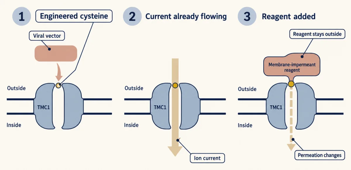
Predict. The reagent changes the permeation of a TMC1-mediated current that was already flowing. What explanation does that timing rule out, and what does it suggest about where the cysteine sits?
Check your prediction
Because the current existed before the reagent was added, a change cannot be explained by altered synthesis or delivery of the protein. And a residue the reagent can reach from outside, whose modification changes the ions that pass, points to a water-filled route, as a pore would provide. Accessibility alone cannot tell a residue that lines the pore from one beside it. The fastest-reacting construct was inhibited within a fraction of a second. The authors called this compelling evidence that TMC1 is a pore-forming component.
The limit is the one the ladder predicts: native partners were still present, and no purified mammalian TMC1 was tested alone.
Without the hair cell: purified and transferred proteins
Jia and colleagues (2020) produced truncated turtle TMC1 and budgerigar TMC2 constructs in insect cells, purified them, put them in liposomes, and found that applied pressure activated channel activity. This supports intrinsic mechanical responsiveness in a reduced system: the protein alone may conduct and be gated by force. The proteins were not mammalian, and it was not a native complex. Activity in proteoliposomes from a single study also carries the usual caution that lipids or contaminating proteins can produce mechanosensitive currents.
Two 2025 studies asked a question left over from the older literature: can TMC1 and TMC2 make a mechanically gated current in a non-hair-cell host? Fu and colleagues reported mechanically activated currents from wild-type human TMC1 and TMC2 in cultured cells after a genome-wide CRISPR interference screen identified a way to raise surface expression. Chen and colleagues obtained mechanosensitive activity from full-length mouse TMC1 and TMC2 expressed in PIEZO1-knockout HEK293T cells after adding a lipidation tag, and TMIE raised the channel activity in that assay. These are recent, heterologous results with assisted targeting. They strengthen the case for the pore, and they leave the native force coupling open. (The CRISPR lesson covers screens of this kind.)
A pore inside a complex: TMIE, LHFPL5, CIB2 and a worm structure
Read the transcript of “A Pore Inside a Complex”
Film transcript: A Pore Inside a Complex
If TMC1 lines the pore, why does the current also depend on other proteins? No native mammalian structure was available, so Jeong, Clark and colleagues turned to a worm. They purified the native TMC-1 complex from C. elegans, and solved it by cryo-EM. The main assembly held two copies each of TMC-1, CALM-1, and TMIE. CALM-1 is the worm relative of the mammalian protein CIB2. The human complex in that paper was a model built on the worm structure, not a measurement. Read the mouse partners through the three factors. Without CIB2, cochlear stereocilia showed no detectable TMC1 or TMC2. So the channel count at the site falls. Without TMIE, tagged TMC1 could not be detected in the stereocilia either.
Predict. Overexpressed TMC1 still reaches the stereocilia there, yet gives no current. What does that tell you? TMIE is needed for more than getting TMC1 there. It may help build, couple or gate a working channel. Deafness-linked TMIE mutations also lowered the unitary current. In cells where TMC2 carried the current, they changed calcium permeability. So TMIE shapes the pore's properties, without being shown to be the pore.
LHFPL5 binds the tip-link protein PCDH15. Without it, Xiong and colleagues saw smaller single-channel events in 2012. Later recordings with TMC1-only channels found no change, so its molecular effect is still open. The current you measure belongs to the complex, not to one protein. Even a complete ladder leaves one question open. How does force from the tip link reach the pore? Of each experiment, ask which factor it tested. The case for the pore rests on several rungs together.
From the film
- The native worm TMC-1 complex solved by cryo-EM held two copies each of TMC-1, CALM-1 and TMIE, and the human complex in that paper was a model.
- In mice, without CIB2 or TMIE, TMC1 was not detected in the stereocilia, and TMIE is needed for more than getting TMC1 there.
- The current you measure belongs to the complex, not to TMC1 alone.
If TMC1 lines the pore, why does the current also depend on other proteins? Start with what sits beside it.
Jeong and Clark and colleagues (2022) purified a tagged native TMC-1 complex from bulk-grown C. elegans and solved it by cryo-EM. The main assembly held two copies each of TMC-1, CALM-1 and TMIE, and each TMC-1 protomer had ten transmembrane helices. CALM-1 is the worm orthologue of mammalian CIB2, so the structure places a CIB2-family protein in the native complex, but it does not resolve mammalian CIB2 itself. The human complex in the paper was a homology model built on the worm structure. The structure listed with this lesson (7USW) is this worm complex.
Now read the mouse partners through the equation. The ages below are postnatal days: P3 is the third day after birth.
CIB2. Mouse cochlear hair cells without functional CIB2 had no detectable normal-polarity transduction at P4 to P7, and CIB2 interacted with TMC1 and TMC2 in biochemical assays (Giese and colleagues, 2017). With endogenous epitope tags, TMC1 and TMC2 could not be detected in stereocilia of Cib2-null cochlear cells (Liang and colleagues, 2021). So working channels at the site are lost or strongly reduced (N falls), although staining is not a channel count, and the result does not say whether CIB2 helps build, deliver or hold the channel there. Liang and colleagues also found that vestibular hair cells, which also express CIB3, keep transduction without CIB2; CIB3 expression rescued Cib2-null cochlear cells, and a co-crystal showed CIB3 bound to a TMC1 intracellular fragment. An earlier antibody study saw no mislocalization (Going deeper).
TMIE. In mouse cochlear outer hair cells, TMC1 and TMC2 did not support transduction without TMIE (Cunningham and colleagues, 2020, P3 explants). Two observations from Tmie-null hair cells:
- With endogenous tags, TMC1 was no longer detected in stereocilia.
- Overexpressed TMC1 and TMC2 still reached stereocilia, and still gave no current.
Predict. If TMIE were needed only to get TMC1 into the stereocilia, what should the overexpressing cells have shown? What does their silence tell you?
Check your prediction
If TMIE only got TMC1 to the stereocilia, forcing TMC1 there should have brought the current back. It did not, so TMIE is needed for more than that: perhaps for assembling a working channel, coupling it to the tip link, or gating or permeation. TMC protein in the stereocilia is not the same as working channels in place, so this result alone does not say which factor fails. The mutation results add more. Deafness-linked Tmie mutations lowered unitary current in P3 to P4 outer hair cells and changed calcium permeability in P5 outer hair cells lacking functional TMC1, so that TMC2 carried the current. Charged residues in the cytoplasmic end of TMIE bound phospholipids including PIP2. So TMIE shapes pore properties without being shown to be the pore.
LHFPL5 (TMHS). It binds the tip-link protein PCDH15. In the neonatal Tmhs-null cochlea, Xiong and colleagues (2012) resolved smaller single-channel events, along with altered activation and adaptation, so i fell in that preparation. That does not make LHFPL5 part of the pore. Later recordings from Lhfpl5-null outer hair cells whose channels contained only TMC1 found an unchanged unitary conductance (Fettiplace, Furness and Beurg, 2022); those authors suggested the earlier smaller events came from channels made with TMC2, and they read LHFPL5’s main role as coupling force from the tip link.
| Partner | Key mouse result | What it shows about N, i and Popen |
|---|---|---|
| CIB2 | TMC1 and TMC2 not detected in stereocilia without it | Working channels at the site fall; staining is not a count |
| TMIE | TMC1 not detected in stereocilia without it; forced there, still no current; mutations lower unitary current and change calcium permeability | Needed for more than localization; its mutations change i and selectivity |
| LHFPL5 | Smaller unitary events without it in 2012; unchanged in later TMC1-only cells | i fell in the 2012 preparation, cause unsettled; also affects coupling and activation |
So the current you measure belongs to the complex, not to TMC1 alone.
Adaptation: the current sags during a held push
During a maintained deflection, the activation curve shifts in the direction of the held deflection. This is adaptation. At fixed voltage and ions, with N and i stable, a held positive deflection that does not saturate the current shows it as a declining current: fewer channels are open at that deflection, a fall in Popen over time. A decline is not seen under every stimulus: a held negative deflection can raise Popen, and saturation or bundle creep can hide the decay. What produces the shift is unsettled. It has a fast and a slow phase, and fast and slow name kinetics, not molecules. In mouse inner hair cells, for example, Corns and colleagues (2014) measured a fast time constant below a millisecond and a slow one of about 11 ms.
Whether calcium entry drives fast adaptation, and whether the motor Myo1c drives slow adaptation, are both disputed, and the way the bundle is pushed seems to matter (Going deeper).
What the ladder settles, and what it does not
The rungs together make TMC1 and TMC2 the best-supported pore-forming proteins: removing and restoring them acts on the whole current, a point mutation and an accessible residue tie TMC1’s sequence to the ions that pass, and purified protein gives force-gated current in a reduced system. Read any new candidate the same way: a lost current speaks only to the product, and the case for a pore rests on converging rungs.
Even a complete ladder says only that the protein forms or lines the pore, not how force from the tip link reaches it.
Design problem
A laboratory reports mechanically gated current from a new candidate protein in a cultured cell line. Design the experiments that would let the field call it the hair-cell transduction channel, and say what each cannot show.
Saved in this browser only.
One way to do it
- Localize it. Tag the endogenous protein, or use a validated antibody with a knockout control, and look at the lower end of the tip link. Cannot show: that it conducts. Antibodies and tags first disagreed in Cib2 mutants.
- Remove it in hair cells that still have working Tmc1 or Tmc2. Ask whether conventional transduction is lost, controlling for genotype and age. Cannot show: that it is the pore, since a trafficking factor would give the same result. A single knockout can also be hidden by a related gene.
- Rescue. Re-express the protein in those cells and ask whether the current returns. Cannot show: pore identity, but it is stronger than a deafness phenotype because it shows function can be restored.
- Change permeation. Introduce a point mutation and measure ion selectivity and unitary current. Cannot show: direct pore lining, because a partner could also change permeation.
- Test acute accessibility. Substitute cysteines, rescue to obtain current, then apply a reagent to cells that already carry current. Cannot show: behaviour of the protein alone, because native partners are still present.
- Purify and reconstitute. Record activity from the protein in liposomes. Cannot show: the native complex or its force coupling, and the species and lipid matter.
Also check that the current is conventional tip-link transduction, not the reverse-polarity PIEZO2-associated current.
Going deeper
The Beethoven single-channel dispute
Pan and colleagues (2013) found that single-channel currents in Beethoven inner hair cells, with Tmc2 removed, were smaller than in cells with one wild-type Tmc1 allele and no Tmc2. The unitary-current result is disputed: Beurg and colleagues (2015) found a similar unitary conductance in Beethoven outer hair cells, while Corns and colleagues (2016) reported reduced conductance. Hair cells carrying Tmc2 without functional Tmc1 showed large single-channel currents. The authors proposed that mixed assemblies of TMC subunits could explain some of the unitary-current differences; that was an interpretation, not a subunit count.
When selectivity changes, compare conductances, not currents
A mutation that changes which ions pass can also move the reversal potential, depending on the ion gradients in the recording. At a fixed holding potential, the unitary current then changes because the driving force changed, even if the conductance did not. A fair comparison between genotypes therefore uses g = i / (V − Erev) with each genotype’s own reversal potential, measured in matched solutions, or compares whole current-voltage curves, not the raw currents at one voltage.
Where TMC1 and TMC2 sit in Cib2 mutants
Giese and colleagues used antibodies and did not detect mislocalization of TMC1 or TMC2 in Cib2 mutants, unlike the endogenous-tag result in the main text. Liang and colleagues showed that their tags did not affect TMC1 or TMC2 function, and a 2025 paper from Giese and colleagues itself describes CIB2 as essential for TMC1 and TMC2 localization. Why the antibody study differed has not been explained.
Other mechanically activated currents
Such currents do not automatically count. A PIEZO2-associated reverse-polarity current in neonatal mouse outer hair cells (Wu and colleagues) is distinct from conventional tip-link transduction, and mouse TMEM63A and TMEM63B, but not TMEM63C, gave stretch-activated currents in HEK cells (Murthy and colleagues, 2018) without any test of native hair-cell necessity or rescue.
No single hair-cell conductance
In the low-calcium estimates of Beurg and colleagues (2006), outer hair cells rose from 145 to 210 pS between sampled low- and mid-frequency positions in the rat cochlea, whereas inner hair cells stayed near the 260 pS of the worked problem at both. Beurg and colleagues (2018) found a conductance gradient in outer hair cells of Tmc2-null mice (P3 to P7), so TMC2 is not needed to produce a gradient, and they proposed that varying numbers of cooperating channels per complex contribute. Neonatal gradients do not give the composition of the adult complex.
Adaptation: the calcium question
A stiff probe imposes a displacement, while a fluid jet applies force, so the bundle can creep under it. In mouse inner hair cells, Corns and colleagues (2014) concluded that calcium entry drives adaptation. Peng, Effertz and Ricci (2013) stimulated rat hair cells (P6 to P10), mainly with a stiff probe and with fluid-jet controls, and found fast adaptation that persisted without calcium entry. The two studies differ in species, stimulus and handling of bundle motion. Caprara and colleagues (2019) showed that step-like force and step-like displacement stimuli give different manifestations of adaptation in rat outer hair cells, so stimulus mechanics could contribute to the disagreement. The authors presented this as reconciling the data in favour of calcium-independent fast adaptation; the lesson leaves the question open.
Slow adaptation and Myo1c
The slow phase has its own dispute. Selective inhibition of an engineered Myo1c blocked adaptation in mouse utricular hair cells (Holt and colleagues). In 2020, Caprara, Mecca and Peng compared hair-bundle creep (the slow continued movement of the bundle during a sustained force step) with slow adaptation in several preparations and found them uncorrelated; cochlear slow adaptation did not require Myo1c activity, and the Myo1c result differed between cochlear and vestibular cells. That challenges the classic motor model of slow adaptation. It does not rule out every function of myosin. In the 2020 work of Caprara, Mecca and Peng, the fast phase ran at about a millisecond or faster and the slow phase at about ten milliseconds.
Preparations and ages in the requirement and accessibility experiments
Kawashima and colleagues (2011) recorded from utricular cells at P1 to P8 and cochlear outer hair cells at P2 to P4 (base) and P5 to P7 (apex). Pan and colleagues (2018) delivered the vector at P1, excised the cochleae at P5 to P7 and cultured them for a further 3 to 13 days. For a D569C construct, the fastest-reacting of the cysteine constructs tested, the current was inhibited with a time constant of 191 +/- 62 ms (n = 7, 2 mM reagent); other constructs reacted far more slowly, up to about 17 s.
Recording details for the conductance problem
The inner hair cells of Beurg and colleagues (2006) were at P6 to P11, in the excised organ of Corti at a low-frequency position, at room temperature (19 to 22 C); the unitary current was 15.0 +/- 1.6 pA (SD, n = 7).
Adaptation measurements in detail
Corns and colleagues (2014) used inner hair cells at P6 to P9 (in the cells of that figure), a fluid jet at -81 mV and room temperature 22 to 24 C, n = 6, mean +/- SEM: time constants of 0.82 +/- 0.31 ms (fast) and 11.4 +/- 4.3 ms (slow).
Your notes
Select any text in the lesson to highlight it or add a note to it. Your highlights and notes are saved in this browser and collected in your notebook.
This browser is not letting the site save anything (a private window or strict privacy settings), so highlights and notes will be lost when you leave the page.
How we measure it
Whole-cell and single-channel recording with bundle stimulation
A pipette records current from a hair cell held at a set voltage while a stiff probe or a fluid jet moves the hair bundle. Comparing genotypes tests requirement, and the size of single events combined with the driving force gives conductance.
Substituted-cysteine accessibility mapping
An amino acid is replaced by a cysteine, and a membrane-impermeant reagent that reacts with cysteines in water is applied to a cell that already has current. A change in permeation after the reagent suggests that the residue lines a water-accessible path.
Proteoliposome reconstitution
A purified protein is inserted into artificial lipid vesicles and recorded by patch clamp. If pressure opens it, mechanical gating does not need the rest of the hair cell, though the lipid and the species of protein still matter.
Cryo-electron microscopy
Thousands of images of frozen, purified complexes are combined into a three-dimensional map. It reveals which proteins sit together and how many copies there are, but not what force the complex feels in a living bundle.
Method films · the methods library
- CRISPR-Cas9 genome editing 2:46
- Cryo-electron microscopy 4:06
- Engineered mutants: site-directed, dominant-negative and constitutively active 4:25
- Family linkage and positional cloning 3:52
- Gene targeting: knockout and knock-in mice 4:13
- Immunohistochemistry and immunofluorescence 4:06
- Patch clamp 4:53
- X-ray crystallography 4:06
Practice questions on this lesson (3) → MCAT-style, original, each with an explanation
Where this lesson fits
Before thisReview first
- Piezo Channels and the Physics of ForceThis course · Lesson 2
It compares candidate mechanically gated channels and the force-to-current logic that this lesson tests against the criteria for a pore.
- Five Ways to Turn a Stimulus into a CurrentThis course · Lesson 1
It introduces the current through a sensory cell and how a stimulus changes channel opening, which the I = N × i × P_open reading builds on.
After thisStudy next
- The Cochlear Amplifier and Otoacoustic EmissionsThis course · Lesson 23
It takes hair-bundle mechanics further, using the transduction current here to drive prestin-based amplification and otoacoustic emissions.
- Hidden Hearing Loss, Gene Therapy and RegenerationThis course · Lesson 24
Gene therapy for deafness depends on knowing which complex members the transduction current needs, as shown here.
Elsewhere on the siteFilms, simulations and method films in other courses
- Where a tone peaksSimulation · Neuroscience
It shows how pushing a hair bundle opens channels, the basic response this lesson dissects.
- Nernst calculatorSimulation · Cell Signaling
It uses the Nernst equation, which you need to get the driving force for a unitary conductance.
Suggested by an AI model from the lessons themselves, then checked. How we use AI
Finish line
Test yourself
8 questions
Practice and MCAT-style, with explained answers.
Next · Lesson 23
The Cochlear Amplifier and Otoacoustic Emissions
A microphone sealed in a healthy ear canal can record sound that your own cochlea made. What does that sound tell us about the amplifier, and what can it not tell us?
Continue →← Lesson 21 · Calcium Feedback Sets Photoreceptor Gain, and When Photoreceptors Die