Sensory Systems · Skim version
Calcium Feedback Sets Photoreceptor Gain, and When Photoreceptors Die
Read the full lesson → Skim the whole course →
The guiding idea
Delete the GCAPs, proteins that turn a rod's gain down, and its response to a single photon becomes about five times larger. Yet cones lacking the GCAPs and recoverin lose sensitivity more steeply than normal cones as a steady background brightens. How can removing gain control make a cell adapt worse in the light?
Calcium feedback turns a photoreceptor's gain down so that a steady background switches off less of its limited dark current, keeping current in reserve and trimming each new response.
From the film: A Current That Light Can Use Up
- Light switches off part of a photoreceptor's dark current, so the current still flowing limits how big the next response can be.
- Calcium feedback turns the gain down as the background brightens, keeping current in reserve and trimming each response, mainly in mouse rods and cones.
- Over a range of bright backgrounds, sensitivity falls roughly as one over the background, so the peak response to a small, brief flash of fixed contrast and duration stays about the same.
From the film: Cut the Loop
- Without the GCAPs, a mouse rod's single-photon response in darkness is about five times larger, and on a background its adaptation is severely compromised.
- The calcium feedback trims each response and keeps current in reserve, so cutting it leaves the cell short of current sooner.
- Some adaptation survives deleting the GCAPs and recoverin, and what supplies it is still unresolved.
From the film: Rods First, Then Cones
- In typical retinitis pigmentosa, cones die too, usually after most nearby rods have gone, although the faulty gene is often not expressed in cones.
- Why cones die is not settled; lost rod support through RdCVF is one candidate, shown in chicken cone cultures and a mouse model, not in people.
- A randomized trial supports a benefit on its mobility test, while a small single-arm study supports observed safety and a preliminary signal only.
-
Why a photoreceptor must turn its own gain down
-
The loop: two sensors, two jobs
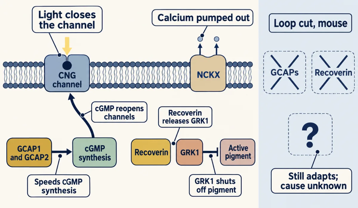
Light reduces calcium entry through outer-segment CNG channels while NCKX continues calcium extrusion. Falling calcium promotes GCAP-dependent cGMP synthesis, releases GRK1 from recoverin inhibition, and relieves calcium-dependent inhibition of channel sensitivity to cGMP. The direct channel effect is small in mouse rods. Removing GCAP1, GCAP2 and recoverin does not eliminate adaptation: the remainder in mouse rods was calcium-dependent, while mouse cones also showed a calcium-independent component. The molecular basis of the unexplained remainder is unresolved. AI-generated illustration -
Reading an adaptation curve: half-sensitivity background and slope
-
Rods and cones divide the range, and rods in daylight
-
A second question: when photoreceptors die
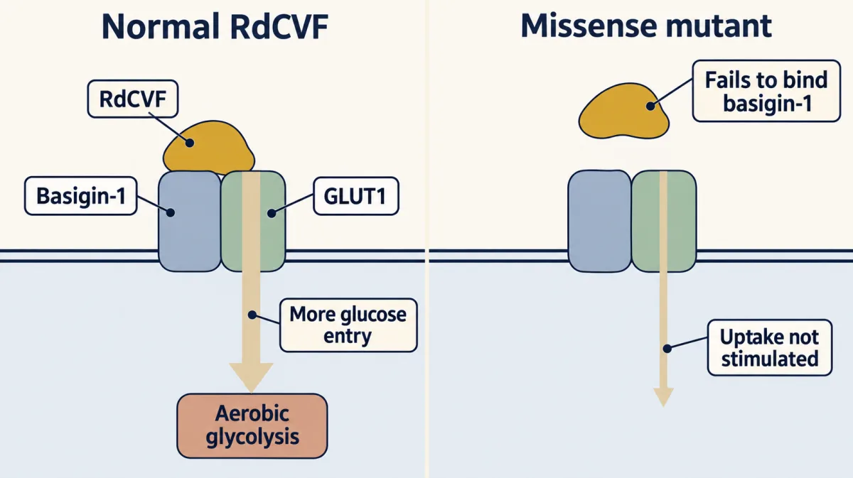
In embryonic chicken cone cultures and the rd1 mouse model, RdCVF binding to basigin-1, which associates with GLUT1, promotes cone glucose entry and aerobic glycolysis. A tested RdCVF missense mutant failed to bind basigin-1 or stimulate glucose uptake; this does not establish that basal glucose transport stops. This experimental pathway is one proposed contributor to rod–cone dependence, not a human trial. AI-generated illustration -
Rescuing vision: what two gene-therapy trials can claim
-
Going deeper
Where this lesson fits
Before thisReview first
- Five Ways to Turn a Stimulus into a CurrentThis course · Lesson 1
It sets out how a stimulus becomes a current, which this lesson assumes when light switches off part of the dark current.
- Parallel Channels Out of the EyeThis course · Lesson 18
It describes the rod and cone outputs whose gain and operating range are compared here.
After thisStudy next
- Restoring Senses: Implants, Prostheses and Optogenetic VisionThis course · Lesson 30
It continues from the gene-therapy and trial claims here to implants and optogenetic vision restoration.
Elsewhere on the siteFilms, simulations and method films in other courses
- Centre and surroundSimulation · Neuroscience
It shows how a rod's output falls as light closes its channels, the dark current this lesson's feedback protects.
- A Fall Can Carry a SignalFilm · Cell Signaling
It shows how light lowers cGMP and closes channels in rods, the dark current this feedback regulates.
- The Mark and the ReaderFilm · Cell Signaling
It shows how rhodopsin kinase and arrestin shut off a receptor, the step recoverin controls through GRK1.
Suggested by an AI model from the lessons themselves, then checked. How we use AI