Neuroscience · Unit 7: Evidence and disease
When Mature Neurons Fail: Neurodegeneration
Rewritten on : new text, figures, flashcards and film. Your notes and highlights stay, but the text around them may have moved.
Lecanemab slowed worsening in early Alzheimer disease by about 27%. The treated group still got worse.
Pause when the reading reaches a film, and offer to play it. Saved on this device.
Watch the film now, or keep listening
Builds on:Memory SystemsDescending Control and the Basal GangliaMotor Units, Reflexes and LocomotionWhy these?
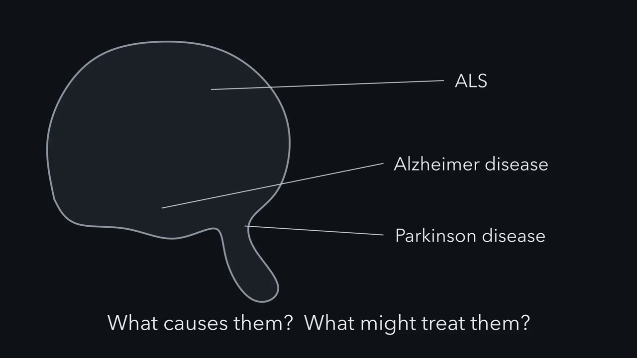 ▶ Watch Three Questions, Three Kinds of Evidence · 2:56
Read the transcript of “Three Questions, Three Kinds of Evidence”
Film transcript: Three Questions, Three Kinds of Evidence
Three diseases each kill a particular set of adult neurons. How do we learn what causes them, and what might treat them? In Alzheimer disease, early losses include neurons of the entorhinal cortex, and of field CA1 in the hippocampus. Both belong to the circuit for new memories. In Parkinson disease, the defining loss is of dopamine neurons in the substantia nigra, in the midbrain. In ALS, motor neurons die. Upper ones sit in the motor cortex. Lower ones send axons to muscle. In each disease, a protein misfolds and clumps. Alzheimer disease has amyloid plaques outside neurons, and tau tangles inside them. In Parkinson disease, α-synuclein is the main protein in the filaments of Lewy bodies. Most ALS has clumps of TDP-43.
Now three questions, each with its own kind of evidence. First, an autopsy tells you what a clump is made of. It does not tell you whether the clump kills the cell. Now look at rare families. A gene variant passes from parent to child along with the illness. That travelling together is called cosegregation. It is strong evidence for a cause. But a harmless variant nearby can travel along too. A second family, and tests of what the variant does, make the case. Here is a puzzle. A family's ALS is caused by a mutation in SOD1. Most people with ALS carry TDP-43 clumps. Will this family's motor neurons have them?
No. In a postmortem series, cases with SOD1 mutations lacked TDP-43 clumps. Their motor neurons died anyway. A disease-causing gene need not encode the protein in the usual deposit. In Alzheimer disease, the three classic dominant genes all affect how amyloid-beta is made or processed. Neither deposits nor disease-causing genes alone show that removing a protein after onset will help. Randomized trials in people give the most direct test, judged on how they think, function or survive. So ask what each finding can show. Autopsy shows what builds up. Families can show what starts it. Trials test whether changing it helps, and what it costs.
Ask two things of every finding. What does it show? And what can it not show?
From the film
- Each kind of evidence supports some claims and not others.
- An autopsy shows what builds up, and families can show what can start the disease.
- Randomized trials in people give the most direct test of whether changing a target helps.
In a large trial, people with early Alzheimer disease received either lecanemab, an antibody that clears amyloid plaques from the brain, or a placebo for 18 months. Both groups got worse. The treated group got worse more slowly.
Amyloid plaques are deposits of a small peptide, amyloid-β (Aβ), that build up between neurons in Alzheimer disease. The gap between the two groups is some of the strongest evidence that a drug that clears amyloid can change the course of the disease. It is also evidence that the drug did not stop it. Reading that result well means knowing what trials, autopsies and family genetics can each show.
Four questions and one idea
This lesson compares three diseases: Alzheimer disease (AD), Parkinson disease (PD) and amyotrophic lateral sclerosis (ALS). For each one it asks four questions.
- Which neurons die?
- What builds up in or around the affected neurons?
- What can start the disease?
- Does removing what builds up help?
The one idea to carry through this lesson. Each kind of evidence supports some claims and not others. A deposit seen at autopsy shows what builds up, not whether it kills. A disease-causing variant found in families shows what can start the disease, not whether removing its product later helps. Whether changing the target helps once the disease is under way is tested most directly by randomized trials in people, judged on clinical outcomes such as thinking, daily function or survival, which also measure the harms. For amyloid in early Alzheimer disease, the answer so far is that decline slows but does not stop.
The rule concerns evidence for cause and treatment. How a committed neuron dies is in death machinery in neurons, and the cell’s protein disposal systems are in quality control and autophagy. After the four questions, the lesson asks why these neurons fail first, which is still open.
Which neurons die: three diseases, three populations
A neurodegenerative disease is a progressive loss of particular neurons. The three diseases here usually begin in adult life. The important word is particular. Selective vulnerability means that the pathology strikes some neuronal populations and spares others, often close neighbours (reviewed by Fu, Hardy and Duff, 2018). Any account of these diseases has to fit this pattern.
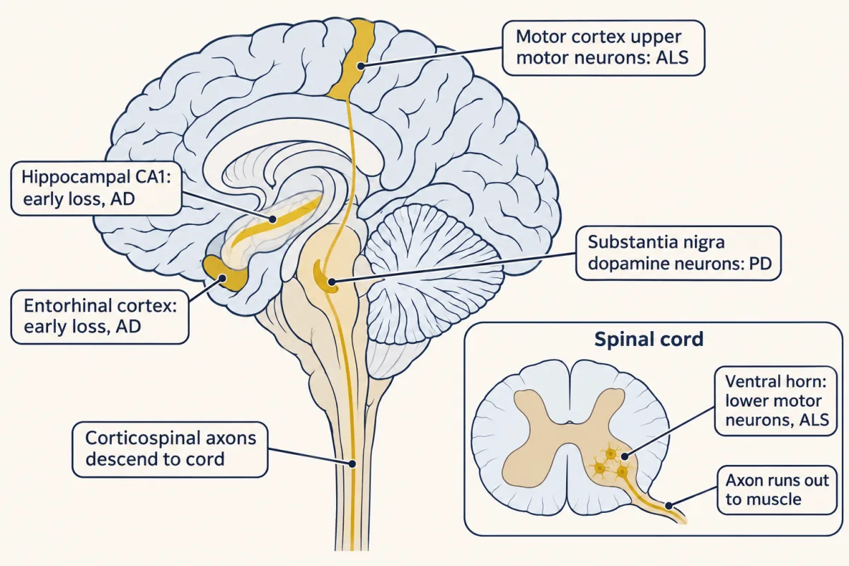
In Alzheimer disease, neurons lost early include neurons of layer II of the entorhinal cortex, the main gateway between neocortex and hippocampus, and pyramidal neurons of hippocampal field CA1. Both belong to the circuit for new declarative memories (memory systems).
In Parkinson disease, the defining loss is of dopamine neurons in the substantia nigra pars compacta (SNpc), the midbrain nucleus whose axons supply the striatum. Their loss unbalances the loops described in the basal ganglia. Even “dopamine neurons” is too broad. When the dopamine neurons of postmortem human substantia nigra were sorted into ten populations by the genes they express, one population, marked by expression of the gene AGTR1, was highly susceptible to loss in PD (Kamath and colleagues, 2022).
In amyotrophic lateral sclerosis (ALS), the targets are motor neurons. Upper motor neurons have their cell bodies in motor cortex and send axons down the corticospinal tract to the spinal cord and the corticobulbar projection to brainstem motor nuclei. Lower motor neurons sit in the brainstem and the ventral horn of the spinal cord and send axons to muscle. Both the cell bodies and their axons degenerate. As lower motor neurons die, whole motor units (one motor neuron and all the muscle fibres it supplies; see motor units and reflexes) drop out.
What builds up: proteins that clump
Each disease has a protein that misfolds and gathers into insoluble deposits seen at autopsy.
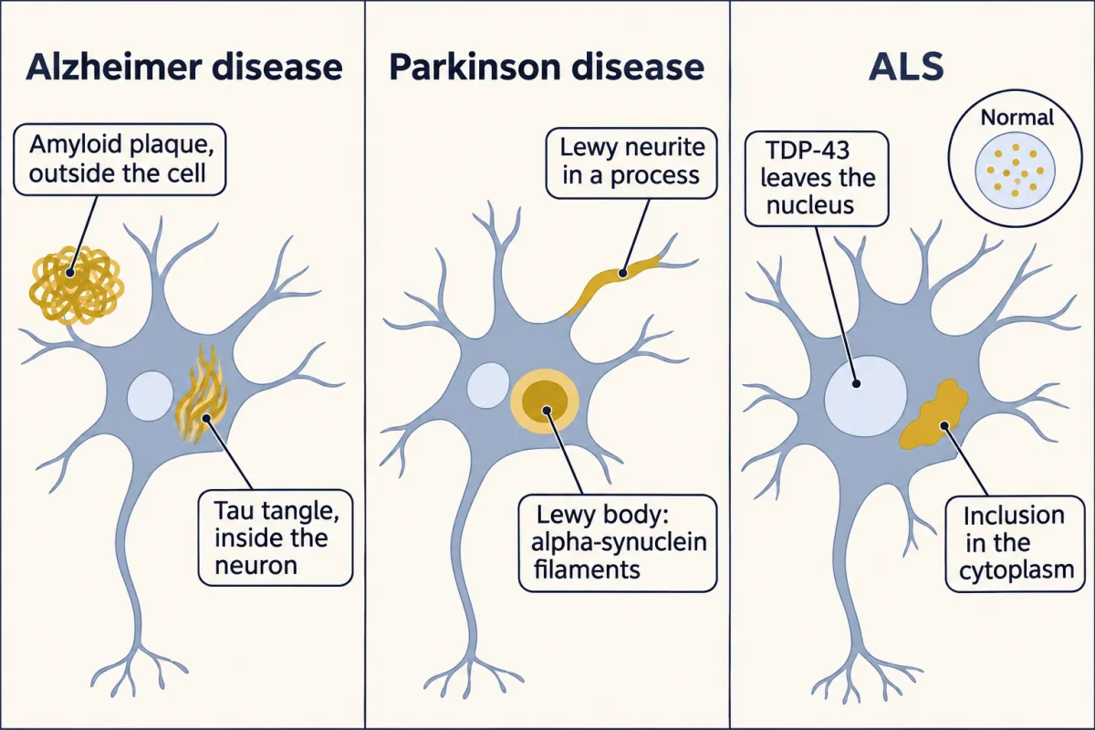
- Alzheimer disease (AD) has two. Amyloid plaques lie outside cells and contain Aβ, peptides cut from amyloid precursor protein (APP) by two protein-cutting enzymes, β-secretase and then γ-secretase. Neurofibrillary tangles lie inside neurons and contain misfolded, hyperphosphorylated tau (carrying extra phosphate groups), a protein that normally binds microtubules.
- Parkinson disease (PD) has Lewy bodies and Lewy neurites, inclusions in neuronal cell bodies and processes. In 1998 Spillantini, Goedert and colleagues found α-synuclein to be the major component of the filaments extracted from Lewy bodies.
- Amyotrophic lateral sclerosis (ALS) in most cases has inclusions of TDP-43, an RNA-binding protein that leaves the nucleus and gathers in the cytoplasm. Neumann and colleagues identified it in 2006. There are exceptions, and the next section uses them.
Predict. Suppose a protein turns up in every lesion of a disease. Does it follow that the protein kills the neuron?
Check your prediction
No. A deposit tells you what a lesion is made of, not that it kills the cell. Small soluble aggregates, loss of the protein’s normal job, or the cell’s response could do the damage. For Lewy bodies, for example, whether the inclusions themselves kill neurons is still argued.
You may also read that misfolded seeds pass from cell to cell within one nervous system and template more misfolding (“prion-like spread”). That is a hypothesis, tested mostly in animal and cell models. These diseases do not pass between people through ordinary contact, although rare transmission of Aβ pathology through contaminated medical material, such as growth hormone taken from cadaver pituitaries, has been reported (Banerjee and colleagues, 2024).
What can start it: genes that cause and genes that raise risk
To ask what starts the disease, look at families. Two kinds of gene variant matter here, and the difference between them carries the rest of the lesson. The background is in pedigrees and complex traits and GWAS.
- A Mendelian variant can cause the disease in the families that carry it. In dominant disease one altered copy can be enough; in recessive disease, such as parkin parkinsonism, both copies of the gene are altered.
- A risk allele only shifts the odds. Many carriers never get the disease, and many patients do not carry it. It is neither necessary nor sufficient.
In a pedigree, a Mendelian disease genotype and the illness travel together: affected relatives carry it, and relatives without it usually stay free of the family’s disease. This travelling together is called cosegregation. It is evidence for a cause, not proof by itself, because a nearby harmless variant can travel with the true one. Finding the same pattern in a second, unrelated family, and showing what the variant does, makes the case much stronger.
Even so, not everyone with the disease genotype falls ill by a given age. The share who do by that age is the penetrance, and it can be incomplete. For LRRK2 G2019S, one consortium estimate was 74% by age 79, and other studies report lower figures.
Mendelian variants are rare, and most people with alzheimer disease (AD), Parkinson disease (PD) or amyotrophic lateral sclerosis (ALS) carry no known Mendelian variant. They matter because each established one names something that can start the disease.
Predict. A family’s ALS is caused by a mutation in SOD1, an antioxidant enzyme. Most people with ALS have TDP-43 inclusions in their motor neurons. Would you expect this family’s motor neurons to have them?
Check your prediction
No. In a 2007 postmortem series, Mackenzie and colleagues found that ALS cases with SOD1 mutations lacked TDP-43 inclusions, yet their motor neurons died. In FUS-linked familial ALS, mutant FUS accumulates in neuronal cytoplasm, and TDP-43 inclusions are also absent (Vance and colleagues, 2009). The table below shows where gene and deposit match and where they part.
The last column says how each gene relates to the usual deposit (Aβ in AD, α-synuclein in PD, TDP-43 in ALS): it may encode that protein, influence how it is processed, or cause disease with different or variable deposits.
| Disease | Mendelian gene (examples) | What the gene makes | Relationship to the usual hallmark deposit |
|---|---|---|---|
| AD | APP (dominant) | amyloid precursor protein | Encodes the precursor from which plaque Aβ is cut |
| AD | PSEN1, PSEN2 (dominant) | presenilin, the catalytic core of γ-secretase, the enzyme whose cut releases Aβ | Influences how Aβ is processed |
| PD | SNCA (dominant) | α-synuclein | Encodes the major Lewy body filament protein |
| PD | LRRK2 (dominant); parkin (recessive, juvenile onset) | a protein kinase; a ubiquitin ligase (an enzyme that tags proteins for disposal) | Encode other proteins; Lewy bodies are variable in LRRK2 disease and absent in most parkin cases studied at autopsy |
| ALS | TARDBP | TDP-43 | Encodes the deposit protein itself |
| ALS | SOD1 | superoxide dismutase 1 | Encodes another protein; typical SOD1 ALS lacks TDP-43 inclusions |
| ALS | FUS | FUS, an RNA-binding protein | Encodes another protein; FUS inclusions occur without the usual TDP-43 inclusions |
| ALS | C9ORF72 (a GGGGCC repeat expansion) | C9ORF72 protein; the disease change is a repeat expansion outside the protein-coding sequence, not a missense change, and expanded-repeat RNA can be translated without a normal start codon into abnormal dipeptide-repeat proteins | Does not encode TDP-43; TDP-43 inclusions are nevertheless typical |
Read down the last column. In Alzheimer disease all three dominant genes act on Aβ. In Parkinson disease and ALS, gene and deposit sometimes part company, so the gene that can start a disease and the protein that builds up need not be the same.
The best-known risk allele is APOE ε4. It raises the risk of late-onset AD, yet even in old age it is not sufficient to produce clinical disease, and many patients carry no ε4.
The AD rows have a pattern: all three dominant genes act on how much Aβ is made or which lengths. Presenilin mutations, for example, shift the cut toward longer forms such as Aβ42, which clump more readily, sometimes while lowering the total. In 1992, building on the APP mutations, Hardy and Higgins proposed the amyloid cascade hypothesis: Aβ accumulation starts a process that leads to tangles, neuron loss and dementia. The presenilin genes, found in 1995, later fit the same pattern. Mendelian genetics supports an initiating role for Aβ in familial disease. It does not show that Aβ alone explains the common late-onset form, and it does not show that removing Aβ from a patient will help. That needs direct evidence of treatment benefit, ideally from randomized trials.
Does removing it help: testing amyloid in trials
Read the transcript of “Slower Worsening Is Still Worsening”
Film transcript: Slower Worsening Is Still Worsening
A drug clears amyloid from the brain. Does that help the person whose brain it is? The antibody lecanemab clears amyloid plaques. In a large trial, people with early Alzheimer disease got it or a placebo. Both groups were followed for a year and a half. The outcome was a clinician's rating of thinking and daily function. It runs from zero to eighteen, and higher means worse. The graph shows each group's change from its starting score. Here is a question. Did the treated group get better?
No. Both lines rise. Both groups got worse. The treated group got worse more slowly. To compare them, take the gap as a share of the placebo group's decline. The gap is a little more than a quarter of the placebo decline. That share is called the relative slowing. That share compares two amounts of decline. It is not a gain in memory. It is not the share of patients helped. It is not anyone's prognosis.
Slower worsening is still worsening. In this trial, a drug that clears amyloid slowed the disease. It did not stop it. A second trial tested a different antibody, solanezumab. It enrolled older adults whose thinking was still normal, but whose brains held extra amyloid. On PET scans, amyloid rose in both groups. It rose more slowly with the drug. Predict. Did their thinking decline more slowly too?
Both groups declined. The trial did not demonstrate slower decline with the drug, despite the smaller rise in amyloid. ALS shows the same thing. In people with S .O .D .1, a L .S., a drug called tofersen lowered SOD1. It also lowered a blood marker of axon damage. But over twenty-eight weeks, the clinical measures did not improve. That is the treatment question. A marker can guide you. But a clinical outcome is what tells you whether changing the target helps. Benefits also have costs. Another antibody, donanemab, was tested in a large trial. Brain swelling or fluid on MRI was far more common with it than on placebo.
Read a trial as two amounts of decline. And keep a change in a marker apart from a change in the person.
From the film
- In the lecanemab trial both groups got worse, and the gap was a little more than a quarter of the placebo decline.
- That share compares two amounts of decline, and slower worsening is still worsening.
- A change in a biomarker is not a clinical benefit, and benefits can come with costs such as brain swelling on MRI.
If Aβ starts the disease, removing it might change the course, though a process already under way may no longer need Aβ to keep going. Antibodies against Aβ have now tested that.
Worked problem: what does “27% slowing” mean? Clarity alzheimer disease (AD) (van Dyck and colleagues, 2023) randomized 1,795 people with mild cognitive impairment or mild dementia due to AD, with amyloid confirmed, to lecanemab or placebo for 18 months. The primary outcome was the change in CDR-SB (Clinical Dementia Rating, Sum of Boxes), a clinician’s rating of cognition and daily function that runs from 0 to 18, higher meaning worse. Adjusted mean changes at 18 months were +1.21 points with lecanemab and +1.66 with placebo. Here we work with these group averages.
Step 1. Absolute difference. 1.66 − 1.21 = 0.45 points less worsening. (The paper subtracts the other way, reporting −0.45 points, 95% confidence interval −0.67 to −0.23.)
Step 2. Relative slowing, using the placebo change as the base:
relative slowing = (placebo change − drug change) / placebo change = 0.45 / 1.66 = 0.271, about 27%
Check: 1.66 × (1 − 0.271) = 1.66 × 0.729 = 1.21.
Step 3, predict first. A headline says the drug “slowed decline by 27%”. Did the treated group still get worse, and what is the 27% a share of?
Check your prediction
Yes. Its mean change was +1.21, a worsening. The 27% is a share of the placebo group’s decline. It is not a 27% gain in memory, not a fraction of patients helped, and not anyone’s prognosis. Slower worsening is still worsening. When the placebo group worsens on average, any relative slowing below 100% still leaves the treated group worsening on average.
In this sketch: group means only, no individual courses; each bar is one number (a reported figure at the published values) drawn from zero, not a trajectory over time. Mechanism-level reading of published trials, not advice.
Rule to keep: a relative figure such as 27% compares two amounts of decline. It is not a 27% gain in memory, a fraction of patients helped or anyone’s prognosis, and setting it beside another trial’s percentage does not rank the drugs, because the scales, populations and durations differ.
What it costs
Safety is part of the result. In TRAILBLAZER-ALZ 2 (Sims and colleagues, 2023), another antibody, donanemab, was tested in 1,736 people with early symptomatic AD and PET evidence of amyloid and tau. ARIA with edema or effusion, amyloid-related imaging abnormalities seen on MRI as fluid or swelling in the brain, occurred in 24.0% of donanemab recipients and 2.1% on placebo.
Report that two ways. As a ratio, 24.0 / 2.1 ≈ 11.4: it was about eleven times as common with the drug. As a gap, it is 21.9 percentage points, about one extra case for every 4.6 people treated over the trial’s 76 weeks: that says how many people it touches. Three deaths with donanemab and one with placebo were considered treatment related.
Donanemab also slowed decline, by 35.1% in people with low or medium tau and by 22.3% in the combined population, but on a different scale (iADRS, the integrated Alzheimer’s Disease Rating Scale) over 76 weeks. Setting 27% beside 35.1% does not rank the drugs: the scales, populations and durations differ.
A biomarker is not a benefit
A slower rise in a biomarker, a lab or imaging measure of the disease process, is not the same as slower decline in the person. In the A4 study (Sperling and colleagues, 2023), cognitively unimpaired adults aged 65 to 85 with elevated amyloid received solanezumab, an antibody to the single-molecule (monomeric) form of Aβ, or placebo for 240 weeks. Amyloid PET is measured in centiloids, a standard scale on which 0 is the average of young adults without plaques and 100 is the average in typical Alzheimer dementia. It rose in both arms, by 11.6 centiloids with drug and 19.3 with placebo. So amyloid built up about 40% more slowly with the drug: (19.3 − 11.6) / 19.3 ≈ 0.40.
Predict. Did these people’s thinking decline more slowly too?
Check your prediction
Not as far as the trial could show. Thinking declined in both groups, and the trial did not demonstrate slower decline with the drug. A biomarker effect alone does not establish clinical benefit.
Amyotrophic lateral sclerosis (ALS) gives the same lesson from another angle. In a 28-week randomized trial in SOD1-mutant ALS, tofersen, an antisense drug that lowers SOD1 production, reduced SOD1 in cerebrospinal fluid and neurofilament light (a protein released from damaged axons) in plasma more than placebo, but clinical endpoints did not improve over those 28 weeks (Miller and colleagues, 2022). Going deeper gives the longer follow-up and the approval that rested on the neurofilament result.
What none of the trials showed
None of these trials showed reversal of decline, return of lost neurons, or benefit beyond the populations and durations studied, and the Clarity AD authors called for longer trials. Many earlier anti-amyloid drugs did not meet their endpoints (reviewed by Selkoe and Hardy, 2016), and verubecestat, which blocks Aβ production, did not help people with early symptoms and left some scores worse than placebo (Egan and colleagues, 2019). Together the results support a contribution of amyloid that treatment can reach, without establishing amyloid as the sole cause.
Why these neurons
Read the transcript of “Protecting Cut Axons Did Not Rescue This ALS Model”
Film transcript: Protecting Cut Axons Did Not Rescue This ALS Model
Why do some neurons fail while their neighbours survive? No one yet knows. So how do you test one idea about it? Start with injury. When an axon is cut, the piece cut off from the cell body breaks into fragments within days. This was long thought of as passive wasting. It is not. The cut-off piece runs an active destruction program. In flies and mice, removing one gene, Sarm1, keeps the cut-off piece intact for weeks. Axons also degenerate in ALS. So the obvious next test is to remove Sarm1 from a mouse model of ALS. These mice make a mutant human SOD1 protein, and they develop motor neuron disease. Predict. Without Sarm1, will these mice live longer?
No. Survival did not improve. Nor did the behavioural, nerve recording or tissue measures. Needed after a cut does not mean needed in this disease. The motor neurons may die by another route. And one mouse model is not human ALS. The result stays inside this model. Does this show axon loss does not matter in ALS? No. That claim goes too far. A better test measures axons, nerve-muscle contacts and cell bodies, as well as survival, in the same mice. The scorers should not know each mouse's genotype. Then the test is repeated in a second model. Here is what each result supports. Losing Sarm1 protects cut axons. In this model, losing it did not improve the disease measures.
State what each result supports, and what it leaves open. Then design the next experiment to test what is still open.
From the film
- Removing Sarm1 protects cut axons, but in mice that make mutant human SOD1 it did not improve the disease measures.
- Needed after a cut does not mean needed in the disease, and one mouse model is not human ALS.
- The result does not show that axon loss is unimportant in ALS, so the next test should measure more outcomes and be repeated in a second model.
No one yet knows why an entorhinal layer II neuron or an SNpc dopamine neuron fails before its neighbours. (In Parkinson disease, some brainstem neurons are affected even earlier than nigral ones.) The rule still applies: ask what each kind of evidence can support. A feature that goes with vulnerability is a correlate. Changing something in an animal model and seeing the disease course change is stronger, but it holds for that model. A mechanism needed after injury may or may not be needed in disease.
A correlate. Nigral dopamine neurons have very large axonal arbors and high energy demand, which may contribute to their vulnerability compared with neighbouring dopamine neurons of the ventral tegmental area.
A manipulation in a model. Selectivity also works within a cell class. Gamma motor neurons, small motor neurons that set the sensitivity of muscle spindles, were completely spared while alpha motor neurons degenerated in SOD1, TDP-43 and FUS mutant mouse models (Lalancette-Hebert and colleagues, 2016).
In the same study, genetically reducing the number of gamma motor neurons, and so the activation of spindle afferents (the sensory fibres from muscle spindles), delayed onset and prolonged lifespan in SOD1G93A mice, which overexpress a mutant human SOD1. So circuit input can change how fast a vulnerable neuron dies, at least in that model.
A mechanism needed after injury. Axons and synapses degenerate in injury and in these diseases alike, and injury offers a well-defined mechanism to compare. After a cut, the part of the axon cut off from the cell body (the distal axon) fragments in Wallerian degeneration, an active program that requires SARM1 in flies and mice (Osterloh and colleagues, 2012); the purified TIR domain of SARM1 has intrinsic NAD+-cleaving activity. The full story is in injury and regeneration. Whether that program drives chronic disease is a separate question.
Design problem
When Peters and colleagues (2018) crossed Sarm1-null mice (mice lacking the gene) with SOD1G93A mice, Sarm1 deletion did not improve survival or the behavioural, electrophysiological or histological outcomes they measured. A colleague concludes that axon degeneration does not matter in amyotrophic lateral sclerosis (ALS). Is that justified, and what would you do next? Predict your answer before you open the box.
Saved in this browser only.
One way to do it
The conclusion goes too far. The 2012 result concerns a cut; it does not show that chronic disease uses the same program. Motor neurons in SOD1G93A mice may die by another route, such as an apoptotic pathway in the cell body, or axons might be saved in part without any gain in survival.
Design: measure separate endpoints in the same mice, with genotype hidden from the scorers. Count axons in ventral roots and peripheral nerve, score neuromuscular junction innervation, estimate motor units electrophysiologically (motor unit number estimation), count spinal motor neuron cell bodies, and record onset and survival.
Include a positive control, a check that the method can detect an effect, in a separate, matched cohort: cut a nerve and confirm that Sarm1-null axons resist degeneration, so a negative result in disease cannot be blamed on a failed knockout. Leave the disease-outcome mice uninjured, because a cut nerve would itself change motor and survival measures. Then test a second model, such as a TDP-43 model, because one SOD1 overexpression line is not human ALS. A clean answer would read: “Sarm1 loss protects cut axons in these mice but does not change the measured course of disease in this model.”
Going deeper
How the dopamine subtypes were found
Kamath and colleagues (2022) sequenced RNA from single nuclei of postmortem human substantia nigra to sort the dopamine neurons, then mapped the populations within the substantia nigra with Slide-seq (a method that reads out RNA at known positions in a tissue section) in macaque midbrain, with human tissue used to confirm the locations.
The founding families
The families behind the table: an amyloid precursor protein (APP) variant cosegregating in a family (Goate and colleagues, 1991); SNCA in Italian and Greek-origin kindreds (1997); parkin deletions in Japanese juvenile parkinsonism (1998); 11 SOD1 missense mutations in 13 families (Rosen and colleagues, 1993); and a GGGGCC repeat expansion in C9ORF72 (2011).
A protective variant: what genetics can say about lifelong change
A rare APP variant, A673T, reduces Aβ production and was found to protect carriers against Alzheimer disease (Jonsson and colleagues, 2012). That is human genetic evidence that supports the idea that lifelong lower Aβ production protects. It does not show that lowering Aβ in someone who is already ill will help, which is the question the trials ask.
Is a 0.45-point gap meaningful?
Whether a gap of this size matters in daily life is openly debated. Andrews and colleagues (2019) estimated the smallest meaningful worsening in one person on this scale at about 1 point in mild cognitive impairment and about 1.6 in mild dementia. Those are thresholds for an individual’s change, not for the gap between two group averages, so setting 0.45 beside them does not settle the question.
A what-if: time saved
This is a what-if, not a trial result. If decline in each arm were a straight line over the 18 months, placebo would worsen by about 0.092 points a month and lecanemab by about 0.067. A drug that changes the rate of decline should then open a gap that grows with time: about 0.15 points at 6 months, 0.30 at 12 and 0.45 at 18. On the same assumption, the 0.45-point gap equals about 4.9 months of placebo decline (0.45 / (1.66 / 18)). Real decline need not be linear, so both figures depend on that assumption.
Tofersen after 28 weeks
In the open-label extension, people who had started tofersen at entry had declined less at 52 weeks than those who started 28 weeks later, a hint that needs confirmation. In 2023 the US Food and Drug Administration granted tofersen accelerated approval on the basis of the fall in plasma neurofilament light, as a marker judged reasonably likely to predict clinical benefit. That is the treatment question in practice: a regulator accepted a biomarker while the clinical answer is still being sought.
Your notes
Select any text in the lesson to highlight it or add a note to it. Your highlights and notes are saved in this browser and collected in your notebook.
This browser is not letting the site save anything (a private window or strict privacy settings), so highlights and notes will be lost when you leave the page.
How we measure it
Pedigree linkage and cosegregation
You follow genetic markers and candidate variants through a family with an inherited disease and ask whether one variant travels with affected relatives and not with unaffected ones. Replication in an unrelated family strengthens the case that the variant causes disease in those families, not in every patient.
Immunoelectron microscopy of extracted filaments
Insoluble filaments are extracted from postmortem brain, labelled with antibodies carrying gold particles, and imaged in the electron microscope. Gold on the filaments identifies a component. It tells you composition, not whether the filaments cause cell death.
Axotomy with genetic loss of function
You cut an identified nerve in flies or mice lacking a gene and compare the distal axon with that of controls over days to weeks. Surviving axons show that the gene is needed for degeneration after injury, in that preparation.
Randomized placebo-controlled trial
Participants are assigned at random to drug or placebo, usually with neither they nor the assessors knowing which, and followed for a fixed time on a prespecified clinical endpoint, with biomarkers and safety measured alongside. The difference between arms estimates the drug's effect in the enrolled population.
Practice questions on this lesson (3) → MCAT-style, original, each with an explanation
Where this lesson fits
Before thisReview first
- Memory SystemsThis course · Lesson 22
You need the memory circuits through the entorhinal cortex and hippocampus from there to see why Alzheimer disease first costs new memories.
- Descending Control and the Basal GangliaThis course · Lesson 16
You need the dopamine loops through the striatum from there to see what losing substantia nigra neurons unbalances.
- Motor Units, Reflexes and LocomotionThis course · Lesson 15
You need the motor unit from there to see why losing lower motor neurons removes whole units of muscle.
After thisStudy next
- Designing a Causal Circuit ExperimentThis course · Lesson 28
It takes the lesson's closing problem, a mouse result that does not carry over, into designing a causal experiment with proper controls.
Elsewhere on the siteFilms, simulations and method films in other courses
- Ras to MAPK: From Membrane to GenesFilm · Cell Signaling
Oncogenes show how stuck switches arise in families and in tumors, like causal versus risk genes here.
Suggested by an AI model from the lessons themselves, then checked. How we use AI
Finish line
Test yourself
8 questions
Practice and MCAT-style, with explained answers.
Next · Lesson 27
Recording Many Neurons: Imaging, Arrays and Atlases
A probe with 960 recording sites does not record one neuron per site. What does it record?
Continue →