Developmental Neuroscience · Skim version
Building the Neuromuscular Junction: Agrin, Lrp4 and MuSK
Read the full lesson → Skim the whole course →
The guiding idea
A mouse without the muscle kinase MuSK dies at birth: its motor axons reach the muscle, but no junction forms.
The muscle proposes; the nerve disposes: in developing mouse muscle, the muscle builds receptor clusters without a nerve, and the nerve decides which stay, agrin protecting them and acetylcholine dispersing the rest.
From the film: The Muscle Proposes, the Nerve Disposes
- A synapse is not built simply because an axon touches a target.
- The chain runs from neural agrin to Lrp4, MuSK with Dok7, rapsyn and then clustered acetylcholine receptors.
- The muscle proposes; the nerve disposes: the muscle builds clusters with its own machinery, and the nerve decides which stay.
From the film: A Band Without a Nerve
- In mouse embryos whose diaphragm receives no motor axons, a band of receptor clusters still forms, so the muscle draws a rough map by itself.
- The muscle's own machinery, including MuSK and rapsyn, is needed to start the band; the nerve's signal is not.
- The nerve still matters: contact makes far more clustering and narrows the band.
From the film: Agrin Protects, Acetylcholine Disperses
- Acetylcholine, released at every terminal, disperses clusters that agrin does not protect.
- In mice lacking both agrin and acetylcholine, clusters return, so a major job of agrin in developing muscle is to protect clusters from acetylcholine.
- In the working model, agrin is anchored in the basal lamina, so its protection stays under the terminal.
-
The sorting problem
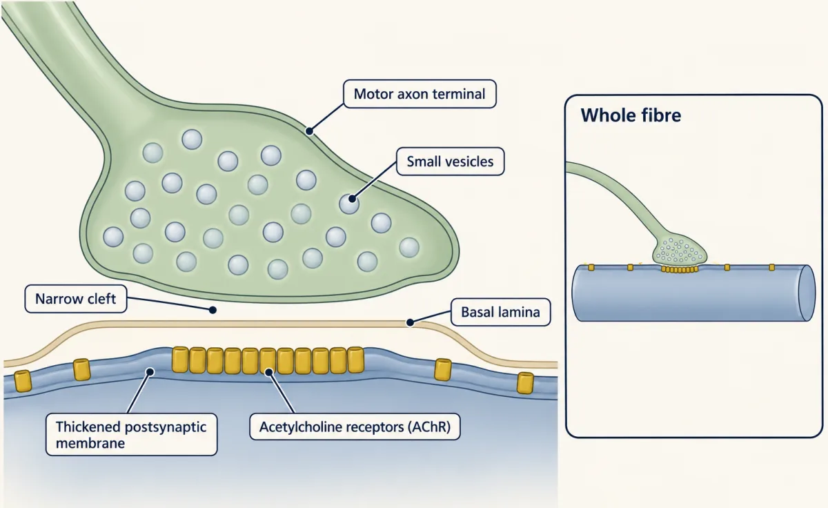
The neuromuscular junction shows all three signs of a synaptic specialization: small vesicles in the terminal, a narrow cleft that holds a sheet of basal lamina, and a thickened postsynaptic membrane where the acetylcholine receptors are concentrated. AI-generated illustration -
How the nerve’s signal was found
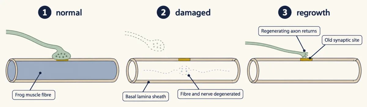
In frog muscle, after the fibres and nerve terminals degenerated, regenerating axons returned to the old synaptic sites on the empty basal lamina sheaths. The basal lamina remembered where the synapse had been. AI-generated illustration -
The muscle starts without the nerve
-
A paradox, and a second force
-
A second effect of activity: the nuclei
-
Signals in both directions
-
NMJ vs. central synapses, and a junction that fails
-
Putting it together
-
Going deeper
Where this lesson fits
Before thisReview first
- The Growth Cone: The Tip That Steers the AxonThis course · Lesson 3
You need how the axon tip reaches and touches its target before asking why contact alone does not build a synapse.
- The Neurotrophic Hypothesis TestedThis course · Lesson 29
The lesson contrasts its rule of making too many and keeping what is supported with the muscle making surplus clusters.
- Neurotrophins, Trk Receptors and p75This course · Lesson 30
Receptor-kinase signaling from a target-derived factor is the model you reuse here for agrin and MuSK.
After thisStudy next
- Synapse Elimination: One Muscle Fiber, One AxonThis course · Lesson 37
A fibre that starts with several axons ends with one, building on the junction formed here.
- Synapses in the Brain: Adhesion, Scaffolds and AstrocytesThis course · Lesson 36
It asks which of this junction's signals carry over to brain synapses, which lack a basal lamina and use other molecules.
- Disorders of the Synapse: Built, Then KeptThis course · Lesson 49
It applies the idea that synapses must be built and then kept to disorders where that fails.
Elsewhere on the siteFilms, simulations and method films in other courses
- Counting failuresSimulation · Neuroscience
The acetylcholine released at the junction acts through the quantal release of transmitter that this simulation explores.
- Blips from a Silent NerveFilm · Neuroscience
It shows how acetylcholine release at the endplate works, which is the transmitter that disperses clusters here.
- Sliding, Not ShrinkingFilm · Organismal Biology
It shows the muscle fiber that the nerve contacts and drives through the junction.
Suggested by an AI model from the lessons themselves, then checked. How we use AI