Neuroscience · Skim version
When Mature Neurons Fail: Neurodegeneration
Read the full lesson → Skim the whole course →
The guiding idea
Lecanemab slowed worsening in early Alzheimer disease by about 27%. The treated group still got worse.
Each kind of evidence about neurodegeneration supports some claims and not others: an autopsy shows what builds up, families show what can start the disease, and randomized trials test whether changing it helps.
From the film: Three Questions, Three Kinds of Evidence
- Each kind of evidence supports some claims and not others.
- An autopsy shows what builds up, and families can show what can start the disease.
- Randomized trials in people give the most direct test of whether changing a target helps.
From the film: Slower Worsening Is Still Worsening
- In the lecanemab trial both groups got worse, and the gap was a little more than a quarter of the placebo decline.
- That share compares two amounts of decline, and slower worsening is still worsening.
- A change in a biomarker is not a clinical benefit, and benefits can come with costs such as brain swelling on MRI.
From the film: Protecting Cut Axons Did Not Rescue This ALS Model
- Removing Sarm1 protects cut axons, but in mice that make mutant human SOD1 it did not improve the disease measures.
- Needed after a cut does not mean needed in the disease, and one mouse model is not human ALS.
- The result does not show that axon loss is unimportant in ALS, so the next test should measure more outcomes and be repeated in a second model.
-
Four questions and one idea
-
Which neurons die: three diseases, three populations
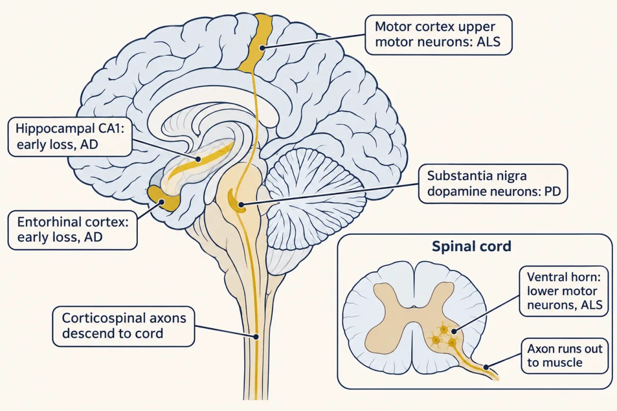
Each disease strikes particular neurons and spares others: entorhinal and CA1 neurons early in Alzheimer disease, nigral dopamine neurons in Parkinson disease, and upper and lower motor neurons in ALS. AI-generated illustration -
What builds up: proteins that clump
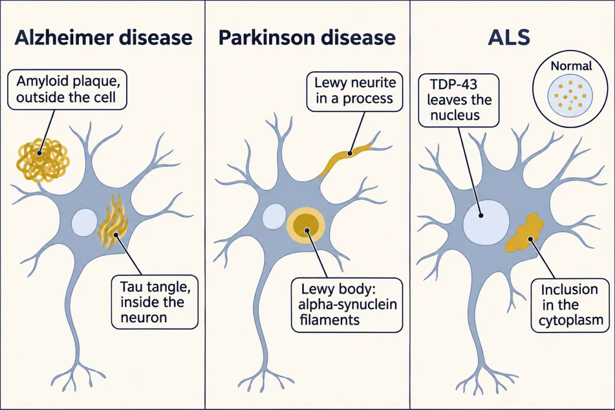
Each disease has its own deposit at autopsy: amyloid plaques outside and tau tangles inside neurons in Alzheimer disease, alpha-synuclein Lewy bodies and neurites in Parkinson disease, and TDP-43 that has left the nucleus for a cytoplasmic inclusion in most ALS. AI-generated illustration -
What can start it: genes that cause and genes that raise risk
-
Does removing it help: testing amyloid in trials
-
Why these neurons
-
Going deeper
Where this lesson fits
Before thisReview first
- Memory SystemsThis course · Lesson 22
You need the memory circuits through the entorhinal cortex and hippocampus from there to see why Alzheimer disease first costs new memories.
- Descending Control and the Basal GangliaThis course · Lesson 16
You need the dopamine loops through the striatum from there to see what losing substantia nigra neurons unbalances.
- Motor Units, Reflexes and LocomotionThis course · Lesson 15
You need the motor unit from there to see why losing lower motor neurons removes whole units of muscle.
After thisStudy next
- Designing a Causal Circuit ExperimentThis course · Lesson 28
It takes the lesson's closing problem, a mouse result that does not carry over, into designing a causal experiment with proper controls.
Elsewhere on the siteFilms, simulations and method films in other courses
- Ras to MAPK: From Membrane to GenesFilm · Cell Signaling
Oncogenes show how stuck switches arise in families and in tumors, like causal versus risk genes here.
Suggested by an AI model from the lessons themselves, then checked. How we use AI