Developmental Neuroscience · Skim version
Myelination: Wrapping the Axon So the Signal Jumps
Read the full lesson → Skim the whole course →
The guiding idea
Wrap an axon in tight layers of glial membrane, leave short bare gaps, and its signal jumps from gap to gap instead of creeping.
Myelin speeds conduction most on large axons, and nerves match the wrap to the axon; in peripheral nerve the axon decides, while in the brain and spinal cord the oligodendrocyte brings much of its own program.
From the film: Why Wrap, and How Much
- Myelin lets the signal jump between nodes of Ranvier, and the speed gain is largest on large axons.
- In peripheral nerve the axon decides how the Schwann cell wraps it; in the brain and spinal cord the oligodendrocyte brings much of its own program.
- Nerves do match the wrap to the axon, so the next question is how.
From the film: The Axon Sets the Dose
- In peripheral nerve the axon decides: the amount of neuregulin-1 type III on its surface is the main signal for whether a Schwann cell wraps it and how thickly.
- Halving the signal thinned the myelin and extra signal thickened it, and extra signal made normally bare sympathetic axons get wrapped.
- The Schwann cell also needs its basal lamina and its receptor Gpr126, so the signal is decisive but not enough alone.
From the film: The Brain Brings Its Own Program
- In the brain and spinal cord the oligodendrocyte brings much of its own program, and in culture it will wrap inert plastic fibers of a suitable diameter.
- Myelin still formed in mice without neuregulin-1 and ErbB signaling in the brain and spinal cord, though extra neuregulin could thicken it.
- Axon cues such as caliber and activity bias the wrap, as when a release-blocked axon in zebrafish larvae was wrapped less often.
-
Why wrap an axon?
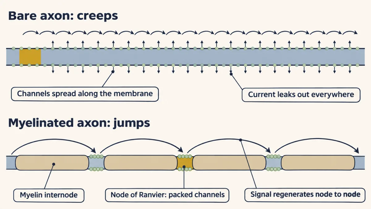
In a bare axon, current leaks out and charges the membrane all along the way, so the action potential is rebuilt on every patch and creeps. Myelin cuts both the leak and the charging, so the current reaches the next node, where sodium channels are packed, and the signal seems to jump from node to node. AI-generated illustration -
Two wrappers
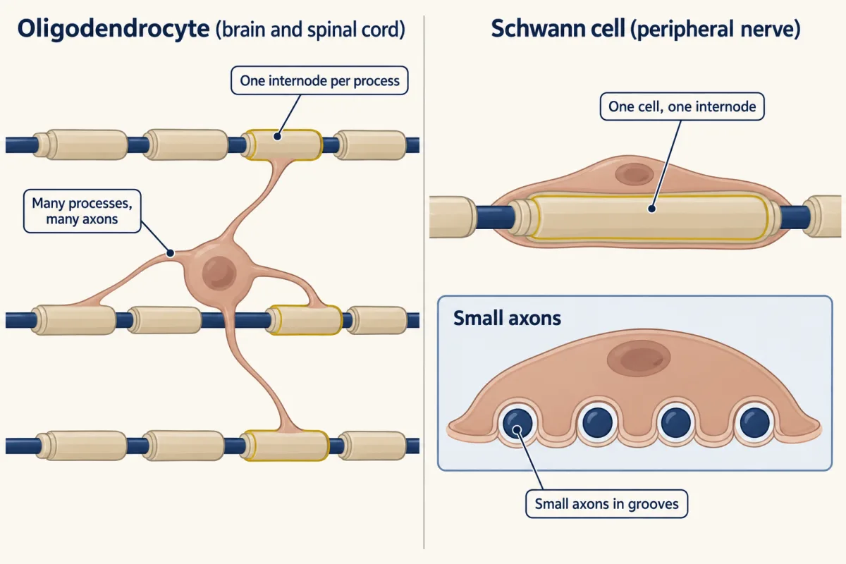
In the brain and spinal cord, one oligodendrocyte makes an internode with each of many processes, usually on different axons. In peripheral nerve, a myelinating Schwann cell makes one internode on one axon. The inset shows a separate, nonmyelinating Schwann cell holding several small axons in grooves without forming myelin. AI-generated illustration -
In nerve, the axon decides
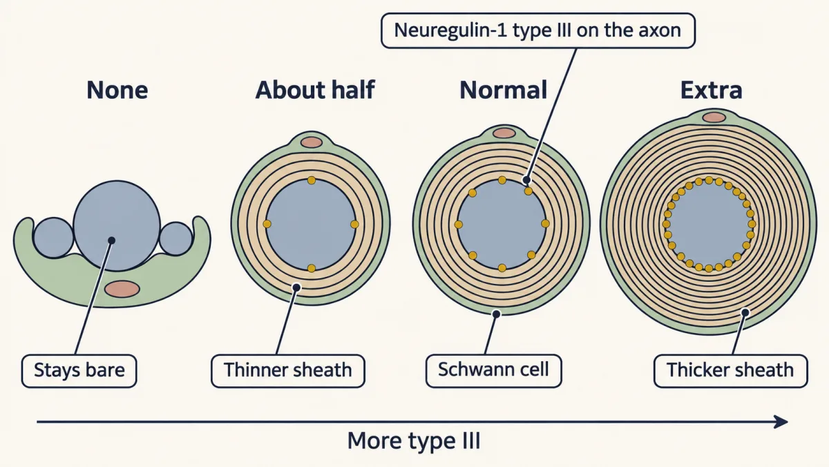
In peripheral nerve, the axon decides. Below a threshold amount of neuregulin-1 type III, an axon stays bare; above it, more type III helps make a thicker sheath, so halving it thins the myelin and adding extra thickens it, on axons of the same size. AI-generated illustration -
In the brain, the oligodendrocyte brings its own program
-
What holds the layers together
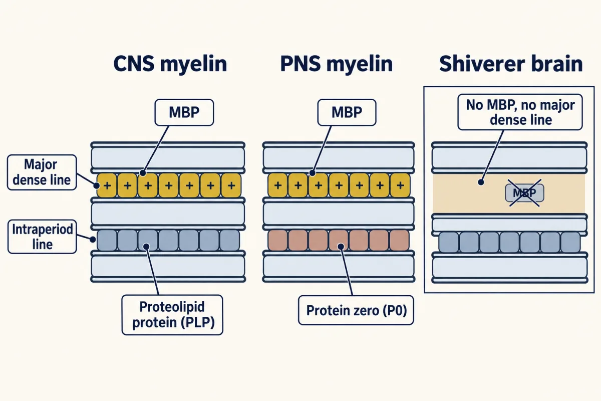
In compact myelin, myelin basic protein pulls the cytoplasmic faces together and squeezes out the cytoplasm, leaving the major dense line. At the outer faces, proteolipid protein in central nervous system myelin and the unrelated protein zero in peripheral nerve help hold the membranes together. In shiverer mice, central myelin has nearly no MBP and lacks the major dense line; peripheral myelin is much less affected. AI-generated illustration -
A second job: feeding the axon
-
Myelin lost, and repaired
-
Putting it together
-
Going deeper
Where this lesson fits
Before thisReview first
- Where Glia Come FromThis course · Lesson 43
It explains where oligodendrocytes and Schwann cells come from, and this lesson assumes you know the two wrappers.
- Neurotrophins, Trk Receptors and p75This course · Lesson 30
It introduces the target-derived signals neurons use, which helps in reading the neuregulin signal an axon presents to Schwann cells.
- The Growth Cone: The Tip That Steers the AxonThis course · Lesson 3
It covers the axon whose surface signals the glial cell reads when deciding whether to wrap.
After thisStudy next
- Feeding the Axon: Glia as Metabolic PartnersThis course · Lesson 45
It takes the oligodendrocyte's second job, passing lactate to axons through MCT1, further.
- Injury and Regeneration: Why the Brain Does Not RegrowThis course · Lesson 46
It uses myelin loss and repair, and axon dependence on glia, to ask why the injured brain does not regrow.
Elsewhere on the siteFilms, simulations and method films in other courses
- Run the 1952 equationsSimulation · Neuroscience
Running the model of the spike shows how the signal travels along an axon, which the node-to-node jump speeds up.
- The Spike and Its PuzzlesFilm · Neuroscience
It shows how sodium channels produce and conduct the impulse that myelin makes jump between nodes.
- A Threshold Is Not a CountFilm · Sensory Systems
It shows that a normal threshold test can hide lost synapses, a lesson about hidden damage like axon loss under normal-looking myelin.
Suggested by an AI model from the lessons themselves, then checked. How we use AI