Cell Biology · Skim version
Membrane Organization and Topology
Read the full lesson → Skim the whole course →
The guiding idea
A membrane has two faces that differ in their lipids and in the parts of each protein they carry. Who builds that difference, and why does vesicle traffic never flip it?
A cell membrane has two faces that differ in their lipids and in the parts of each protein they carry, and the cell keeps it that way.
From the film: Which Side Is Outside?
- Membranes on the ordinary secretory and endocytic routes have two faces: one touches the cytosol, the other a lumen or the outside.
- Lumens on these routes count as outside, because budding and fusing connect them to the outside without crossing a membrane.
- A membrane protein's direction is normally set once, at the ER, as the translocon threads it in.
- A type I protein has its N terminus in the lumen and its C terminus in the cytosol.
- Vesicle traffic never flips a protein, so a sugar added in the ER lumen can face outside after the protein reaches the cell surface.
From the film: Across or Along the Membrane?
- In red blood cells, PS and PE sit mainly on the cytoplasmic leaflet, PC and sphingomyelin mainly on the outer leaflet.
- ATP-driven flippases move selected lipids inward; scramblases let lipids cross both ways without energy.
- Sliding sideways within a leaflet needs no transporter.
- FRAP recovery size reports mobile fraction, while recovery speed helps estimate diffusion under a stated model.
-
Two faces of every membrane
-
The rule: a lumen counts as outside
-
Four arrangements, one rule
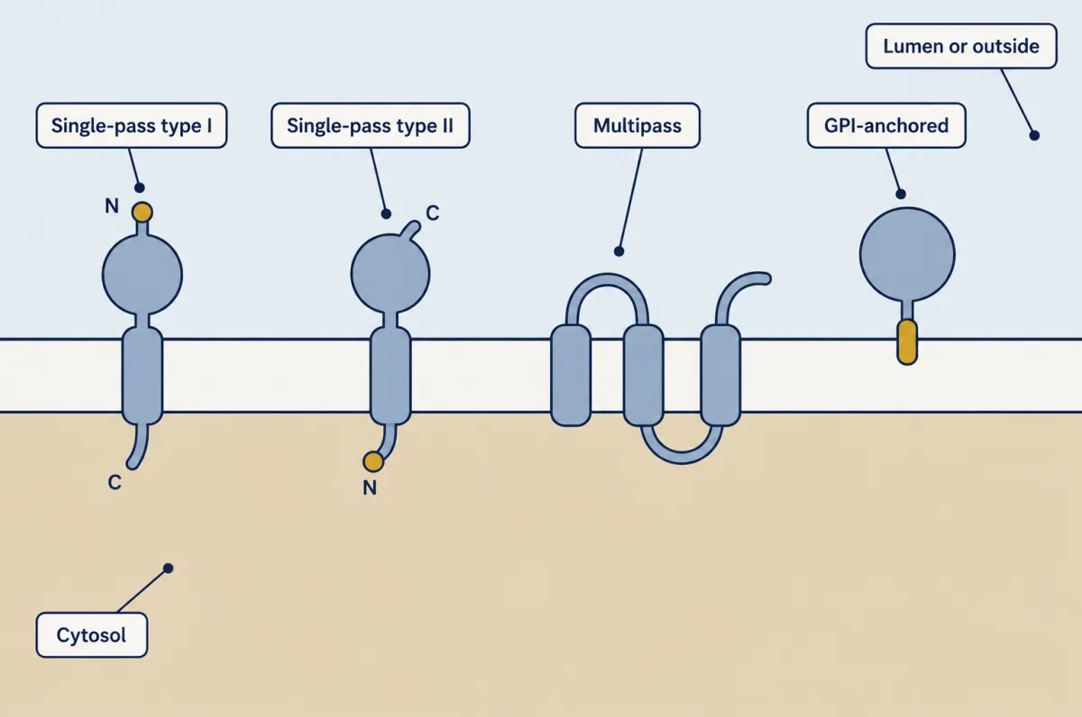
Four common arrangements, drawn with the lumen or outside above and the cytosol below. A single-pass protein's two ends sit on opposite faces, a multipass protein's loops alternate, and a GPI-anchored protein has no spanning segment. AI-generated illustration -
How we know which way a protein faces
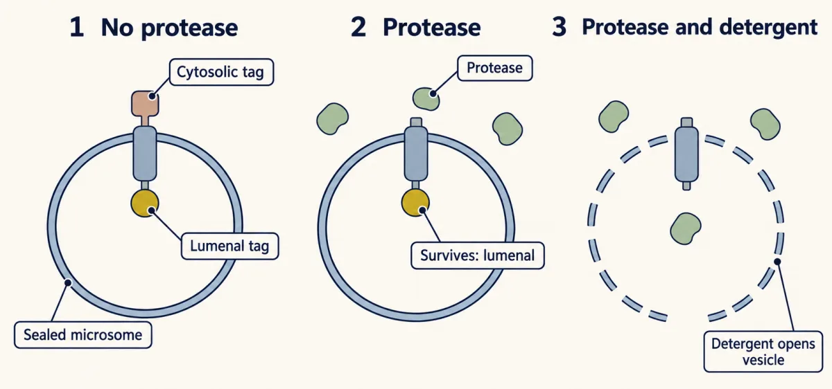
Protease cannot enter a sealed microsome, so the tag that survives is lumenal. Adding detergent first opens the vesicles and both tags are lost, which shows the protease could reach them. AI-generated illustration -
Lipids: the side the cell has to keep pumping
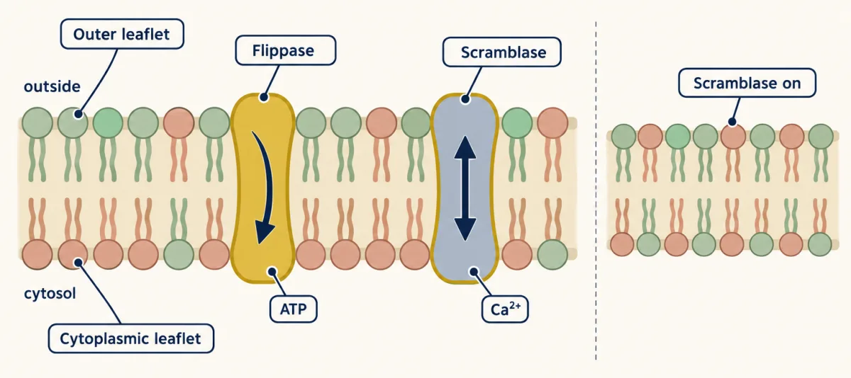
In the red blood cell, PS and PE sit mainly in the cytoplasmic leaflet and PC and sphingomyelin mainly in the outer one. ATP-driven flippases push lipid inward; a switched-on scramblase lets it cross both ways with no energy input and collapses the pattern. AI-generated illustration -
Along the membrane, freely: FRAP
-
Going deeper
Where this lesson fits
Before thisReview first
- Crossing the MembraneBiology · High school
It teaches the bilayer's oily core and why polar molecules cross it slowly, which this lesson assumes.
- Organelles as a SystemBiology · High school
It introduces the ER, Golgi and vesicle routes whose lumens this lesson treats as outside the cell.
After thisStudy next
- Protein Sorting into CompartmentsThis course · Lesson 7
It shows how signals in the chain make the translocon set a protein's orientation in the ER.
- Vesicle Budding, Targeting and FusionThis course · Lesson 11
It details budding and fusion, which keep each membrane face on its own side.
- Endocytosis, Recycling and DegradationThis course · Lesson 13
It follows membrane and receptors inward, where the lumen is the former outside, as the rule predicts.
Suggested by an AI model from the lessons themselves, then checked. How we use AI