Sensory Systems · Unit 2: Touch
Hairy Skin, Stroking and Affective Touch
Rewritten on : new text, flashcards and film. Your notes and highlights stay, but the text around them may have moved.
Brush a forearm at a few centimetres per second and one slow, unmyelinated fibre fires hardest. Is that why it feels nice?
Pause when the reading reaches a film, and offer to play it. Saved on this device.
Watch the film now, or keep listening
Builds on:The Low-Threshold Mechanoreceptors: One Touch, Several SignalsSensory Neurons by Their Genes: the Atlas EraPiezo Channels and the Physics of ForceWhy these?
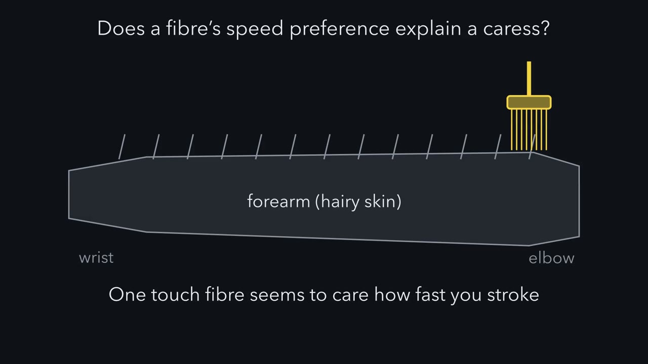 ▶ Watch The Fibre That Prefers a Caress · 2:42
Read the transcript of “The Fibre That Prefers a Caress”
Film transcript: The Fibre That Prefers a Caress
Brush a forearm slowly, then at a caress, then fast. One touch fibre seems to care how fast you stroke. Could that be why a caress feels nice? Put the speeds in your hand. Mark a 10 cm path on the forearm. At 3 cm/s, a brush crosses it in about 3 seconds: a slow caress. Ten times faster is a quick swipe. Ten times slower is a crawl of over half a minute. A low-threshold mechanoreceptor responds to gentle force. Hairy skin has fast, myelinated ones and slow, unmyelinated ones. A C-tactile afferent is a human one with a thin, unmyelinated axon. It conducts slowly. Microneurography records single axons from a skin nerve in an awake person. In 2009, Löken and colleagues recorded this way while a soft brush crossed the forearm at set speeds.
The recorded myelinated touch fibres fired harder the faster the brush moved. Predict. If C-tactile fibres worked the same way, what shape would their curve have? What shape would show a preference? Their firing traced an inverted U. It was highest for strokes between about 1 and 10 cm/s. Slower and faster strokes drove them less.
A separate group of volunteers rated the same strokes. Their average pleasantness had the same shape, highest over the same range. Of the fibre classes recorded, only the C-tactile afferent had a curve of that shape. Why does that matter? Suppose only fibres whose firing rises with speed drove the ratings. If more firing meant more pleasant, ratings would rise with speed. They do not. So the caress-tuned fibre is the obvious candidate. But a candidate is a suspect, not a proven cause. Ratings and recordings came from different people. And other signals combined could, in principle, make the same curve. The one idea: of the fibres recorded in human hairy skin, the C-tactile afferent fired most at caress speeds.
Average pleasantness follows its curve. So its input is the prime candidate for the pleasant side of touch. Only taking the input away can show whether it is needed.
From the film
- In human hairy skin, the C-tactile afferent fires most at caress speeds of about 1 to 10 cm/s, while the myelinated touch fibres recorded fire harder the faster the brush moves.
- Average pleasantness ratings follow the same inverted U, so C-tactile input is the prime candidate for the pleasant side of touch.
- A candidate is not a proven cause: only taking the input away can show whether it is needed.
Brush the hairy skin of a forearm with a soft brush. Go very slowly, then at the speed of a caress, then fast. The myelinated touch fibres recorded in that skin fire harder the faster you go. One unmyelinated class fires hardest at the caress and less at either extreme. Does that preference have anything to do with why a caress can feel nice?
Put the speeds in your hand first. One of the studies below marked a 10 cm path on the forearm. At 3 cm/s a brush crosses it in a little over 3 seconds, about the pace of a slow caress. At 30 cm/s it crosses in a third of a second, a quick swipe. At 0.3 cm/s it takes more than half a minute, a crawl.
Three terms carry the lesson.
- A low-threshold mechanoreceptor (LTMR) is a sensory neuron that responds to gentle mechanical force. The lesson on low-threshold mechanoreceptors introduced the classes. Hairy skin has fast LTMRs, whose axons are myelinated (wrapped in insulating myelin), and slow, unmyelinated ones. An afferent is any sensory fibre carrying signals toward the spinal cord and brain.
- A C-tactile (CT) afferent is a human LTMR with a thin, unmyelinated C-fibre axon, so it conducts slowly. The core lesson on touch and pain noted that some C fibres are low-threshold touch afferents rather than nociceptors; these are the touch ones. Mouse papers use C-LTMR (C-fibre low-threshold mechanoreceptor) for unmyelinated touch afferents. Whether the mouse and human cells are the same is the open question this lesson ends on.
- In microneurography, a fine electrode is passed through the skin into a peripheral nerve to record single axons in an awake person while a controlled stimulus is applied.
The lesson first asks what a CT afferent responds to, whether pleasantness follows it and what happens when the input is taken away, then turns to the mouse: which cells detect a stroke, what switching them on or off does, and which mouse cell, if any, is a CT afferent. The short answer to why a stroke can feel good is that much less is known than popular accounts suggest.
The fibre that prefers a caress
Löken, Wessberg, Morrison, McGlone and Olausson (Nature Neuroscience, 2009) recorded single afferents from the forearm of healthy, awake volunteers by microneurography while a soft brush crossed each fibre’s receptive field (the patch of skin it responds to) at a range of controlled velocities. They treated stroke velocity as a stimulus feature, the way sensory coding treats orientation or frequency, and they recorded myelinated touch afferents as well as C-tactile (CT) afferents.
Predict. A faster stroke deforms the skin faster, so you might expect any touch fibre to fire harder the faster the brush moves. If CT afferents worked that way, what shape would their curve have? What shape would show a fibre that prefers a caress?
Check your prediction
The myelinated afferents’ firing rose with velocity. The CT afferents’ firing traced an inverted U, highest for strokes between about 1 and 10 cm/s and lower on either side. The tuning, not a higher absolute firing rate, set CT afferents apart.
| Myelinated afferents recorded (Löken 2009) | CT afferents | |
|---|---|---|
| Axon | myelinated, fast | unmyelinated, slow |
| As the brush speeds up | firing rises | firing rises, then falls |
| Strongest response | at the fastest strokes tested | at about 1 to 10 cm/s |
| Same shape as the pleasantness curve? | no | yes |
In separate sessions, a separate group of volunteers rated how pleasant the same strokes felt. Their average ratings had the same inverted-U shape, highest over the same range. Why the CT afferent prefers these speeds is not settled by these studies; what matters here is the match, for a simple reason. If ratings were driven only by fibres whose firing rises with speed, and read out in any way that keeps “more firing” meaning “more pleasant”, ratings would also rise with speed. They do not. A fibre that is itself tuned to caress speeds is the obvious candidate.
But a candidate is a suspect, not a proven cause, for two reasons. The ratings and the recordings came from different people, so the 2009 result is a correspondence between two tuning curves. And the same curve could in principle be built by combining other signals: a signal that rises quickly and levels off, minus one that keeps rising, also peaks in the middle. The result does not show that CT firing is needed for pleasantness, or that it is the only input to it.
The one idea to carry through this lesson. Of the touch fibres recorded in human hairy skin, the CT afferent is the one tuned to a preferred stroke speed. It fires most at caress speeds, about 1 to 10 cm/s, while the recorded myelinated fibres fire harder the faster the brush moves. People’s average pleasantness ratings follow the CT curve. That makes CT input the prime candidate for the pleasant side of gentle touch; only taking the input away can show whether it is needed.
The idea covers human hairy skin, the fibre classes recorded, soft-brush strokes over the speeds tested, and group averages. It does not decide how pleasant a particular touch feels to a particular person, which also depends on who is touching, on the situation and on processing in the brain. It does not cover glabrous (hairless) skin such as the palm, where CT afferents are sparse though not absent (Watkins 2021), or the mouse, where “pleasant” can only be a measured outcome.
Take the input away: what people show
Read the transcript of “Take the Input Away”
Film transcript: Take the Input Away
If C-tactile input makes a stroke feel pleasant, what happens when that input is taken away? No one can remove a fibre class from a healthy person. But some people have lost part of their touch pathway. Start with the reverse case. In 2002, Olausson and colleagues studied a patient lacking large myelinated afferents. Stroking that excites C-tactile afferents produced a faint, pleasant sensation. Imaging showed insular activation, and none detectable in primary or secondary somatosensory cortex. One patient shows what this input can do without large fibres, not that it is needed. Next, ten carriers of a nerve growth factor mutation They have fewer thin and unmyelinated fibres.
Predict. How should they rate slow stroking at the speeds C-tactile afferents prefer? They rated it less pleasant than matched controls. And their posterior insula was not modulated by that stroking. The mutation affects more than C-tactile afferents, so this is not a clean removal.
Now cut a route instead. An anterolateral cordotomy cuts part of the spinal cord to relieve intractable pain. In 2019, Marshall and colleagues tested such patients before and after, with a brush at three speeds. Predict. Pain and temperature travel in that tract. If pleasant touch travelled only there, what should the cut do?
Pain, temperature and itch were markedly disrupted. But pleasantness ratings were not significantly changed. With 19 patients, that is not proof of no change. But the ratings did not vanish. The authors proposed a route outside that tract, most likely the dorsal columns. So the two tests ask different questions. With fewer thin fibres, carriers rated caress-speed stroking as less pleasant. Cordotomy cut one route, yet pleasantness survived. Neither test shows which fibres carry the surviving signal. So thin-fibre input seems to contribute, and this route is not needed. C-tactile input remains a candidate, its route unresolved.
From the film
- In carriers of a nerve growth factor mutation with fewer thin fibres, caress-speed stroking was rated less pleasant, though the mutation affects more than C-tactile afferents.
- After cordotomy, pain, temperature and itch were markedly disrupted but pleasantness ratings were not significantly changed.
- So thin-fibre input seems to contribute and the anterolateral route is not needed, while C-tactile input remains a candidate with its route unresolved.
No one can remove a fibre class from a healthy person, but some people have already lost part of their touch input, and their reports and ratings can be compared. Start with the reverse case: a person who kept C-tactile (CT) input but lost the large myelinated afferents.
CT input without the large fibres. In a healthy arm, a brush that excites CT afferents also excites large myelinated touch fibres, so their contributions cannot be separated by asking. In the patient studied by Olausson and colleagues (Nature Neuroscience, 2002), CT stimulation of hairy skin produced a faint, pleasant sensation, and functional MRI showed insular activation with no detectable activity in primary or secondary somatosensory cortex. The authors proposed, as a hypothesis, that CT input may underlie emotional and affiliative responses to caress-like touch. The case shows what CT input can do without large myelinated afferents, not that it is needed. The insular link rests on one patient and one imaging method; emotion and stress discusses what an insular activation can and cannot mean.
Fewer C fibres. Morrison and colleagues (Brain, 2011) tested ten carriers of a nerve growth factor beta mutation (hereditary sensory and autonomic neuropathy type V, HSAN-V), whose density of thin and unmyelinated fibres is reduced.
Predict. If CT input contributes to pleasantness, how should these carriers rate slow arm stroking at the velocities CT afferents prefer, compared with matched controls?
Check your prediction
They rated it as less pleasant than matched controls. Their rating pattern across velocities differed too, and their posterior insular response was not modulated by CT-optimal stroking. That ties hedonic touch to C-fibre density, although the mutation affects more fibres than CT afferents alone, so it is not a clean removal of CT input.
Cut the route. An anterolateral cordotomy cuts the anterolateral spinal cord to relieve intractable pain. The anterolateral tract carries pain and temperature signals. Marshall and colleagues (eLife, 2019) tested adults with refractory cancer pain before and after the operation, rating the pleasantness of brush strokes at three speeds.
Predict. If CT pleasantness travelled only in the anterolateral tract, what should the cut do to the pleasantness ratings? What would you conclude if they survived?
Check your prediction
If the tract were the only route, the cut should have removed pleasantness. It did not: pain, temperature and itch were markedly disrupted, but pleasantness ratings were not significantly changed. With 19 patients, “not significantly changed” is not proof of no change, but ratings that did not vanish rule out the anterolateral tract as the one obligatory route.
The authors proposed that fibres ascending outside the anterolateral funiculus (the anterolateral white matter of the cord), most likely in the dorsal columns (the touch tracts at the back of the cord), carry enough information. The cut did change other judgements: ratings of stroking intensity and of texture fell, while monofilament detection thresholds were spared.
Input versus route. The HSAN-V and cordotomy results can look contradictory, but the two tests ask different questions.
| Test | What it takes away | Pleasantness | What it answers |
|---|---|---|---|
| HSAN-V (Morrison 2011) | some thin and unmyelinated fibres, not only CT afferents | lower | fits a contribution from thin-fibre input |
| Cordotomy (Marshall 2019) | one route, the anterolateral tract | not significantly changed | that route is not needed for the ratings |
Neither test shows which fibres carry the signal that survives the cut: CT afferents, Aβ afferents or both together. CT input remains a candidate whose necessity and route are unresolved.
Worked problem: reading the fit. For the pain-affected side before cordotomy, Marshall and colleagues fitted the group’s ratings with a quadratic in log velocity. On a log axis, equal distances are equal ratios of speed: the tested 0.3, 3 and 30 cm/s sit one tenfold step apart, and Löken’s 1 to 10 cm/s band is one step wide.
y = -2.07x² + 1.88x + 6.22, where x = log10(v in cm/s), R² = 0.22
- Find the peak. dy/dx = -4.14x + 1.88. Set it to zero: x = 1.88 / 4.14 = 0.4541. The second derivative is -4.14, so this is a maximum.
- Convert to velocity. v = 100.4541 = 2.85 cm/s.
- Peak rating. x² = 0.206, so y = -2.07(0.206) + 1.88(0.454) + 6.22 = -0.427 + 0.854 + 6.22 = 6.65.
- Predicted ratings at the tested speeds. At 0.3 cm/s, x = -0.523 and y = -0.566 - 0.983 + 6.22 = 4.67. At 3 cm/s, x = 0.477 and y = -0.471 + 0.897 + 6.22 = 6.65. At 30 cm/s, x = 1.477 and y = -4.517 + 2.777 + 6.22 = 4.48.
- Interpret. The fitted peak sits inside Löken’s 1 to 10 cm/s band, so the patients’ group curve has the familiar shape. But what can the fit show?
- Only three speeds were tested, and a quadratic has three adjustable numbers. If it was fitted by ordinary least squares, as the reported R² suggests, it passes through the average rating at each speed: the step 4 values are those averages.
- So the data show only that the middle speed was rated higher than both the slow and the fast speed. The shape between the tested speeds, including the exact 2.85 cm/s peak, comes from assuming a parabola. The peak sits a little below 3 cm/s only because the slow-speed average (4.67) is a little above the fast-speed one (4.48).
- R² = 0.22 means velocity explains about a fifth of the variance in the ratings that were fitted. Because the curve already passes through the averages, no curve in velocity could do better on these data. The rest of the variance is spread among ratings at the same speed, much of it the difference between patients in how pleasant they found touch overall, not a failure of the curve. Within patients, the negative quadratic fitted better than a line in 14 of the 19 adults in the study.
- No fit to ratings can show that CT afferents are needed.
- x = log10(v)
- 0.000
- Predicted rating y
- 6.22
- Slope dy/dx (points per tenfold change)
- 1.88
dy/dx = −4.14x + 1.88 = 0, so x = 1.88 / 4.14 = 0.454. The second derivative is −4.14, a maximum. v = 100.454 = 2.85 cm/s, and y = −2.07(0.206) + 1.88(0.454) + 6.22 = 6.65.
R² = 0.22: velocity explains about a fifth of the variance in the ratings; most of it lies between people and trials.
In this sketch: one published group quadratic, drawn between the lowest and highest tested speeds (0.3 and 30 cm/s); only 0.3, 3 and 30 cm/s were tested, so the curve between them is the fit's interpolation, for the pain-affected side before cordotomy. No individual ratings and no CT firing curve are drawn.
A group quadratic from the Marshall 2019 cancer-pain patients tested before cordotomy; the correspondence with CT tuning is between curves measured in different people, and no mouse outcome is shown.
Which cells detect a stroke in mice
Read the transcript of “A Net, a Lineage and a Mouse That Cannot Say”
Film transcript: A Net, a Lineage and a Mouse That Cannot Say
A mouse cannot say a touch feels nice. Which of its cells detect a stroke, and what can we measure? In mouse hairy skin, at least three afferent classes are candidates for detecting a stroke. First, unmyelinated touch afferents marked by tyrosine hydroxylase. Their endings run lengthwise along a hair follicle. Second, unmyelinated neurons marked by a receptor called MrgprB4. Their fibres innervate hairy skin only. Third, Aβ beta field afferents. They are myelinated, and their endings wrap around the follicle. A field afferent ignores any single hair. Indenting the skin drives it only at high force.
But one neuron can form as many as 180 endings, around many follicles. Predict. What does a gentle stroke across the skin do to this neuron? It fires. The authors proposed that it sums many weakly sensitive endings. A single hair is a lever. A field afferent is a net.
Now the outcomes. Activating MrgprB4 neurons gave young male mice a place preference. The authors read this as positive reinforcement, reduced anxiety, or both. Adult females whose lineage of these cells was removed during development were less sexually receptive.
A permanent genetic label marks neurons that once expressed the gene, including some that no longer do in adults. And a dopamine-sensor signal in the nucleus accumbens was reduced during mounting. A chamber preference, a mating behaviour, a dopamine signal. Each is an outcome, not a feeling. Back to the one idea. In people, one fibre has a caress-shaped curve that pleasantness follows.
Which mouse cell, if any, matches that human fibre is still unsettled. Keep the human candidate and the mouse outcomes apart until a match is made.
From the film
- In mouse hairy skin, at least three afferent classes are candidates for detecting a stroke.
- A field afferent may respond to gentle stroking by summing many weakly sensitive endings, as the authors proposed.
- Mouse outcomes such as place preference are not feelings, and which mouse cell, if any, matches the human C-tactile afferent is unsettled.
The human evidence names a fibre but cannot open it up. Mouse genetics can label one class of sensory neuron, record from it and switch it on or off. The lesson on low-threshold mechanoreceptors placed C-LTMRs and Aδ endings on mouse hair follicles. In mouse hairy skin, at least three afferent populations are candidates for detecting a stroke, and they are not the same cells.
TH-positive C-LTMRs. Li and colleagues in the Ginty laboratory (Cell, 2011) recorded from sensory neurons in an ex vivo mouse preparation and then identified them. Neurons marked by tyrosine hydroxylase (TH) were C-LTMRs, and they conducted slowly. Their endings were lanceolate: nerve terminals that run lengthwise along a hair follicle, parallel to the hair shaft. They sat on zigzag and awl/auchene follicles (two of the mouse hair types).
MrgprB4 neurons. Liu and colleagues in David Anderson’s laboratory (Nature Neuroscience, 2007) used a genetic tracer to follow sensory neurons expressing the receptor MrgprB4. The fibres were unmyelinated and nonpeptidergic and innervated hairy skin only. That paper was anatomy. Its link to tactile C fibres was a suggestion, not a recording.
Aβ field-LTMRs. Bai, Lehnert and colleagues in the Ginty laboratory (Cell, 2015) used intersectional genetics (Techniques) to reach a class whose endings are circumferential: they wrap around the follicle instead of running along it. A field-LTMR is a large myelinated afferent of this type with a broad receptive field. In vivo recordings from the ganglia showed that these neurons did not respond to deflection of a single hair. Indenting the skin drove them only at forces well above those that drive other Aβ touch afferents, and their firing kept rising as the force rose into the noxious range.
Predict. This neuron ignores any single hair and needs strong indentation. What will it do when a soft brush strokes gently across the skin? A clue from the anatomy: one neuron could form as many as 180 circumferential endings.
Check your prediction
It fired. The authors proposed that the neuron sums many weakly sensitive endings, so a stroke that moves many follicles at once reaches threshold when no single hair can. A single hair is a lever; a field-LTMR is a net. The paper did not test pleasantness or reward.
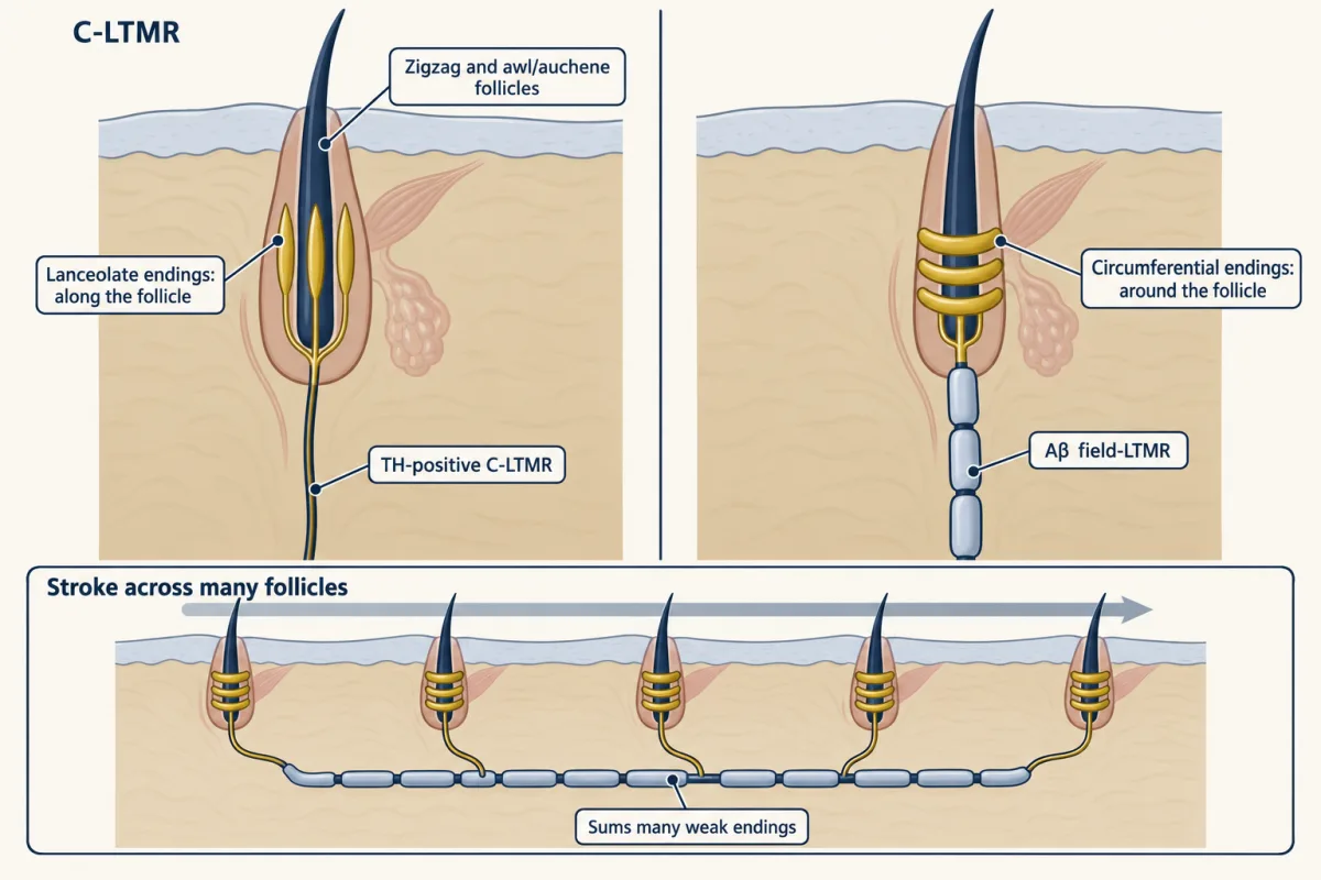
So gentle stroking is not a C-fibre job alone. Any claim that “the stroking receptor” drives a behaviour has to say which of these classes it manipulated.
From skin to reward-like outcomes in mice
Four groups have manipulated mouse touch afferents or their central targets and measured behaviour. Keep each outcome as measured.
Vrontou and colleagues (Nature, 2013) imaged calcium in the spinal terminals of MrgprB4-positive neurons in anaesthetized mice at least 8 weeks old. The terminals responded to massage-like stroking of hairy skin delivered at about 0.5 to 2 cm/s. That is the protocol’s speed, not an estimate of the mouse optimum, and the human 1 to 10 cm/s band should not be carried across. In a separate experiment with male mice about one month old, a DREADD (a designer receptor activated only by a synthetic ligand) was used to activate MrgprB4 neurons, and the mice developed a conditioned place preference: they spent more time in the chamber paired with activation.
Predict. Before reading the authors’ interpretation, list what besides “it felt nice” could make a mouse spend more time in the chamber where its MrgprB4 neurons were switched on.
Check your prediction
The authors read the preference as positive reinforcement, anxiolysis (reduced anxiety) or both, and kept both readings open. Neither is a report of pleasure, and activation tests only sufficiency: it cannot show that the neurons are needed.
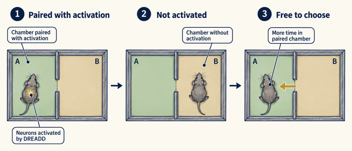
Elias and colleagues in the Abdus-Saboor laboratory (Cell, 2023) worked with adult female mice and a Cre line that labels the Mrgprb4 lineage. Picture the label as a stamp that stays: Cre, an enzyme, permanently switches on a label in cells that recombined after expressing Mrgprb4 at some point, and in their descendants, not only in cells expressing it in adulthood. Optogenetic (light-driven) activation of these afferents in back skin produced place preference and a dorsiflexion resembling lordosis, the arched posture a receptive female mouse takes during mating. Ablating the lineage, a removal that began during development, reduced sexual receptivity in the adults. Stimulating the lineage evoked dopamine release in the brain, and in ablated females a dopamine-sensor signal in the nucleus accumbens, measured by fibre photometry, was reduced during mounting. A dopamine signal is a neuromodulator readout tied to reward prediction and motivation (motivation and reward), not a measure of pleasure.
Huzard and colleagues (Science Advances, 2022) deleted the calcium channel CaV3.2 in the Nav1.8 lineage of male mice, a developmental deletion that the authors took to impair C-LTMR function, and the mice showed fewer social and tactile interactions. Transient chemogenetic enhancement of C-LTMR excitability in adults promoted touch seeking. Two limits apply. Nav1.8 is not restricted to C-LTMRs, so the deletion was not confined to them. In the adult experiment, the authors could not tell whether the DREADD activated the neurons directly or only potentiated them.
Yu and colleagues (Neuron, 2022) started from the brain. Touch-like tactile stimulation increased the firing of oxytocin neurons in the paraventricular nucleus of the hypothalamus (PVH), and Tac1-positive neurons in the lateral and ventrolateral periaqueductal gray sent monosynaptic excitatory projections onto them. Reducing Tac1-neuron activity abolished the touch-evoked oxytocin firing. The stimulus promoted social interaction and place preference. The study did not test which peripheral afferents are needed; it is not evidence about Mrgprb4 neurons. Oxytocin’s wider roles belong to the hypothalamus lesson.
| Study | Mice | Manipulation | Measured outcome | What it does not show |
|---|---|---|---|---|
| Vrontou 2013 | males about one month old | DREADD activation of MrgprB4 neurons | conditioned place preference | pleasure; that the neurons are needed |
| Elias 2023 | adult females | activation, or ablation, of the Mrgprb4 lineage | place preference and lordosis-like dorsiflexion; after ablation, less receptivity and a smaller accumbens dopamine-sensor signal | that adult Mrgprb4 neurons are needed; a feeling |
| Huzard 2022 | males (deletion) | CaV3.2 deletion in the Nav1.8 lineage; adult enhancement of C-LTMR excitability | fewer social and tactile interactions; more touch seeking | an effect confined to C-LTMRs |
| Yu 2022 | mice | reducing Tac1 neuron activity in the periaqueductal gray | loss of touch-evoked oxytocin-neuron firing | which peripheral afferents are needed |
Which mouse neuron is a CT afferent, and what “pleasant” can mean in a mouse
Return to the one idea. On the human side, one recorded fibre class, the C-tactile (CT) afferent, has a caress-shaped tuning curve that average pleasantness follows; reduced thin-fibre input fits a contribution from that input, and the cordotomy shows the anterolateral tract is not needed, but neither identifies the fibres or the route. On the mouse side there are several cell classes and several outcomes, and the bridge between the two is missing.
The human term CT afferent is often used interchangeably with the mouse term C-LTMR, but whether the two name the same cells is unresolved. TH-positive C-LTMRs do not express MrgprB4, so the two are different populations, and Huzard and colleagues described the identity of the mouse population best matching human CTs as controversial. Vrontou and colleagues found that adult MrgprB4 fibres responded to stroking and not to pinch. A 2025 study by Du and colleagues (iScience) imaged the Mrgprb4 lineage in mice and found responses to both mechanical and thermal stimuli; mice lacking the lineage lost a conditioned preference for mild pressure. A polymodal population would be a poor fit for a pure “pleasant touch” line, but the lineage is broader than the adult population, because Mrgprb4 expression starts in a broader set of progenitors, so mixed responses in it say little about adult Mrgprb4 neurons.
The sensory neuron atlas lesson explains why a transcriptomic class is not yet a function. Settling the match needs human molecular data tied to physiologically identified units, and that work has begun (Yu, Nagi and colleagues, 2024). It has not yet matched a human CT afferent to a mouse type.
The second gap is the word itself. In people, “pleasant” is a rating. In a mouse it can only be an outcome: time in a chamber, a posture, a dopamine-sensor signal, contact with cage mates. Each is useful; none is a report of feeling, and none licenses a claim about human health or wellbeing. The forward lessons on touch in autism models and itch return to these afferents from other angles, and circuit experiment design sets out the logic of necessity and sufficiency used below.
Design problem
Elias and colleagues used a developmental Mrgprb4 lineage. Design experiments to test whether Mrgprb4-expressing afferents in adult mice are needed for rewarding touch, and say what each result would and would not show.
Saved in this browser only.
One way to do it
Access in adults. Use an inducible driver for Mrgprb4, such as a CreER line if a validated one exists (otherwise it must be built and validated first), and give tamoxifen to adult mice, so that only cells expressing the marker then are targeted. Confirm recombination in the ganglia with a reporter and check which cells are labelled, since the lineage and the adult population may differ. Include tamoxifen-treated mice lacking the driver.
Two separate arms. Sufficiency: activate the adult population (optogenetically or chemogenetically) and measure place preference. Necessity: ablate or silence it in adults (diphtheria toxin receptor, or an inhibitory DREADD), then test touch-conditioned place preference, the same reward readout the sufficiency arm uses. Measure receptivity to a male and the accumbens dopamine-sensor signal during mounting by fibre photometry as separate social and neural outcomes.
Controls for other explanations. Measure locomotion and posture so a motor change is not read as receptivity, and run an anxiety assay so anxiolysis is not read as reward. Test both sexes if the claim is general.
Check the modality. Image the targeted neurons to stroking, pressure and temperature. If they respond to more than touch, call the result “required for this behaviour,” not “a pleasure line.”
Human counterpart. A mouse marker cannot stand in for a human CT afferent. A human claim is best tested by relating identified CT activity to pleasantness ratings, ideally within the same participants, and eventually by molecular identification of the recorded fibre class. Necessity in people needs a loss-of-function case instead, such as the reduced C-fibre density studied by Morrison and colleagues.
Going deeper
Before 2009
Unmyelinated touch afferents had been recorded in human hairy skin before 2009 (Johansson 1988, Nordin 1990, Vallbo 1999), but what they were for was still a hypothesis.
Glabrous skin: sparse, not absent
Watkins and colleagues (Journal of Neurophysiology, 2021) recorded three C-tactile (CT) afferents in human glabrous hand skin, about 1 for every 80 myelinated mechanoreceptors recorded, and estimated CT density there at around seven times lower than in hairy skin. Three units are a small sample, and the estimate carries that limit.
The cordotomy study in detail
A twentieth recruit could not be tested after the operation. The brush crossed the marked 10 cm path on the dorsal forearm, and patients rated pleasantness from -10 to +10.
On the route, mouse work points elsewhere: Choi and colleagues (Nature, 2020) described Gpr83-positive spinoparabrachial neurons, part of the anterolateral system, that take low-threshold mechanosensory input and carry positive or negative valence. The species gap is unresolved.
Preparation details for the mouse populations
The ex vivo preparation of Li and colleagues combined skin, nerve, dorsal root ganglion and spinal cord from mice aged 2 to 3 months. The TH-positive C-LTMRs had a mean conduction velocity of 0.58 ± 0.02 m/s in that preparation. Low-threshold mechanoreceptors (LTMRs) innervating the same or neighbouring follicles sent central projections into narrow dorsal horn columns, which the authors read as support for local integration, the topic of the dorsal horn lesson.
Vrontou and colleagues declined to call MrgprB4 neurons C-LTMRs by electrophysiological criteria, because von Frey filaments (calibrated fibres pressed onto the skin) did not activate them.
Lineage labelling and the human atlas
Du and colleagues used mice of both sexes aged 8 to 16 weeks. About 45% of Mrgprd+ neurons are labelled in the Mrgprb4 line they imaged. Yu, Nagi and colleagues (Nature Neuroscience, 2024) sequenced individual human ganglion neuron somata and identified 16 types of human ganglion neuron. They found divergence from mouse and likely human-specific types, and used the molecular profiles to guide microneurography.
Your notes
Select any text in the lesson to highlight it or add a note to it. Your highlights and notes are saved in this browser and collected in your notebook.
This browser is not letting the site save anything (a private window or strict privacy settings), so highlights and notes will be lost when you leave the page.
How we measure it
Microneurography with psychophysics
A fine electrode inserted through the skin into a peripheral nerve records single afferent axons in an awake person while a controlled stimulus is applied. Paired with ratings, it lets you compare a fibre's tuning with what people report, though in Löken 2009 the two came from separate groups.
Intersectional and lineage genetics (Cre, CreER, Flp)
Recombinase lines give access to a sensory neuron class for labelling, recording, ablation or activation. A constitutive Cre permanently labels cells that recombined after expressing the driver, and their descendants (a lineage); an inducible CreER given tamoxifen in adulthood marks cells expressing it then; combining two drivers narrows the class. Bai and colleagues combined TrkC and Ret genetics in this way to reach Aβ field-LTMRs.
In vivo calcium imaging of afferent terminals
A genetically encoded calcium indicator in a defined afferent population is imaged at its spinal terminals in an anaesthetized mouse while the skin is stroked or poked. It shows which stimuli drive the population, at the level of the terminals imaged.
Fibre photometry with a dopamine sensor
An optical fibre over a brain region collects fluorescence from a genetically encoded dopamine sensor during behaviour. It reports changes in extracellular dopamine signal there, not a feeling.
Practice questions on this lesson (3) → MCAT-style, original, each with an explanation
Where this lesson fits
Before thisReview first
- The Low-Threshold Mechanoreceptors: One Touch, Several SignalsThis course · Lesson 4
It introduces the LTMR classes, including C-LTMRs and the fast myelinated fibres, whose stroke responses this lesson compares.
- Sensory Neurons by Their Genes: the Atlas EraThis course · Lesson 3
You need its point that a gene-defined neuron class is not yet a function to judge which mouse cell matches a CT afferent.
- Piezo Channels and the Physics of ForceThis course · Lesson 2
It explains how touch force opens channels, which underlies why field-LTMRs need strong indentation before they respond.
After thisStudy next
- Where Touch Is First Processed: Dorsal Horn and Dorsal Column NucleiThis course · Lesson 8
It follows where stroke-driven input from these afferents is first processed in the spinal cord and brainstem.
- Touch Over-reactivity in Autism ModelsThis course · Lesson 9
It returns to these mouse touch afferents and tests how changes in them alter touch reactivity and behaviour.
- Itch: A Sense of Its OwnThis course · Lesson 13
It looks at related Mrgpr-expressing sensory neurons and how their identity is tied to what the animal does.
Elsewhere on the siteFilms, simulations and method films in other courses
- Cut half the cordSimulation · Neuroscience
It shows how a touch afferent's response to light stroking and pain input is read in the spinal cord.
Suggested by an AI model from the lessons themselves, then checked. How we use AI
Finish line
Test yourself
8 questions
Practice and MCAT-style, with explained answers.
Next · Lesson 8
Where Touch Is First Processed: Dorsal Horn and Dorsal Column Nuclei
The brainstem can hear about one touch of the paw twice: directly, and again by a route through the spinal cord that, in a simple conduction model, arrives a few milliseconds later. What does the second report add?
Continue →← Lesson 6 · Meissner and Pacinian Corpuscles: Flutter and Vibration