Developmental Neuroscience · Unit 3: Coordinates: patterning the tube
Head to Tail: Hox Genes, Retinoic Acid and Rhombomeres
Rewritten on : new text, flashcards and film. Your notes and highlights stay, but the text around them may have moved.
Rhombomeres r2 and r4 start out looking alike. What tells one to build jaw motor neurons and the other facial ones?
Pause when the reading reaches a film, and offer to play it. Saved on this device.
Watch the film now, or keep listening
Builds on:Neural Induction: The Default ModelNeurulation and Neural Tube DefectsThe Organizer: Spemann and MangoldWhy these?
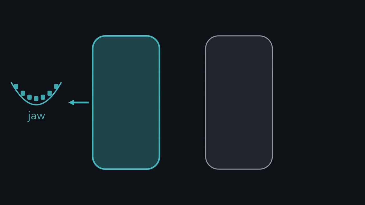 ▶ Watch Two Segments, Two Jobs · 2:28
Read the transcript of “Two Segments, Two Jobs”
Film transcript: Two Segments, Two Jobs
Two hindbrain segments look almost alike. One builds the neurons that work the jaw. Another builds those that move the face. What tells them apart? From above, the early hindbrain is a row of bulges called rhombomeres. They run from r1 to r7, front to back. In 1989, Andrew Lumsden and Roger Keynes mapped where these motor neurons arise, in chick embryos. Trigeminal neurons, for jaw muscles, came from r2 and r3. Facial neurons came from r4 and r5. Swallowing neurons came from r6 and r7. Are the bulges real units?
Label one cell before boundaries appear, and one after. Where do their descendants go? In 1990, Scott Fraser, Keynes and Lumsden did it with a tracer dye. Clones labelled early could span two rhombomeres. Clones labelled after boundaries formed stayed inside one. Each rhombomere is a compartment: its progenitors do not cross into the next. Some neurons born later still migrate across.
Units that keep their cells need a label: a set of transcription factors, mostly from Hox genes. In mouse, Hox domains have front edges at rhombomere boundaries. Read the code column by column. Every rhombomere differs. Rhombomeres r3 and r4 share Hoxa2 and Hoxb2. What separates them is Krox20 in r3. And later, strong Hoxb1 in r4. One idea to carry through: more early retinoic acid, a more posterior code; the code helps set the neurons. Early in the hindbrain, more retinoic acid, a vitamin A product, pushes a segment toward a more posterior identity. That combination, sharpened by segment genes such as Krox20, helps decide which motor neurons it builds.
So lookalike segments are not alike. Each carries its own code.
From the film
- Rhombomeres look alike but each carries its own code of transcription factors, mostly from Hox genes.
- Neighbouring segments can share many Hox genes, and segment genes such as Krox20 help tell them apart.
- More early retinoic acid gives a more posterior code, and the code helps set which neurons a segment makes.
Early on, the segments of the embryonic hindbrain look alike. Yet one will make the motor neurons that work the jaw, and its neighbour two doors down the ones that move the face.
What tells them apart is a code of transcription factors that each segment carries. The code helps set which neurons the segment makes and where their axons go, and a signal made from vitamin A helps write it. This lesson shows the code, tests whether it causes a segment’s identity, and then follows it back to the signal. That signal is one likely reason a retinoid taken in early pregnancy can disturb the embryo’s face and brain.
Two segments that look alike
The neural tube is the hollow rod that forms when the edges of the neural plate fold up and fuse. It becomes the brain and spinal cord. Its front end soon swells into three vesicles: the forebrain, the midbrain and the hindbrain. The hindbrain goes on to make the pons, the cerebellum and the medulla.
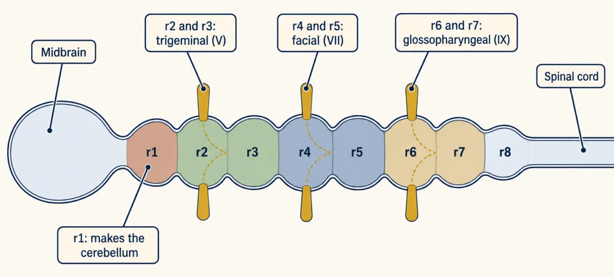
Look at the early hindbrain from above and you see a row of small bulges called rhombomeres, numbered r1 to r7 from front to back, with a less distinct r8 behind. They look like repeats of one design, like a new street where every house is built to the same plan.
They do not behave like repeats. In 1989 Andrew Lumsden and Roger Keynes mapped, in chick embryos, where the branchiomotor neurons arise: the motor neurons for the muscles of the jaw, face and throat. The motor neurons of each cranial nerve came from a pair of rhombomeres.
| Rhombomeres | Motor neurons they make | Muscles they supply |
|---|---|---|
| r2 and r3 | trigeminal (nerve V) | first-arch muscles: the jaw |
| r4 and r5 | facial (nerve VII) | second-arch muscles |
| r6 and r7 | glossopharyngeal (nerve IX) | third-arch muscles: the throat, for swallowing |
Each nerve left the hindbrain from the even-numbered member of its pair. (Cranial nerves are numbered I to XII from front to back.) Birds have no muscles of facial expression. In mammals the matching second-arch neurons move the face, which is why the mouse experiments below speak of facial movement.
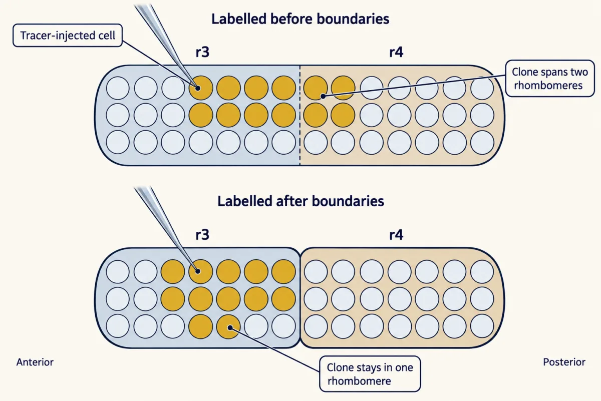
Predict. A pattern that repeats every two bulges suggests the bulges are real units. If they are, where should the descendants of one hindbrain cell end up if it is labelled with a dye before the boundaries between rhombomeres appear? And if it is labelled after?
Check your prediction
In 1990 Scott Fraser, Keynes and Lumsden injected single cells with a tracer, a dye that cannot pass to neighbouring cells. Clones labelled before boundaries appeared could span two rhombomeres. Clones labelled after boundaries formed stayed inside one.
Once its boundaries form, each rhombomere is a compartment: a block of progenitor cells (dividing cells that go on to make neurons and glia) whose descendants do not cross into the next. Some neurons born later do migrate across; the facial motor neurons of the mouse, met below, are one.
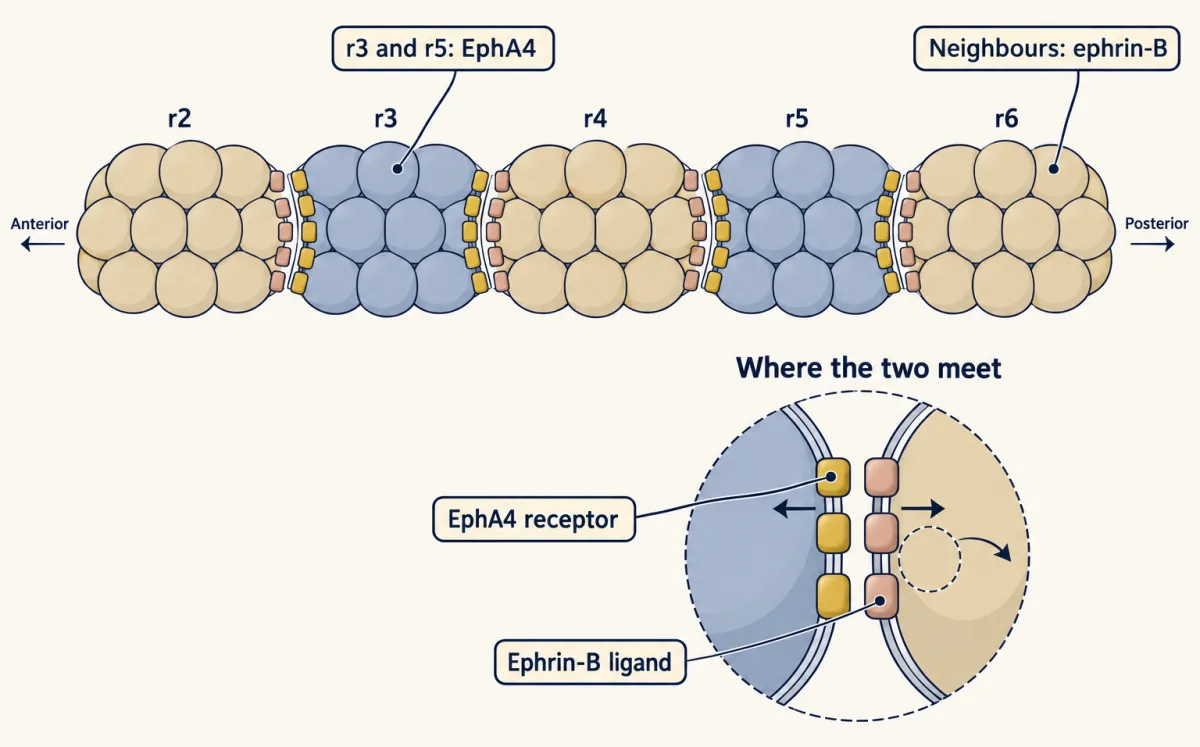
How are the cells kept apart? One reason is contact repulsion. In fish and frog embryos, r3 and r5 make the receptor EphA4, while their neighbours make ephrin-B ligands, and cells sort where the two meet.
So the hindbrain is built of units that keep their cells. That makes the question sharp. Like each house on that street, each unit needs a number before anyone moves in: some label that tells its cells what to become. What is it?
The code each segment carries
The label is a set of transcription factors, proteins that switch other genes on and off. Most come from Hox genes, a family met properly further on. For now, two facts are enough. Hox genes are numbered in paralog groups, 1 to 13, by where they sit in a cluster on the chromosome: group 1 at the end called 3′, group 13 at the 5′ end (3′ and 5′ name the two ends of a DNA strand). And in the hindbrain, the Hox genes switched on in domains with sharp front edges are mainly those of groups 1 to 4.
In 1989 David Wilkinson, Robb Krumlauf and colleagues reported that mouse Hox genes have expression domains whose front edges fall at rhombomere boundaries, often two rhombomeres apart. The table is a simplified summary of the early mouse neuroepithelial code around the time boundaries form, built on that mapping and later work. It leaves out expression levels, differences between dorsal and ventral cells, and later changes; a dash means absent in this schematic, not absent for life. The motor row is the chick map from above.
| r1 | r2 | r3 | r4 | r5 | r6 | r7 | |
|---|---|---|---|---|---|---|---|
| Hoxa2 (group 2) | — | ● | ● | ● | ● | ● | ● |
| Hoxb2 (group 2) | — | — | ● | ● | ● | ● | ● |
| Hoxb1 (group 1) | — | — | — | ● strong | — | — | — |
| Hoxa3, Hoxb3 (group 3) | — | — | — | — | ● | ● | ● |
| Hoxb4, Hoxd4 (group 4) | — | — | — | — | — | — | ● |
| Krox20 (segment gene) | — | — | ● | — | ● | — | — |
| MafB, or Kreisler (segment gene) | — | — | — | — | ● | ● | — |
| Motor neurons (chick) | — (r1 contributes to the cerebellum) | V | V | VII | VII | IX | IX |
Read it column by column. Every rhombomere has a different combination, but neighbours share many genes. Rhombomeres r3 and r4 share Hoxa2 and Hoxb2, so those two cannot tell them apart. What does is a segment gene, Krox20 in r3, and one Hox gene that, later in development, is strong in r4 alone: Hoxb1.
The segment genes are transcription factors too, each made in particular segments: Krox20 in r3 and r5, and MafB (Kreisler) in r5 and r6. They switch on particular Hox genes in those segments. Together, Hox genes and segment genes give each rhombomere its character, including the motor neurons it makes.
The first column is empty. No Hox gene is expressed in r1, and the front edge of Hoxa2 sits at the r1/r2 boundary. That edge is held, not merely inherited: in chick, a signal from the boundary between midbrain and hindbrain keeps Hoxa2 out of r1, and changing that signal changes where Hoxa2 is found. That signalling centre has its own lesson.
The one idea to carry through this lesson. More early retinoic acid, a more posterior code; the code helps set the neurons. Early on, over the range tested, more retinoic acid, a product of vitamin A, shifts a stretch of hindbrain toward the code of segments farther back, and less shifts it the other way. The code a rhombomere ends up with, sharpened by segment genes such as Krox20, helps decide which motor neurons it builds.
What the rule covers.
- Which genes. The Hox genes of paralog groups 1 to 4 in the hindbrain, early, before the segments sharpen.
- Which claim. The direction of change, often by moving Hox domains forward. It does not give the exact edges or how many Hox genes a segment carries, so the table above does not break it, and retinoic acid can switch some Hox genes off as well as on. Segment genes and local signals refine the code.
- Helps, not decides alone. The code is needed for a segment’s identity, as the knockout below shows, but it works with segment genes, not by itself.
Remove one letter: Hoxb1
Read the transcript of “Remove One Letter”
Film transcript: Remove One Letter
If the Hox code sets a segment's identity, what happens when one gene is taken away? A conventional knockout is a mouse line with one gene disrupted in every cell. In a normal mouse, facial motor neurons are born in r4 and migrate back to r6. There they form the facial motor nucleus. In the Hox code, Hoxb1 is the clearest difference between r4 and r3. Predict. Knock out Hoxb1. Does r4 disappear? Or does it stay and become something else?
In 1996, two groups reported Hoxb1 knockouts. One was led by Studer and Krumlauf, the other by Goddard and Capecchi. Rhombomere four was still there. Its progenitors still divided, and still made motor neurons. But r4 lost parts of its identity. Its motor neurons behaved like the trigeminal neurons of r2 and r3. In both mutants, the facial motor neurons failed to form or to migrate normally. No normal facial motor nucleus formed.
Compare a different gene, Krox20. Without it, r3 and r5 do not persist as distinct segments. Some of their cells survive, mix with neighbours, and take on their character. In kreisler (MafB) mutants, r5 is missing. So there are two different outcomes. Without Krox20, distinct territories are lost. Without Hoxb1, r4 remains but changes. That is the code acting as a cause, not just a marker.
Next: where do the code's edges come from? The takeaway: without Hoxb1, r4 survives but loses key parts of its identity. Other Hox knockouts differ, gene by gene.
From the film
- Without Hoxb1, rhombomere 4 stays in the mouse but loses parts of its identity.
- Its motor neurons then behave like trigeminal neurons, and the facial motor nucleus fails to form normally.
- Without Krox20, by contrast, r3 and r5 do not persist as distinct segments, so the code acts as a cause, not just a marker.
A conventional knockout is a mouse line in which one gene has been disrupted in every cell. What goes wrong shows what the gene is needed for.
In a normal mouse, facial branchiomotor neurons are born in r4 and migrate backward to r6, where they form the facial motor nucleus. That nucleus supplies the muscles of facial expression through nerve VII.
Predict. Knock out Hoxb1. Does r4 disappear, or does it stay and become something else? Use the table to decide, and say what r4’s motor neurons should do.
Check your prediction
Start from the code. Hoxb1 is the clearest Hox difference between r4 and r3. Take it out, and in this simplified table r4 is left with r3’s Hox genes, Hoxa2 and Hoxb2. But the code is a network, not a list. Hoxb1 helps keep Hoxb2 on in r4, so in the mutant Hoxb2 drops there too. And r4 never had Krox20, the r3 segment gene. So r4 should stay, but take on trigeminal character closer to that of r2.
The motor neurons follow. Neurons born in r4 should no longer behave like facial neurons. They may fail to make the facial program, or make a trigeminal-like one and stop migrating backward to r6. Without a working facial motor nucleus, the muscles of facial expression lose their motor supply, so you would predict weak or absent facial movement.
In 1996 two groups reported Hoxb1 knockouts, one led by Studer and Krumlauf and one by Goddard and Capecchi. In both mutants the facial branchiomotor neurons failed to form or to migrate normally, and the facial motor nerve was lost. Studer and colleagues found that r4 lost parts of its identity and that its motor neurons behaved like trigeminal neurons of r2 and r3. Goddard and colleagues reported that the motor nucleus of nerve VII failed to form.
Rhombomere 4 is still there. Its progenitors still divide, and they still make motor neurons, now with a different identity.
Not every patterning gene works this way. There are two outcomes to tell apart:
- Territory lost. In mice lacking Krox20, r3 and r5 fail to persist as distinct territories, and r5 is missing in kreisler (MafB) mutants. Fate mapping shows that some Krox20-lacking cells survive, mix with neighbours and take on the identity of even-numbered segments. Krox20 couples a segment’s identity to keeping its cells apart.
- Identity changed, segment kept. Without Hoxb1, r4 stays recognizable but loses important parts of its identity.
These are different outcomes, not two separate classes of gene. Either way, the code acts as a cause, not just a marker. The next question is where its edges come from.
Where the edges come from: a tap at the back, a drain at the front
Read the transcript of “A Tap at the Back, a Drain at the Front”
Film transcript: A Tap at the Back, a Drain at the Front
What helps set the front edges of the Hox code? A signal made from vitamin A. Retinoic acid is made from retinol, which is vitamin A, in two steps. Raldh2, the main enzyme for the last step, is made in mesoderm beside the posterior hindbrain and spinal cord. Cyp26 enzymes destroy it. Cyp26a1 is strong toward the front of the neural plate. Picture a bath, with the tap at the back and the drain at the front. Retinoic acid ends up high at the back and low at the front. In the nucleus, it binds the retinoic acid receptor, RAR. RAR pairs with its partner, RXR, on short DNA sequences called response elements. Hoxa1 and Hoxb1 carry these elements. In cell culture, 3′ Hox genes respond to lower doses than 5′ genes.
In a simple threshold model, then, fewer Hox genes come on toward the front, where the signal is lower. Predict. Give mouse embryos extra retinoic acid at an early stage. Which way should the Hox edges move? Forward. In 1992, Marshall, Krumlauf, Lumsden and colleagues found the hindbrain Hox domains changed. Rhombomeres two and three shifted toward the identity of four and five. Too much signal shows a response, not the normal role. Predict. Now block the drain. Mice lacking Cyp26a1 clear less retinoic acid at the front. What changes?
The front shifts: the front of the hindbrain looked partly transformed toward a more posterior character. Remove the tap instead, in mice lacking Raldh2, and the posterior hindbrain is poorly patterned. Changing the signal sets the direction, not the exact edges. In the hindbrain, group 2 genes reach farther forward than group 1. Yet 3′ prime genes need less signal. So other inputs help set those edges. Retinoids are teratogens, agents that cause birth defects. In mouse and chick, extra retinoic acid early shifts the Hox code. It also disturbs the neural crest cells that build much of the face. In humans, this mechanism is inferred from animal work.
More early retinoic acid gives a more posterior code; the code helps set the neurons. Other inputs help draw the final edges.
From the film
- Retinoic acid, made from vitamin A, is made toward the back of the hindbrain and destroyed toward the front by Cyp26 enzymes.
- Extra early retinoic acid shifts Hox domains forward, and less of it leaves the posterior hindbrain poorly patterned.
- The signal sets the direction of the code, not the exact edges, which other inputs help draw.
- Retinoids are teratogens in mouse and chick, and in humans this mechanism is inferred from animal work.
Retinoic acid is made from retinol (vitamin A) in two oxidations, through retinaldehyde. In the early embryo the main enzyme for the second step is Raldh2, made in the mesoderm (the middle tissue layer) beside the posterior hindbrain and the spinal cord. A family of enzymes called Cyp26 oxidizes retinoic acid to inactive products, and Cyp26a1 is strongly expressed toward the front of the neural plate.
Picture a bath with the tap at one end and the drain at the other. Supply behind and destruction in front leave retinoic acid high toward the back of the hindbrain and low toward the front. The picture works for something that spreads slowly, like dye running in with the tap water, which is darkest near the tap. It does not work for the water level, which evens out.
Retinoic acid is small and oily, so it crosses membranes and reaches the nucleus. There it binds the retinoic acid receptor (RAR), which works as a pair with the retinoid X receptor (RXR) on short DNA sequences called retinoic acid response elements (RAREs). Both are nuclear receptors, ligand-activated transcription factors of the kind covered in that lesson. Hoxa1 and Hoxb1 carry RAREs.
In cell culture, 3′ Hox genes respond to lower doses of retinoic acid than 5′ genes. In a simple threshold model, then, fewer Hox genes would come on toward the front, where the signal is lower or arrives for a shorter time, and those that did would lie toward the 3′ end of the cluster. The real hindbrain bends this model, as shown below. Retinoic acid mainly drives paralog groups 1 to 5, which include the hindbrain’s groups 1 to 4. The more 5′ Hox genes of the spinal cord depend mainly on fibroblast growth factor (FGF) and Wnt signals.
If that is right, changing the amount of signal should move the code.
Predict. Expose mouse embryos to extra retinoic acid at an early stage. Which way should the Hox edges move, and what should happen to r2 and r3?
Check your prediction
The edges should move forward, so the front segments carry a code that normally sits farther back. In 1992 Heather Marshall, Krumlauf, Lumsden and colleagues reported that exposing mouse embryos to retinoic acid at an early stage changed the hindbrain Hox domains and transformed r2 and r3 toward the identity of r4 and r5.
Extra signal shows how the embryo responds to too much. It does not by itself show what the normal signal does.
Predict. Now leave the tap alone and block the drain. Mouse embryos lacking Cyp26a1 cannot destroy retinoic acid at the front as they normally would. What should change, and where?
Check your prediction
Retinoic acid should persist farther forward, so the front of the hindbrain should shift toward a more posterior character. Cyp26a1 is strongest at the front, so the front is most at risk; effects elsewhere depend on stage and on the other Cyp26 enzymes. Mouse embryos lacking Cyp26a1 showed a rostral (front) hindbrain that looked partly transformed toward a more posterior character (Abu-Abed et al., 2001).
Less signal pushes the other way. In mouse embryos lacking Raldh2, and in quail embryos deprived of vitamin A, the posterior hindbrain is poorly patterned: its rhombomeres, roughly r4 to r7 in quail, are reduced, lost or misspecified, so posterior character is lost (Niederreither et al., 2000; Maden et al., 1996). Blocking synthesis like this is the better guide to what the normal signal does.
What the gradient cannot do
The table shows where the rule stops.
- Edge order. Hoxa2 and Hoxb2, in group 2, reach farther forward than Hoxb1, in group 1, though 3′ genes need less signal. Other inputs help set the group 2 edges: the midbrain–hindbrain signal that keeps Hoxa2 out of r1 in chick, and in mouse Krox20 and Hoxb1, which regulate Hoxb2 in r3, r5 and r4.
- Narrowing. A threshold rule in which retinoic acid only switches genes on predicts that each gene stays on from its edge all the way back. Hoxb1 instead ends up in r4 alone. Loops by which Hox genes and segment genes regulate one another do that, along with a second RARE in Hoxb1 that represses it in r3 and r5 (Studer and colleagues, 1994). So the signal takes part in the narrowing too.
- Sharpness. A smooth signal does not by itself say how sharp or stable an edge will be. Segment genes, feedback between genes and cell sorting (shown in fish and frog) make the boundaries in the table sharp and keep them that way.
Whether cells read how much signal they get or how long they get it is still argued (the Graduate explanation on this page says more).
Retinoids as teratogens
A teratogen is an agent that causes birth defects when an embryo or fetus is exposed to it. Retinoids qualify because the early embryo uses this very signal to set its axis. In mouse and chick embryos, extra retinoic acid at an early stage shifts the Hox code of the hindbrain and disturbs the neural crest cells that leave it to build much of the face. In human embryos this mechanism is inferred from the animal work rather than shown directly, but it fits the ears, jaw and heart in the pattern of defects described in Going deeper. Too little vitamin A harms the embryo as well. The dose has to be right in both directions.
Order on the chromosome, order along the body
Why are Hox genes switched on in an ordered series at all? The answer begins in the fruit fly.
A homeotic mutation turns one body part into the likeness of another: legs where antennae should be, or a second pair of wings in place of the small balancing organs called halteres. Edward Lewis spent decades on one cluster of such genes, the bithorax complex. In 1978 he proposed that its genes lie along the chromosome in the same order as the body segments they control, from front to back.
In 1984 two groups found what these genes share: a short stretch of DNA common to several fly homeotic genes. It was named the homeobox. It encodes the homeodomain, a part of the protein that later work showed binds DNA, with one helix lying in the major groove. One of the two groups, Gehring’s in Basel, soon detected related sequences in vertebrate DNA. Body-plan genes belong to a shared animal toolkit (see that lesson).
Hox genes are one branch of the much larger homeobox family, and they come in clusters. Mice and humans have 39, in four clusters called Hoxa, Hoxb, Hoxc and Hoxd, each on a different chromosome. Genes that sit at the same position in different clusters, such as Hoxa3, Hoxb3 and Hoxd3, form a paralog group. There are 13 groups, and no cluster has all 13. Paralogs have similar sequences and often similar expression, so they can stand in for one another, and knocking out a single Hox gene often gives a milder defect than you might expect.
The clusters obey the rule Lewis saw, called colinearity.
- Spatial colinearity. Along the body as a whole, genes at the 3′ end of a cluster (group 1, then group 2 and so on) have the most anterior front edges of expression, reaching into the hindbrain. Genes farther toward the 5′ end have edges farther back, and group 13 is expressed near the tail.
- Temporal colinearity. In mouse and chick, 3′ genes are also switched on first and 5′ genes later, as the body axis grows backward. This is a vertebrate observation. In the fly the timing rule is weak or absent.
Predict. By the whole-body rule, which should reach farther forward in the hindbrain: Hoxb1, in group 1, or Hoxb2, in group 2? Check the table before you open this.
Check your prediction
The rule says Hoxb1. The table says otherwise: Hoxb2 starts at r3 and Hoxa2 at r2, while Hoxb1 starts at r4. In the hindbrain the order bends, because other inputs help set the group 2 edges. Colinearity is a trend along the whole body, not a law for every boundary.
What this example teaches about every axis
The hindbrain shows three principles that will come back in the lessons on the tube’s other axes.
- Particular genes are switched on at particular places and particular times.
- Signals that spread from a source, and are destroyed or opposed elsewhere, form graded concentrations, and those gradients help set the pattern. Here the source is Raldh2 and the drain is Cyp26.
- The signals restrict where certain transcription factors are made, and those factors then regulate the genes that give each region its character.
In the hindbrain, the signal is retinoic acid, working alongside fibroblast growth factor (FGF) and Wnt signals, and the transcription factors are the Hox proteins, together with segment genes such as Krox20.
Putting it together
| Experiment | Animal and age | Observed result | What it supports | What it does not establish |
|---|---|---|---|---|
| Map motor neuron origins (1989) | chick embryo | nerve V from r2–r3, VII from r4–r5, IX from r6–r7; each leaves from the even member | segments have distinct jobs, in pairs | that the segments are units of lineage |
| Label single cells (1990) | chick embryo | clones cross boundaries if labelled before they form, not after | rhombomeres are compartments for progenitors | what label each carries; later neurons can still migrate |
| EphA4 and ephrin-B patterns | fish and frog embryos | EphA4 in r3 and r5, ephrin-B next door; cells sort at contact | one way the cells are kept apart | that it is the only way |
| Map Hox expression (1989) | mouse embryo | front edges of Hox domains at rhombomere boundaries | each segment carries a code | that the code causes identity |
| Knock out Hoxb1 (1996) | mouse | r4 stays; its neurons behave like trigeminal neurons; facial motor nucleus fails | a Hox gene is needed for r4 identity | that Hoxb1 works alone |
| Knock out Krox20 | mouse | r3 and r5 fail to persist as distinct segments; some of their cells survive with neighbouring identities | some genes keep a segment as a distinct territory | that the cells were deleted |
| Extra retinoic acid, early (1992) | mouse embryo | Hox domains changed; r2 and r3 toward r4 and r5 | more early signal pushes front segments toward a posterior identity | what the normal signal does |
| Knock out Cyp26a1 (2001) | mouse embryo | rostral hindbrain partly more posterior | the drain keeps the front low | exact edges |
| Knock out Raldh2; vitamin A deficiency (2000; 1996) | mouse embryo; quail embryo | posterior hindbrain poorly patterned; posterior rhombomeres reduced, lost or misspecified | the normal signal is needed for the posterior code | exact edges |
| Isotretinoin exposure (1985) | human children exposed early in pregnancy | ears, face and jaw, heart and brain affected | fits the animal mechanism | that the human defects come from a Hox shift; this is inferred |
The guiding idea holds up within its scope: more early retinoic acid, a more posterior code; the code helps set the neurons. Extra signal or a blocked drain pushes the front toward a posterior identity, and losing the tap costs the back its posterior character. Segment genes, local signals, repression and cell sorting shape the exact edges.
One link to the neurotrophic hypothesis lesson, held loosely. The retinoid experiments show that the signal is needed (Raldh2 loss) and that changing it changes identity (extra retinoic acid, Cyp26a1 loss). In that lesson’s terms, this points toward an instructive signal. Showing which features of the embryo’s own gradient carry position would need experiments that control where and when the signal acts.
Design problem
An embryo has extra Cyp26 activity in the posterior hindbrain. Using the guiding idea and its scope, predict which way the Hox edges move and say what the rule cannot tell you about which genes go first. Say what you would expect for the segments and motor neurons of r4 to r7. Then say how you would tell a segment that has changed identity from one that is missing.
Saved in this browser only.
One way to do it
Direction. If the extra drain acts during the early patterning window, it should lower retinoic acid in the posterior hindbrain. By the rule, posterior segments should shift toward a more anterior identity, the direction seen when Raldh2 or vitamin A is lacking.
What the rule cannot tell you. It cannot say which genes go first or how far each edge moves. Even in a simple threshold model, lowering the signal by a factor k moves every edge back by the same distance only if the signal falls off exponentially (the shift is then λ ln(1/k), where λ is the distance over which the signal falls e-fold); with other profiles the shifts differ, and edges in front of the region with extra Cyp26 need not move at all. Nor does the gradient set every edge: the midbrain–hindbrain signal holds Hoxa2 out of r1, and loops and repression confine Hoxb1 to r4, so some Hox domains may move less than the gradient alone predicts. To find out, you would measure retinoic acid activity and Hox expression over time.
Segments and neurons. If the code shifts back, posterior segments would carry a code more like that of the segments in front of them. By the logic of the Hoxb1 knockout, their motor neurons might then take a more anterior program. That is a prediction from the rule, not a result in this lesson.
Identity or lost territory. Ask first whether each segment is there: its boundaries, its width, dividing progenitors and segment genes such as Krox20 in r3 and r5. A missing boundary or marker shows that a recognizable territory has failed, but not why. Cells may have changed identity, mixed with neighbours or died. To tell these apart, combine the shape of the tissue with several independent markers, lineage tracing, and counts of dividing and dying cells. If the segment is there, ask what it makes: which motor neurons, where they migrate, and which even-numbered rhombomere their axons leave from. A segment that is present but makes the wrong neurons has changed identity, as r4 does without Hoxb1.
Going deeper
The regions of the early tube
The three vesicles have formal names: the forebrain (prosencephalon), the midbrain (mesencephalon) and the hindbrain (rhombencephalon). The forebrain then divides into the telencephalon, which makes the cerebral hemispheres, and the diencephalon, which makes the thalamus and its neighbours and pushes out the optic vesicles that become the retinas. The hindbrain divides into the metencephalon (pons and cerebellum) and the myelencephalon (medulla).
Repeated units such as rhombomeres are called neuromeres. The question this lesson answers for the hindbrain can be put for all of them: how is the nervous system specified within neuromeres?
The homeobox in numbers
The homeobox is about 180 base pairs long and encodes a homeodomain of about 60 amino acids. It was found in 1984 by the laboratory of Walter Gehring in Basel (William McGinnis, Michael Levine, Ernst Hafen, Atsushi Kuroiwa and Gehring) and by Matthew Scott and Amy Weiner in Thomas Kaufman’s laboratory at Indiana University. The fly has eight Hox genes, in two complexes on one chromosome.
The somites keep time too
The somites, blocks of mesoderm beside the tube that form one pair at a time (roughly every 90 minutes in chick), show temporal colinearity too, but that story belongs to them.
Two Hox genes lost together
Mice lacking both Hoxa1 and Hoxb1 have been described as losing r4 and r5. The published descriptions differ in their wording, and some describe a severe reduction and remodelling of the r4–r5 region rather than a clean loss of both segments.
Isotretinoin and the first reports
Isotretinoin, a synthetic form of retinoic acid (13-cis-retinoic acid), went on sale in the United States in 1982 as a treatment for severe acne. By 1985, Edward Lammer and colleagues had described a pattern of defects in children exposed to it early in pregnancy: small or missing ears, an altered face and jaw, heart defects, and abnormalities of the brain. They called it retinoic acid embryopathy. A pill taken for the skin had reached the embryo while its head and face were being laid out.
The 1996 Hoxb1 knockouts
The two 1996 Hoxb1 knockout reports were by Michele Studer, Lumsden, Laila Ariza-McNaughton, Allan Bradley and Krumlauf in Nature, and by J. M. Goddard, Mireille Rossel, Nancy Manley and Mario Capecchi in Development.
Your notes
Select any text in the lesson to highlight it or add a note to it. Your highlights and notes are saved in this browser and collected in your notebook.
This browser is not letting the site save anything (a private window or strict privacy settings), so highlights and notes will be lost when you leave the page.
How we measure it
In situ hybridization
A labelled strand of RNA or DNA that matches a gene's message is applied to a whole embryo or a section, and it sticks where that message is present. It shows where and when a gene is switched on. Here it revealed Hox domains whose edges sit at rhombomere boundaries.
Single-cell lineage tracing
One cell is injected with a dye that cannot pass to its neighbours, and its descendants are found later. Where the clone ends up tells you whether its cells were free to cross a boundary. In the chick hindbrain, clones labelled after boundaries formed stayed in one rhombomere.
Gene knockout by targeting
A gene is disrupted in mouse embryonic stem cells, and the cells are used to make a line of mice that lacks the gene in every cell. What goes wrong shows what the gene is needed for. Hoxb1 knockouts showed that one Hox gene is needed for the identity of rhombomere 4.
Timed retinoid exposure
Embryos are given extra retinoic acid, or a drug that blocks its synthesis, at a chosen stage. Shifts in gene expression and anatomy show how the embryo responds to too much or too little signal at that stage; blocking synthesis is the better guide to what the normal signal does. Extra retinoic acid pushed anterior hindbrain segments toward a more posterior identity.
Practice questions on this lesson (3) → MCAT-style, original, each with an explanation
Where this lesson fits
Before thisReview first
- Neural Induction: The Default ModelThis course · Lesson 6
You need the neural plate and its default posterior-to-anterior patterning from there to follow how the hindbrain gets an address.
- Neurulation and Neural Tube DefectsThis course · Lesson 7
This lesson assumes the closed neural tube and its early regions, which that lesson builds.
- The Organizer: Spemann and MangoldThis course · Lesson 5
Graded signals from a source that pattern the neural plate are introduced there, and retinoic acid extends that logic.
After thisStudy next
- Local Organizers: The Midbrain-Hindbrain BoundaryThis course · Lesson 9
It shows how a boundary signal holds Hoxa2 out of r1, an edge only mentioned here.
- Transcription Factor Codes: Building a Motor NeuronThis course · Lesson 11
It takes the idea that a transcription factor code sets neuron identity and works it out for spinal motor neurons.
Elsewhere on the siteFilms, simulations and method films in other courses
- Why Far Signals Ride the BloodFilm · Cell Signaling
A retinoic acid gradient is a morphogen-style signal whose range depends on how far it travels.
Suggested by an AI model from the lessons themselves, then checked. How we use AI
Finish line
Test yourself
8 questions
Practice and MCAT-style, with explained answers.
Next · Lesson 9
Local Organizers: The Midbrain-Hindbrain Boundary
How does one protein on a bead make a second midbrain grow, built back to front?
Continue →