Cell Biology · Skim version
Cytoskeletal Polymers and Dynamics
Read the full lesson → Skim the whole course →
The guiding idea
A microtubule in a dish of pure tubulin grows for minutes, then collapses many times faster. Why would a polymer be built to fail? Because a polymer that switches by itself lets a cell explore space, rebuild its scaffolding and organize division.
Cells keep their cytoskeleton in constant, regulated turnover so they can explore space, change shape, move and build the division spindle.
From the film: Tight Ends, Loose Ends
- Each filament end has a critical concentration: above it the end grows, below it the end shrinks.
- If subunits never hydrolysed their nucleotide, both ends would share one critical concentration and the polymer would simply settle.
- Hydrolysis after joining gives a polymer tight ends, with a low critical concentration, and loose ends, with a high one.
- At treadmilling steady state, actin grows at its tight barbed end and shrinks at its loose pointed end.
- A microtubule end is tight while it keeps its GTP cap and collapses many times faster when it loses it.
From the film: When a Drug Becomes a Test
- A drug tests a mechanism only when its target, dose and test system stay attached.
- The same change in polymer amount can begin at free subunits, filament ends or the lattice.
- Latrunculin A sequesters actin monomers and can also speed loss at the ends of freshly built filaments.
- Nocodazole effects depend on dose and on the cultured cell system.
- A second measurement can separate mechanisms that produce the same outcome.
-
Three polymers built for different jobs
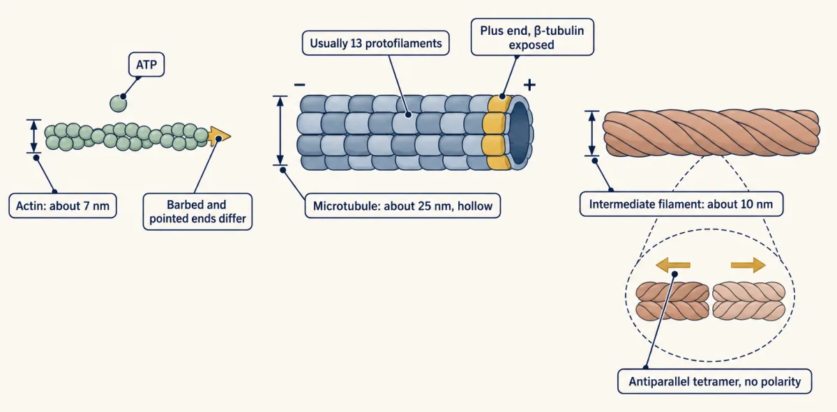
Three polymers drawn to the same scale. Actin and microtubules have two different ends; the intermediate filament's building block points both ways, so its two ends are alike. AI-generated illustration -
One end: the doorway and its critical concentration
-
Two ends: why hydrolysis makes them differ
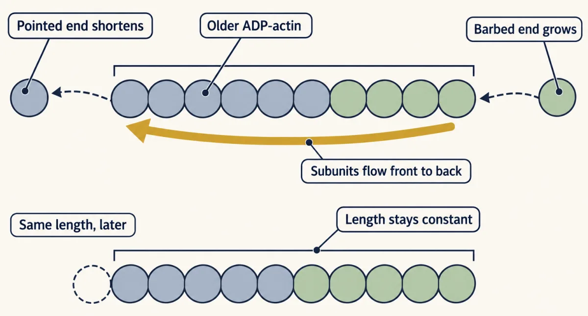
Treadmilling: at steady state the barbed end gains subunits as fast as the pointed end loses them, so the filament keeps its length while subunits flow through it. AI-generated illustration -
One end, two states: dynamic instability
-
Drugs as tests
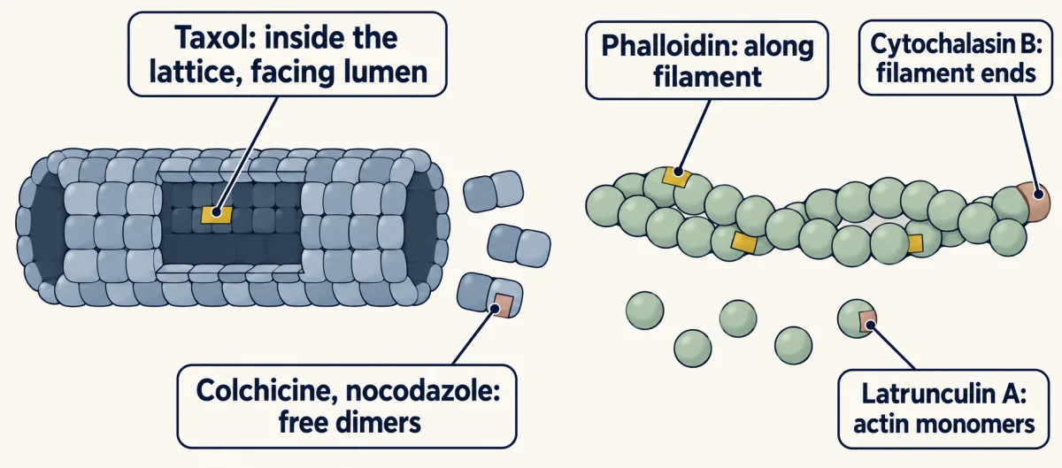
Where each drug binds: taxol inside the assembled lattice, facing the lumen, colchicine and nocodazole on free tubulin dimers, phalloidin along actin filaments, cytochalasin B at their ends, latrunculin A on actin monomers. Effects depend on dose and system. AI-generated illustration -
Going deeper
Where this lesson fits
Before thisReview first
- Transport Across MembranesThis course · Lesson 5
You need rate constants and the idea of a balance point, which this lesson reuses for the filament end.
- Experiments That Distinguish MechanismsThis course · Lesson 3
The drug section relies on tying a perturbation to its target, dose and test system.
After thisStudy next
- Molecular Motors and CargoThis course · Lesson 15
Motors walk along polar filaments, so the plus and minus ends and their dynamics set the tracks' direction.
- Cell Shape, Migration and CiliaThis course · Lesson 16
Actin treadmilling and network turnover drive protrusion and migration, and microtubules build cilia.
- Mitosis and Chromosome SegregationThis course · Lesson 19
Dynamic instability lets microtubules search for chromosomes and build the spindle.
Elsewhere on the siteFilms, simulations and method films in other courses
- Optical trap (optical tweezers)Method film
This lesson's polymer growth and shortening can be measured as single-filament forces and steps.
Suggested by an AI model from the lessons themselves, then checked. How we use AI