Life Science · Unit 1: Cells
Inside a Cell
Rewritten on : new text, flashcards and film. Your notes and highlights stay, but the text around them may have moved.
Why is a heart muscle cell crowded with mitochondria, while a fat cell is mostly one big drop of fat?
Pause when the reading reaches a film, and offer to play it. Saved on this device.
Watch the film now, or keep listening
Builds on:Cells Are EverywhereWhy these?
Where this lesson sits
- Virginia SOL LS.2 b
- Practices LS.1 c, LS.1 d, LS.1 e
Essential question: What are the parts inside a cell, and how do they work together to keep it alive?
-
Life Science (grade 7) LS.2 b You are here
This lesson
-
Biology
Preview it at high school level
In Biology you will follow one product all the way through a cell. The instructions stay in the nucleus. Ribosomes, tiny protein builders, sit on the rough endoplasmic reticulum and build the protein. The protein then moves to the Golgi apparatus, which sorts and packs it. Small bubbles of membrane called vesicles carry it to the cell membrane, join with it and release the protein outside the cell. Mitochondria supply ATP, a small molecule that carries energy to the work. You will also meet lysosomes, which break down worn-out parts, and compare cells that have a nucleus with bacteria that do not.
-
Biology II Biology II: Advanced Survey of Biology Topics, BIOII.2 a
Preview where it goes next
Virginia's Biology II: Advanced Survey of Biology Topics course (BIOII.2 a) studies the cell as a system that keeps conditions inside it steady. Students look at the nucleus, mitochondria, ribosomes, endoplasmic reticulum, Golgi apparatus and cytoskeleton, a network of fibers that gives the cell shape and moves things inside it. They ask how each part depends on the others, and they design their own investigations of how cells respond to change.
-
College
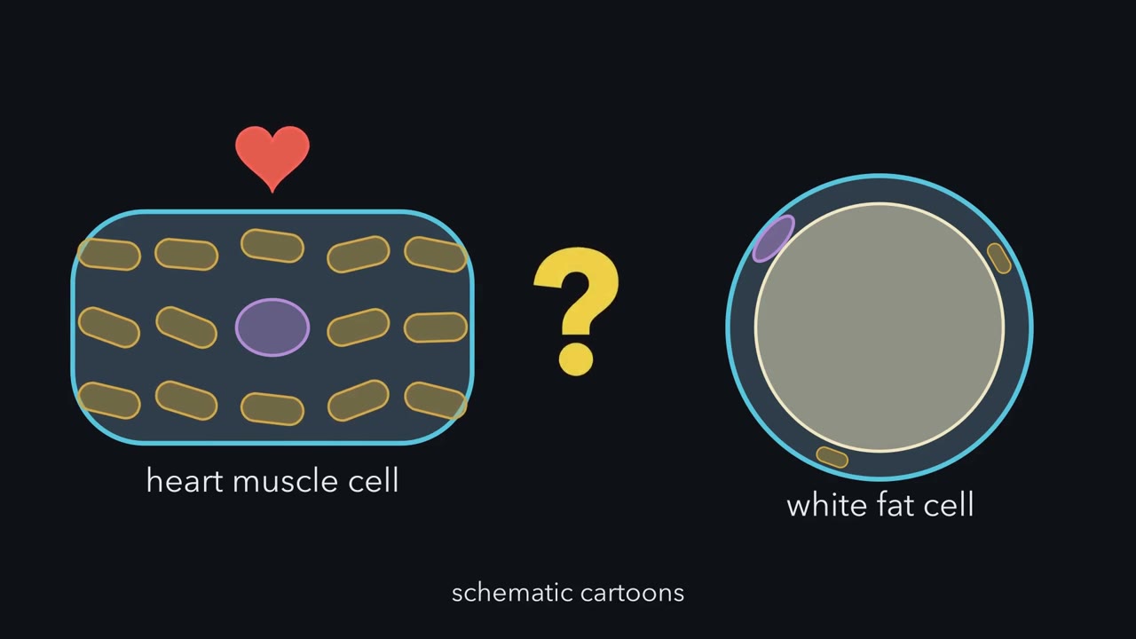 ▶ Watch One Cell, Many Jobs, Shared Space · 3:18
Read the transcript of “One Cell, Many Jobs, Shared Space”
Film transcript: One Cell, Many Jobs, Shared Space
Your heart muscle cell and a white fat cell share many of the same kinds of parts. So why do they look nothing alike inside? Let's build a cell and find out. Start with the outline. The cell membrane is the thin boundary around the cell. It controls what enters and leaves.
Inside is the cytoplasm: a jelly-like fluid and the parts in it, except the nucleus. The parts inside are called organelles. An organelle is a small part that does a special job. The nucleus holds the cell's instructions. The ER is a network of folded membranes. It helps make and move many proteins and fats.
Mitochondria release energy stored in food, such as sugar and fats. They release it in a form the cell can use. A vacuole is a sac that stores water, food and wastes. Plant cells have one large vacuole. Many animal cells have only small sacs. Now give the cell one job: replacing a worn-out protein. The nucleus holds the instructions. The ER helps make the new protein and move it. The mitochondria release the energy that building uses. The membrane lets in food and oxygen.
Four parts, one job. Every life job needs a team like this. Growth, repair and response each need several parts working at once. So what if only the mitochondria stop? Does only energy use slow down, or do other jobs slow down too? Pause and decide.
Other jobs slow down too. The nucleus and the ER are still fine. But building needs energy, so the repair path slows and stalls. Growth and response slow down with it. No part decides anything; each one simply follows chemistry. Here is the second idea. Picture the cell from above, as a floor plan. Each part is a room. Together, the rooms make up the whole cell. So when one room takes a big share, the others take smaller shares.
Cells with different jobs often hold their parts in different amounts. A heart muscle cell works without a break. It releases a lot of energy, mostly from fats. So mitochondria fill a large share of its space. A white fat cell stores fat. So one big fat drop fills most of its space. The other parts sit in a thin rim around the edge. Many of the same parts, very different floor plans. A part's size alone does not tell you a cell's job. But comparing parts with jobs can show a pattern.
From the film
- An organelle is a small part inside a cell that does a special job.
- Every life job, such as replacing a worn-out protein, uses several parts working together.
- If the mitochondria stop, many jobs slow down, not just energy use.
- Cells with different jobs often hold their parts in different amounts. A part's size alone does not tell you a cell's job.
The first film opened with a puzzle: a heart muscle cell and a white fat cell share many kinds of parts, yet their floor plans look nothing alike. At rest, your heart beats about 60 to 100 times a minute, so its muscle cells release a lot of energy, mostly from fats. A white fat cell has almost the opposite job: it stores fat for later. How much of each cell goes to each part, and does that match the job?
What do you think? Decide before you read.
-
Scientists disagree: The parts depend on one another. In a 1968 experiment, described below, chemicals that blocked energy release in the mitochondria also left new proteins made for export stuck in the ER.
What scientists think
Scientists disagree: The parts depend on one another. In a 1968 experiment, described below, chemicals that blocked energy release in the mitochondria also left new proteins made for export stuck in the ER.
-
Scientists disagree: Measurements show big differences. In the mammals measured, mitochondria fill about a fifth to over a third of a heart muscle cell. In white fat cells one fat drop fills most of the cell. A mature red blood cell has no mitochondria at all.
What scientists think
Scientists disagree: Measurements show big differences. In the mammals measured, mitochondria fill about a fifth to over a third of a heart muscle cell. In white fat cells one fat drop fills most of the cell. A mature red blood cell has no mitochondria at all.
-
Scientists disagree: Energy cannot be made, only changed from one form to another. Mitochondria do not make energy; they release the energy stored in food, in a form the cell can use. Scientists measure this by tracking the food and oxygen that cells take in and use.
What scientists think
Scientists disagree: Energy cannot be made, only changed from one form to another. Mitochondria do not make energy; they release the energy stored in food, in a form the cell can use. Scientists measure this by tracking the food and oxygen that cells take in and use.
The one idea. A cell is one team of parts. Every life job needs several parts working at once, and cells with different jobs often hold their parts in different amounts.
Where it holds: plant and animal cells, though a few, like mature red blood cells, lack whole parts. No part plans anything; each follows chemistry. Heart and fat cells show a pattern, not a rule: a part’s size alone does not tell you a cell’s job.
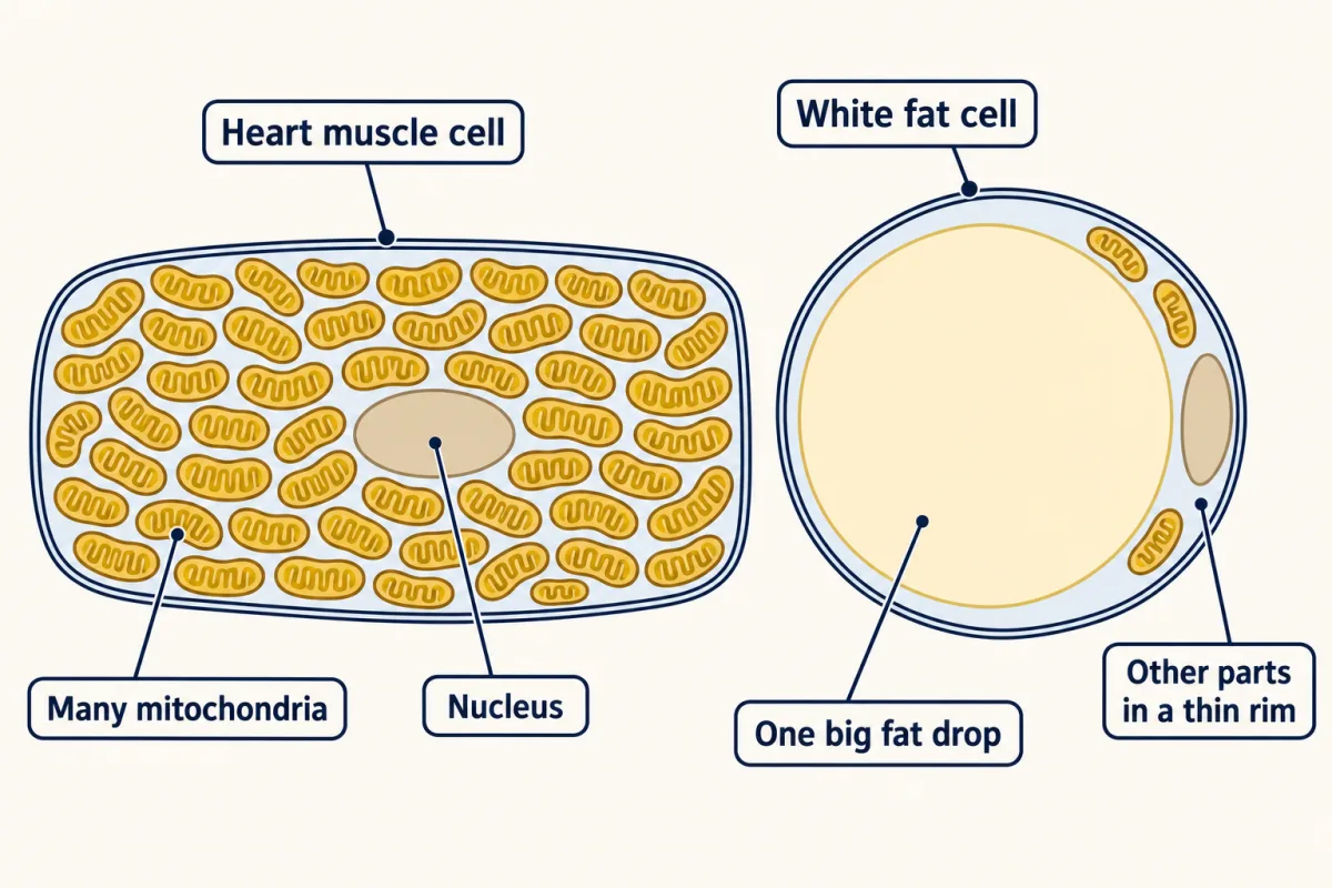
Explain: the parts and their jobs
- organelle
- nucleus
- cell membrane
- cytoplasm
- endoplasmic reticulum
- mitochondria
- vacuole
- metabolism
- photosynthesis
- cellular respiration
For reference, here are the first film’s six parts, with details it left out. An organelle is a small part inside a cell that does a special job.
- Nucleus: holds the instructions that guide what the cell makes and does.
- Cell membrane: controls what enters and leaves, and lets the cell sense its surroundings.
- Cytoplasm: everything inside the membrane except the nucleus, fluid and organelles together. Many reactions happen in the fluid.
- Endoplasmic reticulum (ER): folded membranes that help make and move many proteins and fats.
- Mitochondria: release much of the energy stored in food such as sugar (glucose) and fats, in a form the cell can use.
- Vacuole: stores water, food and wastes. Many animal cells have only small sacs, and sacs called lysosomes break down wastes and worn-out parts.
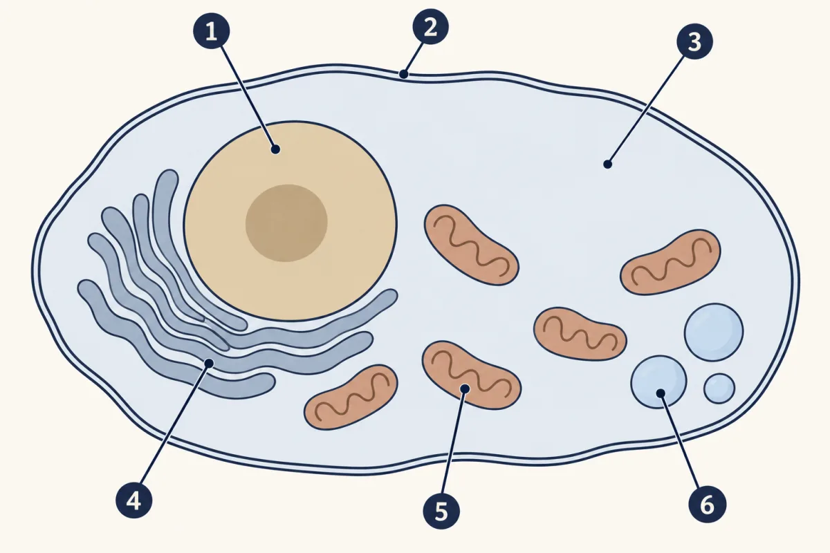
- nucleus
- cell membrane
- cytoplasm
- endoplasmic reticulum
- mitochondria
- vacuole
- nucleus
- cell membrane
- blank
- blank
- blank
- blank
Word bank: cytoplasm, endoplasmic reticulum, mitochondria, vacuole
- blank
- blank
- blank
- blank
- blank
- blank
Check your labels
- nucleus
- cell membrane
- cytoplasm
- endoplasmic reticulum
- mitochondria
- vacuole
The first film followed one job; the table maps the others.
| Job | Parts that help |
|---|---|
| Using energy | Mitochondria, cytoplasm |
| Making molecules | Nucleus (instructions), ER, cytoplasm |
| Removing wastes | Cell membrane, vacuoles and other small sacs |
| Responding to the environment | Cell membrane, nucleus |
Not every cell has every part. A mature human red blood cell has no nucleus and no mitochondria. Bacteria have no nucleus (that lesson). Plant cells share the first five parts and add a cell wall, a large central vacuole and, in green cells, chloroplasts (that lesson).
Explore: switch off one part of a cell
Now test the team: switch off one part at a time and watch the six life-process bars.
In this sketch the bars are relative lengths, not measurements. The more a process uses a part and the bigger that part’s share, the more its bar drops. Reproduction also loses part of growth’s drop, because a cell grows before it divides. Block widths show small, ordinary or large only, not to scale. Only the mitochondria shares and the leaf cell’s large vacuole come from the lesson. A leaf cell’s ordinary mitochondria share is a guess here. Every other part is ordinary size. Heart and fat cells are drawn with a vacuole only to keep the three cells alike. A leaf cell also has a cell wall and chloroplasts, which this sketch leaves out. Nothing is chosen by chance.
Rule to keep: the parts work as one system, so trouble in most parts slows several life processes.
Try this.
- Pick the heart muscle cell and switch off the mitochondria. Count how many of the six bars drop.
- Turn the mitochondria back on. Switch off the cell membrane instead. Predict which bars drop, then check.
- Pick the fat storage cell and switch off the mitochondria again. Predict whether its bars drop as much as the heart cell’s did. Then compare.
Explain: the parts work as a team
The first film’s stalled repair job was a cartoon. Scientists saw a real case in 1968. They added chemicals that stopped energy release in the mitochondria to slices of guinea pig pancreas. New proteins made to be sent out of the cell got stuck in the endoplasmic reticulum (ER) and could not move on toward the cell’s exit. The same teamwork runs every life process.
- Growth and repair use the path the first film followed: nucleus, ER and mitochondria.
- Reproduction. Before a cell divides, it grows and copies its instructions, so it needs the same team (cell division).
- Gas exchange. In a cell using oxygen, oxygen moves in across the cell membrane, and carbon dioxide moves out. Mitochondria use the oxygen. In bright light, a green leaf cell can give out more oxygen than it takes in. That happens when its photosynthesis runs faster than its respiration (how gases cross).
- Response. A signal reaches the cell, often at the cell membrane, and the cell changes what it makes or does.
- Metabolism means all the chemical reactions inside the cell, such as building molecules up and breaking them down. Two key examples: in photosynthesis, plant cells use radiant energy (light) to make sugar (glucose) (photosynthesis). In cellular respiration, cells break down sugar and release its stored energy (releasing energy).
Every job needs the team. But how much of a cell is each part? The second film shows how scientists measure it.
Read the transcript of “Counting Dots to Measure a Cell”
Film transcript: Counting Dots to Measure a Cell
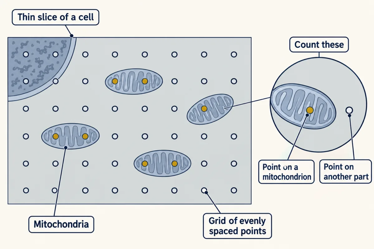
Every job needs the team. But how much of a cell is each part? You can't pour a cell into a measuring cup. So how do scientists find out how much of a cell is mitochondria? First, they preserve the cell and cut a very thin slice. A transmission electron microscope sends a beam of electrons through the slice. It shows small organelles that a school microscope cannot. Because the cell was preserved, each picture is a still view, not a living cell. Next comes a simple trick called point counting. Lay an even grid of dots over the picture. Then look at where each dot lands. Dots that land on mitochondria turn gold. One slice gives only a rough guess.
Over many fairly chosen pictures, the share of gold dots estimates the share of the cell's volume that mitochondria fill. Try it on a small grid. Count the gold dots, then count all the dots. The share appears on the screen. This toy grid lands inside the real range for heart muscle. Real studies used many pictures and many more dots. Look at heart muscle cells from ten kinds of mammals.
Mitochondria filled about a fifth to over a third of the cell, depending on the kind of mammal. Now drop the same grid on a white fat cell. Before the dots turn gold, predict. Will its share be bigger or smaller than the heart cell's?
Much smaller. Almost every dot lands on fat. One team laid a grid of 682 points over pictures of white fat cells from chicken embryos. Fat droplets filled about nine tenths of the cell. Mitochondria filled less than one percent. The fat takes most of the floor. Everything else, mitochondria included, sits in a thin rim around it. Is that rim short of mitochondria? The lesson lets you work it out. So in the heart cell, mitochondria fill a large share. In the fat cell, stored fat fills most of the space. Many of the same parts, different floor plans. One caution. These cells came from different animals and different studies. They show a pattern across cells with different jobs.
They do not prove what causes it. Still, simple counting, repeated over many pictures, turned images into numbers. Those numbers show a pattern: cells with different jobs have different floor plans.
From the film
- Scientists lay an even grid of dots over a picture of a thin cell slice and count the dots on each part.
- Over many fairly chosen pictures, the share of dots on mitochondria estimates the share of the cell's volume they fill.
- Mitochondria filled about a fifth to over a third of heart muscle cells in ten kinds of mammals.
- In white fat cells from chicken embryos, fat droplets filled most of the space, and mitochondria less than 1%.
- These measurements show a pattern across cells with different jobs, not proof of what causes it.
Elaborate: same parts, different amounts
The second film compared reported measurements for heart muscle and white fat cells; here are six more. First, test the pattern on one.
Predict. A guinea pig pancreas cell builds digestive enzymes, which are proteins, and sends them out of the cell. Will its mitochondria fill as large a share as in heart muscle, or less?
Check your prediction
Less: about 8.1%, below the whole heart range. Its job leans on making and moving protein, and the same study found that rough ER, the part of the ER where many proteins are made, filled about 22% of these cells.
Each block is about 2 percentage points of the cell’s volume (one percentage point is one step on the percent scale, such as 8% to 9%). In the heart row, solid blocks show the low end of the range, and pale blocks reach the high end. The bars are rounded, but the numbers are exact.
| Cells measured | Mitochondria (% of cell volume) | Graph |
|---|---|---|
| Hummingbird flight muscle (rufous hummingbird) | 34.5 | █████████████████ |
| Heart muscle, 10 kinds of mammals | 22 to 37 (range) | ███████████░░░░░░░░ |
| Human arm muscle, elite skiers | 9.0 | ████▌ |
| Human leg muscle, elite skiers | 8.6 | ████▌ |
| Guinea pig pancreas cell that makes digestive enzymes | 8.1 | ████ |
| Chicken brown-like fat cell, grown in a dish for 22 days | 4.2 | ██ |
| Chicken white fat cell, embryo | 0.7 | ▏ |
| Human red blood cell, mature and healthy | 0 (none) | (no bar) |
- Looking only at rows with a single value, which measured cell had the largest share of mitochondria? Which had the smallest share above zero? The longest single bar is ___. The shortest bar above zero is ___.
- Find the mean of the two skier values (add them, then divide by 2). Skier muscle averages ___%, which is ___ than the heart range.
- How many percentage points apart are the hummingbird muscle and the pancreas cell? The hummingbird muscle share is ___ percentage points higher.
- Which listed cells have the longest bars? Describe one pattern, then give one reason the table alone cannot show that a cell’s job caused it. The longest bars belong to ___. The table cannot prove cause because ___.
The studies measured the space mitochondria filled, not their number. Why is the white fat cell’s bar so short? Going deeper works it out.
Claim, evidence, reasoning
Make a claim that answers the essential question: do a cell’s parts match its job? Use the table.
Start with: The data show that cells with blank jobs have blank amounts of mitochondria.
Start with: Mitochondria filled blank% of blank but only blank% of blank.
Start with: This makes sense because mitochondria blank, and a cell that blank needs more of that.
Compare with a model answer
Cells with different jobs give different amounts of space to mitochondria. Mitochondria filled 34.5% of hummingbird flight muscle and 22% to 37% of heart muscle. They filled only 0.7% of chicken white fat cells. Heart muscle never stops, and hummingbird flight muscle powers very fast wingbeats. Both release a lot of energy from food, which is the mitochondria's job. Elite skiers' muscles work hard but not nonstop, and they had far less, about 9%. Fat cells mostly store fat. The fat cell share is small partly because fat droplets take most of the space. One limit: the rows come from different studies, species and methods. So the data show a pattern, but they do not prove what causes it.
Saved in this browser only.
Design problem
Does the spacing of a counting grid change your answer? Plan a test using a printed electron microscope picture of a cell and clear grids you draw yourself.
Saved in this browser only.
One way to do it
- Question: Does grid spacing change the measured share of a cell filled by mitochondria?
- Independent variable: spacing between grid points, such as 10 mm, 5 mm and 2 mm.
- Dependent variable: percent of points that land on mitochondria.
- Constants: the same printed picture, the same rule for a point on an edge, and the same lighting.
- Control: the finest grid (2 mm) is the comparison for the others.
- Repeated trials: place each grid in three different spots on the picture and count each time. Find the mean for each spacing.
- Safety: use a ruler and pencil only; no sharp tools needed.
- What it means: if the coarse grid gives values far from the fine grid, it needs more points to be trusted.
Evaluate: check your thinking
Back to the hook: the films and the table answer it with the same pattern, many shared parts in very different amounts. Two examples show the pattern; they do not make it a rule for every cell.
Science practices you used: using a model and naming its limits, reading a data table and a graph, finding a mean and a difference, and planning a fair test with repeated trials.
Going deeper
The fat cell’s thin rim
Predict. In the white fat cells, fat droplets filled about 90.9% and mitochondria about 0.7%. In the part that is not fat, do mitochondria fill closer to 1% or to the pancreas cell’s 8%?
Check your prediction
Closer to 8%. The part that is not fat is about 9.1% of the cell, and 0.7 out of 9.1 is about 7.7%. This is a rough estimate that assumes both numbers come from the same cells. The tiny share mostly reflects the fat taking the space.
Where the numbers come from
Sources: Mathieu-Costello and colleagues, 1992 (hummingbird); Barth and colleagues, 1992 (heart, ten species including humans); Ørtenblad and colleagues, 2018 (skiers, Norway); Bolender, 1974 (pancreas, five guinea pigs); Mezentseva and colleagues, 2008 (chicken cells). The red blood cell row is a textbook fact, not a measurement. Most rows come from different studies; the two skier rows share one study, and so do the two chicken rows.
Your notes
Select any text in the lesson to highlight it or add a note to it. Your highlights and notes are saved in this browser and collected in your notebook.
This browser is not letting the site save anything (a private window or strict privacy settings), so highlights and notes will be lost when you leave the page.
How we measure it
Transmission electron microscope
This microscope sends a beam of electrons through a very thin slice of a cell, so it shows small organelles and details of organelles that a school microscope cannot show. The cells are preserved first, so each picture is a still view, not a living cell.
Point counting
A grid of evenly spaced points is laid over a microscope picture, and you count how many points land on each part. Over many fairly chosen pictures, the share of points that land on mitochondria estimates the share of the cell's volume they fill.
Where this lesson fits
Before thisReview first
- Cells Are EverywhereThis course · Lesson 1
It introduces cells as the basic units of living things, which this lesson opens up to look at their parts.
After thisStudy next
- Plant Cells and Animal CellsThis course · Lesson 3
It uses the organelles you just met to show what plant cells add, like the cell wall and chloroplasts.
- Diffusion and OsmosisThis course · Lesson 5
It takes the cell membrane's job of controlling what enters and leaves and explains how water and gases cross.
- Releasing Energy from FoodThis course · Lesson 10
It takes the mitochondria's job of releasing energy from food and shows how cellular respiration does it.
Elsewhere on the siteFilms, simulations and method films in other courses
- One Cell, Many JobsFilm · Biology
It shows how a single cell does every life job using its own parts.
- Division of labourSimulation · Biology
It lets you see cells sharing out jobs among their parts.
Suggested by an AI model from the lessons themselves, then checked. How we use AI
Finish line
Test yourself
8 questions
Practice and MCAT-style, with explained answers.
Next · Lesson 3
Plant Cells and Animal Cells
Why does a limp stalk of celery get crisp again in water, when drinking water does not make you any stiffer?
Continue →