Life Science · Unit 1: Cells
Cells Are Everywhere
Rewritten on : new text, flashcards and film. Your notes and highlights stay, but the text around them may have moved.
How can a drop of pond water look empty to your eyes and still be full of living things, all made of cells?
Pause when the reading reaches a film, and offer to play it. Saved on this device.
Watch the film now, or keep listening
Builds on:Alive or Not?Why these?
Where this lesson sits
- Virginia SOL LS.2 a
- Practices LS.1 b, LS.1 c, LS.1 d, LS.1 f
Essential question: How do we know something is alive if it is too small to see?
-
Life Science (grade 7) LS.2 a You are here
This lesson
-
Biology
Preview it at high school level
In Biology you will learn more ideas that are added to the cell theory. Three of them say that the chemical reactions of life, called metabolism, happen inside cells; that DNA, the molecule that carries traits, is passed from cell to cell; and that all cells are built from the same basic kinds of materials. You will also learn about resolution, the smallest gap at which two points still look separate. The eye resolves about 0.1 mm, the best standard light microscopes about 0.2 µm, and an electron microscope much finer. Those numbers explain why some cell parts were seen only with newer tools.
-
Biology II Biology II: Advanced Survey of Biology Topics, BIOII.2 a
Preview where it goes next
Virginia's Biology II: Advanced Survey of Biology Topics course (BIOII.2 a) treats the cell as a system. Its parts, such as the nucleus, mitochondria, ribosomes, endoplasmic reticulum, Golgi apparatus and cytoskeleton, work together and help keep conditions inside the cell steady. Students study how each part's job depends on the others. Some of these parts, and the fine details inside the rest, are too small for a light microscope, so much of what we know about them came from electron microscopes and other newer tools.
-
College
 ▶ Watch The Size Test · 3:03
Read the transcript of “The Size Test”
Film transcript: The Size Test
This drop of pond water looks empty. Is it? And if tiny living things swim in it, why can't your eyes see them? To answer, we need one test of size. Start with a metric ruler. Its smallest marks are one millimetre apart. Now split one millimetre into a thousand equal parts. Each tiny part is one micrometre (µm). Your eyes have a limit. Most people cannot make out anything smaller than about 100 µm, a tenth of a millimetre. We will draw that limit as a blue line, the eye line. Now build a size ladder. Each rung is ten times bigger than the rung below it. We place real cells on the ladder at their real sizes. A frog egg is one cell, often about a millimetre across.
It sits above the eye line, so you can see it. A human egg, about one hundred micrometres, sits right on the line. A green alga, a red blood cell and a bacterium all sit below it. Most cells are somewhere between about one and one hundred micrometres. That puts most of them below the eye line, too small to see without help. A microscope uses lenses to make small things look larger. Magnification is how many times larger the picture looks than the real thing. So the picture looks as big as the real size times the magnification. Turn the dial up, and every picture slides up the ladder together. The cells do not change. Only their pictures grow.
Watch the red blood cell. At 40x, its picture is already over 300 µm. It has crossed the eye line. Now the green alga, about 20 µm wide. Set the dial to just 10x. Will its picture cross the eye line? Yes. Its picture is 200 µm, twice the eye line. The red blood cell, at the same 10x, still sits below the line. A school microscope magnifies 40, 100 or 400 times. At 400x, most cells pass the eye line. Then you can see their shapes, if the lens shows them clearly and they stand out. So a clear drop is not always empty. Most living things in pond water sit below the eye line, where our eyes cannot reach. That is why most cells stayed hidden until microscopes were made.
From the film
- Most people can make out a thing only when it looks bigger than about 100 µm, a tenth of a millimetre. That limit is the eye line.
- Most cells are about 1 to 100 µm across, so most are too small to see without help.
- A microscope makes things look bigger: picture size = real size × magnification.
- At 400x, a school microscope lifts most cells past the eye line. You see their shapes if the lens shows them clearly and they stand out.
A spoonful of pond water can look completely clear. Put one drop under a microscope, and you may see tiny living things swimming and turning. Why did your eyes miss them? And how can anyone know something is alive when it is too small to see?
What do you think? Decide before you read.
-
Scientists disagree: Clear water can hold living things too small for your eyes, and a school microscope often shows them. Not seeing something does not prove it is absent.
What scientists think
Scientists disagree: Clear water can hold living things too small for your eyes, and a school microscope often shows them. Not seeing something does not prove it is absent.
-
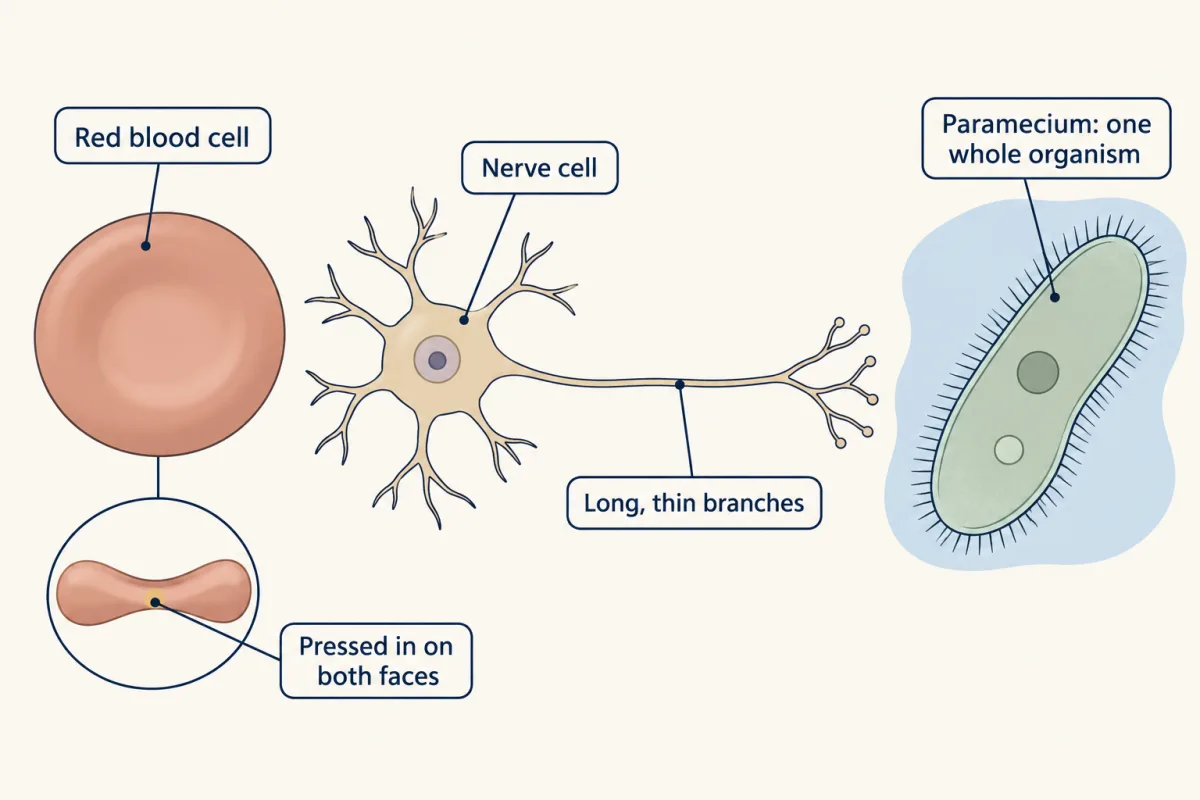
Scientists disagree: A human red blood cell is a disc about 8 micrometres (thousandths of a millimetre) across, pressed in on both faces. A Paramecium, a one-celled pond organism, is about 200 micrometres long. A frog egg is one cell, about 1 millimetre across in many kinds of frogs.
What scientists think
Scientists disagree: A human red blood cell is a disc about 8 micrometres (thousandths of a millimetre) across, pressed in on both faces. A Paramecium, a one-celled pond organism, is about 200 micrometres long. A frog egg is one cell, about 1 millimetre across in many kinds of frogs.
-
Scientists disagree: In science, a theory is a well-tested explanation backed by a great deal of evidence. The cell theory rests on hundreds of years of observations, and it was corrected when new evidence appeared.
What scientists think
Scientists disagree: In science, a theory is a well-tested explanation backed by a great deal of evidence. The cell theory rests on hundreds of years of observations, and it was corrected when new evidence appeared.
The one idea. A microscope makes things look bigger: picture size = real size × magnification, where picture size is how big a thing looks. At 40x, a cell looks about as big as something 40 times wider seen by eye alone. At a normal reading distance, most people can make out something only when it is bigger than about 100 micrometres (µm), or 0.1 mm. We call that limit the eye line.
Scope: passing the eye line is needed, but it is not enough. To see a cell’s shape, the lens must also show the detail clearly, and the cell must stand out from its background (a stain helps). Light itself sets a limit too; see Where light runs out.
How small is too small to see?
The first film left out the lenses that do the multiplying. A school light microscope has an eyepiece, the lens you look into, that magnifies 10 times (10x), and objective lenses just above the slide, often 4x, 10x and 40x. Multiply the two to get the total magnification: 10 × 4 = 40x, 10 × 10 = 100x and 10 × 40 = 400x. The slide sits on the stage, the light source shines up through it, and the focus knob sharpens the picture.
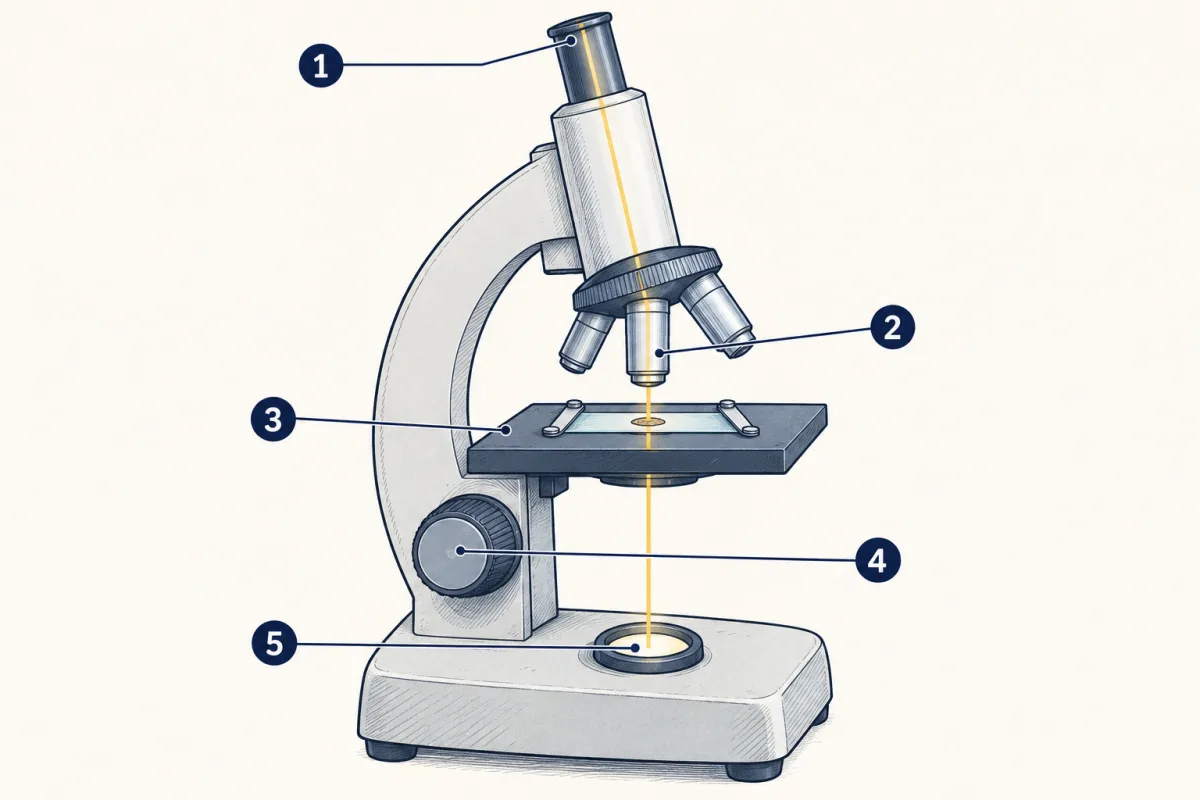
- eyepiece
- objective lenses
- stage
- focus knob
- light source
- eyepiece
- blank
- stage
- blank
- blank
Word bank: focus knob, light source, objective lenses
- blank
- blank
- blank
- blank
- blank
Check your labels
- eyepiece
- objective lenses
- stage
- focus knob
- light source
Use it safely and well:
- Carry it with two hands, one under the base and one on the arm.
- Start with the lowest power objective and focus.
- Move what you want to see to the center, then switch to a higher power.
- At higher power you see a smaller circle of the slide. This circle is the field of view.
Try baker’s yeast, a cell about 5 µm wide. At 40x its picture is 5 × 40 = 200 µm. It passes the size test but is only a speck; its oval shape shows at 400x.
Predict. A bacterium is about 2 µm long. Will its picture pass the eye line at 40x? At 100x?
Check your prediction
At 40x: 2 × 40 = 80 µm, under the eye line, so no. At 100x: 2 × 100 = 200 µm, so it passes the size test, and the simulation shows it. In a real microscope an unstained bacterium is still hard to make out at 100x; that is the “not enough” part. Bacteria are usually viewed at higher power, often stained.
Predict. A Paramecium is about 200 µm long. By the size test, can you see it with your eyes alone? Then why did the drop look clear?
Check your prediction
The size test says yes: 200 µm is twice the eye line. But a Paramecium is nearly colourless, a faint speck that is easy to miss: the “stand out” part of the rule. Most other living things in the drop are under 100 µm, too small for the eye at all.
Explore: zoom into a drop
The simulation zooms from your eye through a hand lens to a light microscope at 40x, 100x and 400x, on four samples, using only the size test. Watch the field of view and the scale bar, a line that shows a length in micrometres.
First, pick a sample. Then press a bigger tool to zoom in.
- Total magnification
- Circle of view, across (in this sketch)
- Smallest object shown (in this sketch)
In this sketch: an object shows when its size × the magnification is at least 100 µm, the eye’s limit. For cells, the narrower side is used. Circle sizes, the 10x hand lens, the onion, cork and cheek cell sizes, and the spots shown are sketch values. Pond contents are a sketch. There is no focusing or staining.
Keep this rule: an object shows only if it is bigger than the smallest size the tool can show, and higher power shows a smaller circle of the slide.
Try this.
- Pick pond water and look with your eye only. Predict how many kinds of living thing you will see. Then zoom to 100x and count again.
- Before you switch from 100x to 400x, predict what will happen to the field of view and the scale bar. Then test it.
- Look at onion skin, then cork, at the same power. Both show boxes. Note one way they differ inside.
What the microscope showed: living things are made of cells
- organism
- cell
- unicellular
- multicellular
- cell theory
- microscope
- magnification
- micrometre
- theory
An organism is one living thing, such as a tree, a mushroom, a dog or a bacterium. A cell is the smallest unit that can carry out the processes of life.
Some organisms, such as a Paramecium, are just one cell: they are unicellular. Others, such as you and an oak tree, are multicellular, made of many cells. Tiny does not always mean one cell: some animals in pond water are too small to see well, but they are made of many cells. You will sort living things this way in that lesson.
Cells are not all alike: a red blood cell is a small disc, and a nerve cell can have long, thin branches.
Most cells also have smaller parts inside, called organelles; you will meet them in that lesson.
The cell theory sums up what scientists have learned about cells in three parts:
- All living things are made of one or more cells.
- The cell is the basic unit of life.
- All cells come from cells that already exist.
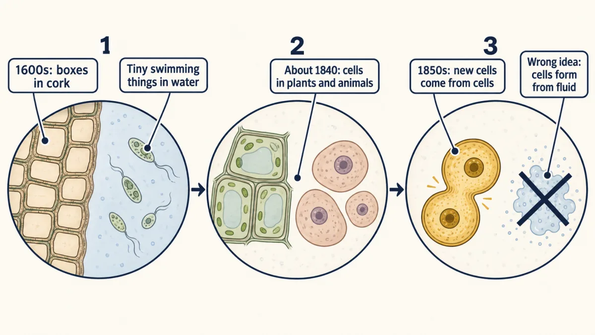
Learning this took about two hundred years and better tools, starting with cork boxes like the ones you just zoomed into.
Read the transcript of “A Theory Built in Steps”
Film transcript: A Theory Built in Steps
You now know the three parts of the cell theory. But how did people come to know them? Seeing cells was only the first step. It started with a slice of cork. In 1665, Robert Hooke published a book of drawings made with his microscope. He cut a very thin slice of cork and shone light on it through a lens. He saw tiny spaces with thin walls between them, like a honeycomb. He called them cells. Under his lens, the boxes crossed the eye line. Cork comes from the bark of a tree. Look closely at the empty boxes. Were Hooke's boxes living cells, or something else?
Something else. The cork cells were dead. Only their walls were left, so Hooke saw the outside of cells, not the living parts inside them. In a letter dated 1676, Antonie van Leeuwenhoek described drops of water. He used small microscopes with a single tiny lens. Water from his well looked clear, yet he reported many tiny living things in it. He checked one dish of rainwater again and again. At first he found nothing alive in it. Then one day he saw a few tiny creatures, and the next day more. When a view was not clear, he said so and did not count it. His careful records showed that seeing nothing is not the same as nothing being there. Microscopes kept getting better.
By about 1840, scientists had looked at many plants and animals, and found cells in them. That was strong evidence for the first part of the theory. But where do new cells come from? At first, some scientists thought new cells formed out of a clear fluid, a bit like growing crystals. Then a new cell would need no parent cell at all. That idea had to be tested by looking harder. More observations, including work published in the 1850s, showed that new cells form when a cell divides. One cell becomes two. The theory dropped the wrong idea and kept the rest. The three parts did not arrive all at once. Many steps came after a better tool or a more careful look.
Each one added evidence, and the cell theory grew from it.
From the film
- In 1665 Robert Hooke saw tiny boxes in a thin slice of cork. They were the empty walls of dead cells.
- Leeuwenhoek's careful records showed that seeing nothing does not prove nothing is there.
- By about 1840, scientists had found cells in many plants and animals, strong evidence for the first part of the theory.
- Later work, including work published in the 1850s, showed that new cells form when a cell divides, not from a clear fluid. The theory dropped the wrong idea and kept the rest.
What makes it a theory
The second film followed the evidence; why call the result a theory? In everyday talk, a theory can mean a hunch. In science, a theory is a well-tested explanation backed by a great deal of evidence. A theory can still change if strong new evidence appears; the clear-fluid idea in the second film is one example.
You will see how one cell becomes two in that lesson.
Where light runs out
Predict. Both films stopped at the light microscope. If you could keep magnifying far past 400x, would it keep showing new, smaller parts of a cell?
Check your prediction
Not forever. Light itself blurs details that are very close together. Even the best standard light microscope has ordinary glass lenses. It cannot show two details apart if they are closer than about 0.2 µm. Past that point, more magnification only makes a bigger blur, not new detail. Special super-resolution microscopes get around this limit.
So the size test is only the first check. A school microscope shows most whole cells, but not the smallest parts inside them. For those, scientists built the electron microscope, which uses a beam of electrons instead of light; it added new evidence about what cells are made of.
How big are cells?
Here are real cells in size order, with the two limits drawn in as rows.
| Cell, or line | Size (µm) | What was measured |
|---|---|---|
| Frog egg | about 1,000 | width, textbook estimate |
| Paramecium caudatum | about 200 | length, a usual size |
| Human egg cell | about 100 | width, textbook estimate |
| Eye line | about 100 | smallest thing you can make out |
| Green alga (Haematococcus), swimming form | 20 | width |
| Human red blood cell | 7.7 | width |
| Baker’s yeast cell | about 5 | width, a typical value |
| Bacterium (E. coli) | about 2 | length |
| Best standard light microscopes | about 0.2 | smallest gap at which two details still look separate |
Treat the values as examples of scale, not exact numbers to memorize.
- Which living things in the table sit above the eye line? Which sit below it? Which is about equal?
- At 40x, which cell in the table still has a picture smaller than 100 µm?
- How many times wider is a red blood cell than the 0.2 µm light-microscope limit? Divide 7.7 by 0.2.
- A frog egg is about 1,000 µm across. How many millimetres is that?
Claim, evidence, reasoning
Make a claim that answers the question: how do we know something is alive if it is too small to see? Use the table.
Start with: Most cells in the table are blank than the eye can see, so blank.
Start with: For example, a blank is blank µm, but the eye can make out only things larger than about blank µm.
Start with: This means scientists needed blank to study these cells, because blank.
Compare with a model answer
Most cells in the table are too small for the eye, so we know about them from microscopes. A red blood cell is 7.7 µm wide and a bacterium is about 2 µm long. Both are far below the eye's limit of about 100 µm. Both are bigger than the 0.2 µm limit of the best standard light microscopes. So a strong enough lens can show them, especially if a stain makes them stand out. A few cells, like a frog egg, are big enough to see. One limit: the values come from different labs and books, not one sample, and some are rough estimates.
Saved in this browser only.
Evaluate: check your thinking
In two sentences: why did the pond drop look clear, and how do we know the things in it are alive?
One good answer
Most living things in the drop are smaller than about 100 µm, and the few larger ones, such as a Paramecium, are nearly colourless and easy to miss. Careful study of such living things shows that each one is made of one or more cells, and better microscopes gave the evidence the cell theory grew from.
Design problem
Compare samples from living things, which should show cells, with materials that are not made of cells. Plan an investigation with a school light microscope.
Saved in this browser only.
One way to do it
- Question: Do the samples from living things show cells, while the others do not?
- Independent variable: the type of sample. From living things: onion skin, a thin slice of potato, and a drop of pond water that holds living things. Not made of cells: a few grains of clean sand, a few pure salt crystals and a scrap of plastic wrap.
- Dependent variable: whether you see cells, and how many cells you can count in one field of view.
- Constants: the same microscope, magnification (100x, then 400x) and light for every slide.
- Blank control: a clean slide with only the distilled water used on the other slides, so you can tell what comes from the slide or water.
- Repeated trials: three slides of each sample, each counted by two students.
- Measure: find the field of view width in mm with a clear metric ruler at low power, and change it to µm. Divide that width by the number of cells that fit across the field to estimate one cell’s size.
- Safety: slides and cover slips are glass; tell your teacher about any broken glass. Wash your hands after touching pond water. Wear goggles if a stain is used. Do not taste anything.
- One surprise to think about: cork and most paper are not alive now, yet cork shows boxes and paper shows long fibres. Both are walls left by plant cells. Not alive now is not the same as never alive.
Going deeper
Where the cell sizes come from
The bacterium, yeast, blood cell and alga values come from the BioNumbers database at Harvard Medical School. Its values come from lab studies, a cell size database and a textbook, published from 1982 to 2009. None come from pond samples. The egg and eye values come from an OpenStax textbook (2016). The Paramecium length is the usual size of Paramecium caudatum in species descriptions. The light microscope limit comes from a University of Arizona biology tutorial (2004).
The exact measurements
The E. coli length in the study was 1.94 to 2.72 micrometres. A second kind of yeast, fission yeast, was about 4 micrometres wide (3.74 micrometres in the study). The usual length of Paramecium caudatum ranges from 170 to 330 micrometres.
Science practices you used: using a microscope safely and measuring in metric units, reading a data table and doing math with it, planning a fair test with a control, and explaining why a theory is not a guess.
Your notes
Select any text in the lesson to highlight it or add a note to it. Your highlights and notes are saved in this browser and collected in your notebook.
This browser is not letting the site save anything (a private window or strict privacy settings), so highlights and notes will be lost when you leave the page.
How we measure it
Light microscope
Glass lenses bend light to make a thin sample look many times larger, usually 40 to 400 times in a school microscope. The best standard light microscopes can show two details apart down to about 0.2 micrometres; special super-resolution microscopes go finer. A school microscope shows less fine detail, but enough to see most whole cells.
Electron microscope
A beam of electrons, not light, forms the image. It shows parts of cells far too small for an ordinary light microscope, so it added new evidence about what cells are made of.
Where this lesson fits
Before thisReview first
- Alive or Not?Biology Together · Ages 5–10, with a grown-up
You need to know what living things do before asking whether a tiny speck in pond water is alive.
After thisStudy next
- Inside a CellThis course · Lesson 2
It takes you inside the cell to the organelles this lesson only mentions.
- How Cells DivideThis course · Lesson 4
It shows how one cell becomes two, the evidence for the third part of the cell theory.
- From Cells to OrganismsThis course · Lesson 6
It sorts living things into one-celled and many-celled and shows how cells share jobs in a body.
Elsewhere on the siteFilms, simulations and method films in other courses
- What a Microscope Can ShowFilm · Biology
It shows magnification and resolution, which set how small a thing a light microscope can show.
Suggested by an AI model from the lessons themselves, then checked. How we use AI
Finish line
Test yourself
8 questions
Practice and MCAT-style, with explained answers.
Next · Lesson 2
Inside a Cell
Why is a heart muscle cell crowded with mitochondria, while a fat cell is mostly one big drop of fat?
Continue →