Organismal Biology · Skim version
Blood Vessels, Blood and Lymph
Read the full lesson → Skim the whole course →
The guiding idea
Blood moves slowest in the narrowest vessels in your body. How can narrower mean slower?
Fluid flows down a pressure difference, at a rate set by the resistance in the way; across a capillary wall, the difference that counts is blood's push outward minus the plasma proteins' pull inward.
From the film: Downhill in Pressure
- Along a level vessel with no pump inside, flow equals the pressure difference divided by the resistance in the way.
- Equal pressure at both ends, however high, drives no flow.
- The arterioles hold most of the resistance, and their smooth muscle lets them change it: in an ideal tube, resistance rises with the fourth power of a falling radius.
- A single capillary can resist more than an arteriole, but so many lie side by side that together they resist less.
- In a simplified steady-state tree where no blood bypasses the capillaries, the same flow crosses every level, so blood is slowest where the total cross-section is largest: the capillaries.
From the film: Landis's Blocked Capillary
- Across a capillary wall, the driving pressure is the blood's push outward minus the plasma proteins' pull inward, each a difference between inside and outside the wall.
- Landis blocked single frog capillaries and timed the drift of trapped red cells to measure fluid crossing the wall.
- Filtration rose roughly in proportion to capillary pressure and reversed near the oncotic pressure of frog plasma.
- His experiment did not show that fluid keeps returning at the venous end; in most tissues, lymph returns it.
-
Each wall fits its job
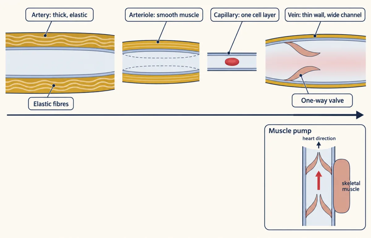
Each wall fits its job: arteries recoil to keep pressure up between beats, arterioles set resistance with smooth muscle, capillaries exchange across one layer of endothelial cells, and compliant veins hold most of the blood, with valves and muscles pushing it toward the heart. AI-generated illustration -
Numbers on the 1/r4 lever
-
Starling’s balance in numbers
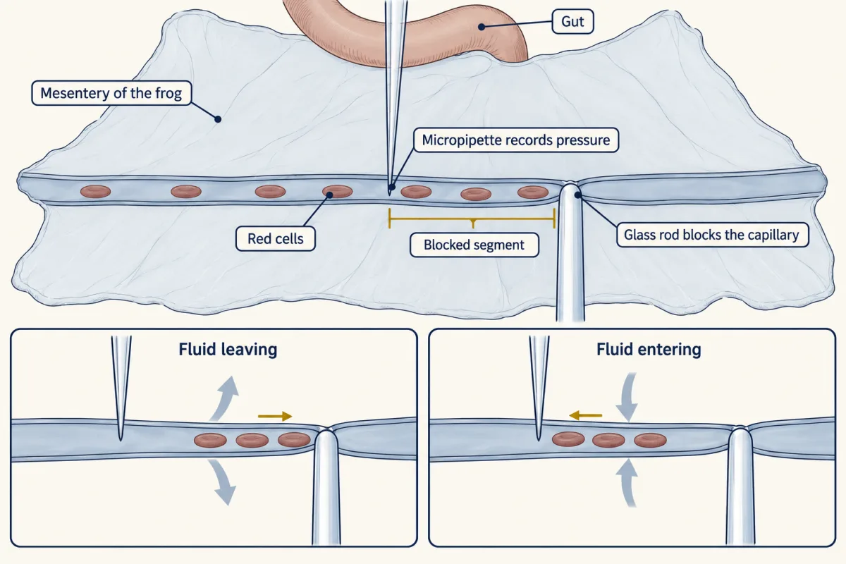
Trapped red cells work as a flow meter: their drift gives the rate of filtration at the pressure the pipette records. AI-generated illustration -
Lymph: the return route
-
Blood and stopping a leak, compared across animals
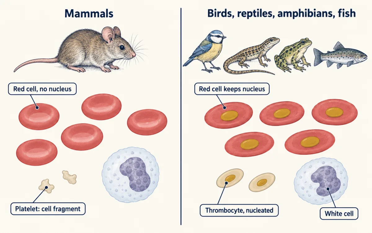
Mammals have red cells without a nucleus and platelets; almost all other vertebrates keep the red-cell nucleus and use nucleated thrombocytes instead of platelets. AI-generated illustration -
Going deeper
Where this lesson fits
Before thisReview first
- Circulation and the HeartThis course · Lesson 10
You need the closed circuit and the heart's output from there to follow flow through arteries, capillaries and veins.
- Size, Scale and SurfaceThis course · Lesson 2
Surface area and volume reasoning from there explains why a huge total capillary area suits exchange.
- Tissues, Organs and HomeostasisThis course · Lesson 6
The tissue types, including smooth muscle and endothelium, come from there and are used in each vessel wall here.
After thisStudy next
- Gas Exchange and BreathingThis course · Lesson 12
Gases cross thin capillary walls in the lungs, using the slow flow and large area shown here.
- The KidneyThis course · Lesson 16
The Starling forces here reappear as the filtration pressure across the glomerular capillary wall.
- Carrying Oxygen and Carbon DioxideThis course · Lesson 13
It picks up the red cells and hemoglobin introduced here and shows how blood carries oxygen.
Suggested by an AI model from the lessons themselves, then checked. How we use AI